Indian Leishmaniasis
Susmita Ghosh,aPriyanka Banerjee,bAvijit Sarkar,aSimanti Datta,band Mitali Chatterjeea
Department of Pharmacologyaand Centre for Liver Research, School of Digestive and Liver Diseases,bInstitute of Post Graduate Medical Education and Research, Kolkata, India
Leishmania donovaniis considered the causative organism of visceral leishmaniasis (VL) and post-kala-azar dermal leishmania-sis (PKDL). Testing of 4/29 DNA samples from VL and PKDL patients as well as 2/7 field isolates showed an aberrant internal transcribed spacer 1 (ITS1) restriction fragment length polymorphism (RFLP) pattern, which upon sequencing strongly matchedLeptomonas seymouri, thus confirming its presence in Indian leishmaniasis.
V
isceral leishmaniasis (VL) is a vector-borne disease caused byreplication of parasites of theLeishmania donovanicomplex
(L. donovaniandL. infantum) within the macrophage-phagocytic system. In the Indian subcontinent and parts of Africa, its trans-mission is anthroponotic (2), with post-kala-azar dermal leish-maniasis (PKDL) being a sequel of VL, and is characterized by a macular, maculo-papular, or nodular rash (11).
Generally, in the Indian subcontinent, patients presenting with clinical features suggestive of VL/PKDL have the diagnosis con-firmed by the presence of parasites in Giemsa-stained smears and/or culture positivity, serological diagnosis (enzyme-linked immunosorbent assay [ELISA] or rK39 strip test), and, rarely, by molecular approaches. The causative parasites are assumed to be
L. donovani, although in recent years, studies have shownL. don-ovanicausing cutaneous leishmaniasis in Sri Lanka (18).
Addi-tionally, a lower trypanosomatid,Leptomonas seymouri, has been
detected in isolates from patients with VL, but not in clinical spec-imens (19).
Molecular diagnosis of leishmaniasis is often by PCR that typ-ically targets the internal transcribed spacer 1 (ITS1), separating the genes coding for small subunit (SSU) rRNA and 5.8S rRNA (7). Additionally, isolates have been characterized by restriction fragment length polymorphism (RFLP) analysis of the ITS1 re-gion (9) or the gene fragment encoding the 70-kDa heat shock protein (hsp70) (13), the latter being among the first kinetoplastid genes to be cloned and characterized due to their conserved nature (10). Upon routine diagnosis of patients with suspected VL or PKDL by ITS1 PCR, a different band pattern was reported that did
not match the classicalL. donovaniWHO reference strain DD8
(MHOM/IN/1980/DD8) (7). Accordingly, this study was under-taken to study the RFLP patterns of clinical specimens sourced from patients with VL or PKDL along with archived parasite iso-lates from a different patient population.
The study population included 29 patients from 1 January 2010 to 31 January 2012 who were admitted to the School of
Trop-ical Medicine, Kolkata, with clinTrop-ical features of VL (n⫽23) or
PKDL (n⫽6). Clinical materials included peripheral blood from
patients with VL or lesional skin biopsy specimens from patients with PKDL after obtaining informed consent. The diagnosis of VL/PKDL was confirmed by rK39 strip test (20), ELISA for
anti-leishmanial antibodies, and PCR of the ITS1 region ofLeishmania
sp (7). The study received approval from the Institutional Ethical Committee of the School of Tropical Medicine, Kolkata, India,
and Institute of Postgraduate Medical Education and Research, Kolkata, India.
In addition, our study included archivedLeishmaniaisolates
(n⫽7; V1 to V5, P1, and P2), obtained from patients with VL
(n⫽5) or PKDL (n⫽2); all except V5 presented at the School of
Tropical Medicine between 2006 and 2011. In patients with VL, parasites were isolated from spleen/bone marrow aspirates (7), while for PKDL, a 3-mm punch biopsy specimen from a nodule was collected in medium 199 (M199) supplemented with 20% heat-inactivated fetal calf serum (FCS), penicillin G (50 IU/ml),
and streptomycin (50g/ml). The material was passed through a
230-m sterile iron mesh and finally resuspended in 1.5 ml of the
same medium, and after incubation at 24°C, culture growth was evident after 5 to 10 days. After transformation from amastigotes to promastigotes, they were gradually adapted into M199 supple-mented with 10% FCS, penicillin G (50 IU/ml), and streptomycin
(50g/ml) and subcultured every 2 to 3 days, the inoculum being
1⫻106/ml. When parasites reached the range of 107, they were
cryopreserved (approximately 1⫻107parasites per cryo vial) in
freezing medium (M199 containing 30% FCS and 7.5% dimethyl sulfoxide [DMSO]).
All of the archived strains were typed by ELISA using
species-specificL. donovanimonoclonal antibody (5) and PCR-RFLP (9).
For PCR, DNA following isolation from peripheral blood, skin biopsy specimens, and isolates (QIAamp DNA minikit; Qiagen,
Hilden, Germany) was eluted in 200l elution buffer. Different
parts ofLeishmaniawere amplified, namely (i) ribosomal ITS1 (9)
and (ii) hsp70 (13). Amplification reactions were performed in 25
l of mixture (JumpStart REDTaq ReadyMix reaction mix;
Sig-ma-Aldrich Chemicals, St. Louis, MO) in a Master cycler (Eppen-dorf, Hamburg, Germany). The amplified ITS1 and hsp70 regions were digested using HaeIII (Fermentas, Glen Burnie, MD); briefly,
reactions were carried out using 1 U of HaeIII, 1⫻buffer, and 5l
Received15 April 2012Returned for modification7 May 2012 Accepted10 May 2012
Published ahead of print23 May 2012
Address correspondence to Mitali Chatterjee, ilatim@vsnl.net, or Simanti Datta, seemdatt@gmail.com.
Copyright © 2012, American Society for Microbiology. All Rights Reserved.
doi:10.1128/JCM.00966-12
on May 16, 2020 by guest
http://jcm.asm.org/
of the amplicon (approximately 100g of DNA) and incubated at 37°C for 3 h (for ITS1) or overnight (for hsp70). The digested product was analyzed by electrophoresis (3% agarose, 5 V/cm for 1.5 h) along with a 100-bp DNA ladder or GeneRuler low-range DNA ladder (Fermentas, Glen Burnie, MD) and visualized in a G-BOX Gel Doc system (Syngene, Cambridge, United Kingdom) using Gene Tools software (version 4.01.04).
For sequencing of archivedLeishmaniaisolates, PCR products
of the ITS1 region were purified (QIAquick gel extraction kit; Qiagen, Hilden, Germany) and then cloned into the pJET1.2 vec-tor by blunt end ligation (CloneJET PCR cloning kit; Fermentas, Glen Burnie, MD). Recombinant plasmid DNA was used to trans-formEscherichia coliDH5␣; eight colonies with an ITS1 insert were selected for each sample. Plasmid DNA was purified from colonies using a Qiagen plasmid minikit (Qiagen, Hilden, Ger-many) and sequenced (BigDye Terminator v3.1 cycle sequencing kit; Applied Biosystems, Foster City, CA) on an automated DNA sequencer (ABI Prism 3130, Foster City, CA). DNA sequence ed-iting and analysis were performed using Seqscape V2.5 software (Applied Biosystems, Foster City, CA).
The reference sequences of the ITS1 gene from several trypano-somatid species were retrieved from GenBank and aligned with the sequence determined in this study (http://www.ncbi.nlm.nih .gov/GenBank/index.html) using ClustalW software and a phylo-genetic tree constructed by the neighbor-joining method using MEGA version 5.0 (21).
Blood was sourced from patients with VL (n⫽23) and lesional
skin biopsy specimens from patients with PKDL (n⫽6) (Table 1);
58.62% of patients hailed from Bihar, India (17/29), and among them, 11 (64.70%) were from zones with antimonial resistance (17). Of the remaining 12 patients, 11 were from West Bengal, India, and one was from Chhattisgarh, India, whose areas of anti-monial resistance, if any, have not been defined.
Analysis of the ITS1 PCR products of these 29 patients showed two distinct trends, namely (i) a single 320-bp amplicon in 86.2% (19 VL and 6 PKDL) of samples that matched the reference strain
DD8 and (ii) dual bands of 320 and 418 bp in 13.8% (4 patients with VL). Examination of the RFLP pattern of the 320-bp product revealed a pattern similar to that of DD8, having 3 fragments with sizes of 191, 75, and 54 bp, defined as “pattern A.” With regard to the 4 samples having a dual band pattern, each band was gel ex-tracted, purified, and digested separately with HaeIII; the 320-bp product had an RFLP profile similar to that of DD8 (i.e., pattern A), while the larger PCR product of 418 bp remained undigested by HaeIII and was defined as “pattern B.”
Among the seven archived isolates studied, five were obtained from bone marrow/splenic aspirates of patients with VL (V1 to V5), while two were from dermal tissue of patients with PKDL (P1 and P2). The majority of these patients (5/7, except V1 and V3) hailed from Bihar, the main zone of endemicity for VL in India; among them, two (V2 and P1) were from areas of antimonial resistance and three (V4, V5, and P2) were from an area having no antimonial resistance (17). The remaining two archived isolates (V1 and V3) were isolated from a patient each from West Bengal and Assam, respectively, whose patterns of antimonial resistance, if any, are yet to be defined.
All archived strains showed strong binding with D2, anL.
don-ovanispecies-specific monoclonal antibody (12), and the absor-bances obtained were comparable with that obtained with DD8
(MHOM/IN/1980/DD8), the L. donovani reference strain;
ac-cordingly, they were typed asL. donovani. To further characterize
these archived isolates, we performed ITS1 RFLP and found two variations in the PCR products (Fig. 1, inset) that were verified by HaeIII digestion. RFLP data showed that pattern A was dominant, being present in 5/7 isolates (71.4%), and pattern B was present in 2 isolates (28.6%) (Fig. 1), akin to the profile obtained in clinical specimens. This lack of digestion by HaeIII has not been reported previously in leishmaniasis and suggests unusual variations in the
[image:2.585.40.287.76.295.2]sequence of the ITS1 region amongLeishmaniastrains. Although
TABLE 1Clinical features of the study population
Feature
Result for patients with:
VL (n⫽23) PKDL (n⫽6)
Age (yr)
Mean⫾SD 30.7⫾19.2 31.6⫾16.6 Median (range) 25.5 (1–70) 25 (13–57)
Male/female ratio 16/7 5/1
History of VL (%) NAa 83.3
Interval between cure of VL and onset (yr)
Mean⫾SD NA 7.2⫾2.1 Median (range) NA 6 (5.5–10)
Spleen size (cm)
Mean⫾SD 10.1⫾7.9 NA
Range 3–29 NA
Liver size (cm)
Mean⫾SD 4.4⫾2.9 NA
Range 2–10 NA
a
NA, not applicable.
FIG 1RFLP analysis of the ITS1 region amplified fromLeishmania donovani isolates. Lanes: 1, DD8; 2, V1; 3, V2; 4, V3; 5, V4; 6, V5; 7, P1; 8, P2; M, low-range DNA ladder. (Inset) PCR assay of the ITS1 region fromLeishmania donovaniisolates. Lanes: M, 100-bp ladder; 1, PCR control (water); 2, DD8; 3, V1; 4, V2; 5, V3; 6, V4; 7, V5; 8, P1; 9, P2.
on May 16, 2020 by guest
http://jcm.asm.org/
[image:2.585.303.541.456.675.2]some sequence variations in the ITS1 region between strains ofL. donovanihave been reported, (9,16), a difference of 100 bp in the PCR product has not been reported to date.
To substantiate our findings, we performed hsp70 PCR-RFLP with our archived isolates. Once again, two patterns emerged: i.e.,
5 isolates matched theL. donovanireference strain, whereas 2
iso-lates (V5 and P2 which showed pattern B for ITS1 RFLP) showed another pattern (data not shown). The latter pattern was not
com-parable with any otherLeishmaniaspecies (13), but it was similar
to that reported in 9 Indian isolates (19). In the clinical specimens, the hsp70 PCR did not yield any product (data not shown).
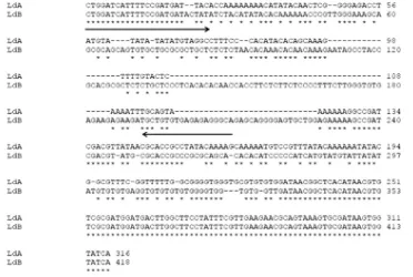
In order to identify the organism from which this aberrant ITS1 gene was being amplified, the nucleotide sequence of the 418-bp fragment was determined, wherein we selected DD8 and P2 as representatives of patterns A and B, respectively; P2 was selected as it had undergone very few passages following transfor-mation. We aligned two sequences denoting one sequence, “LdA,” forL. donovaniDD8, representative of pattern A and denoting the other, “LdB,” for P2, representative of pattern B; the ClustalW alignment of the two sequences showed several mismatches and deletions (score, 73), indicating there were significant differences between them (Fig. 2A). To determine whether these sequence
variants were due toTaqpolymerase errors, two colonies of the
variant were selected for a second round PCR using the same primers and resequenced; they were all identical to the original sequence, confirming that the observed sequence variant was not a technical error. Furthermore, two sequences were BLAST searched independently, where LdB showed a strong match with
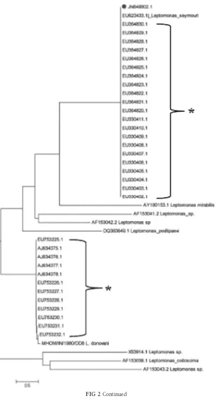
Leptomonas seymouri(accession no.EU623433.1). Based on the sequence of the ITS1 region of P2 and DD8, a neighbor-joining
tree was constructed that included 33L. donovaniIndian isolates,
available at National Centre for Biological Information (http: //www.ncbi.nlm.nih.gov), and an extended set of organisms (Fig.
2B). The tree showed that P2 was most closely related to
Leptomo-nas seymouri(score, 99.0) (Fig. 2B) along with 21L. donovani
Indian isolates. The remaining 12L. donovaniIndian isolates were
phylogenetically closely related to DD8 (Fig. 2B) (http://www .ncbi.nlm.nih.gov/nuccore/EU364830). Additionally, sequence analysis of these 33 isolates showed that the size of the ITS1 PCR
product of the 21 isolates that matchedLeptomonas seymouriwas
418 bp, while the size of the 12 Indian archived isolates that matched with DD8 was 320 bp. Importantly, no HaeIII restriction site was present in the 418-bp sequence, corroborating our obser-vations (Fig. 1).
To confirm the presence ofLeptomonas seymouriin the clinical
specimens, we designed a reverse primer from a 30-bp portion (nucleotide positions 197 to 227) unique to this organism. It was selected based on it being the inserted sequence present in the ITS1 region of P2 following alignment of P2 and DD8 (Fig. 2A).
The forward primer (5=CTGGATCATTTTCCGATGATACTAT
3=) was designed from the common sequence (bp 1 to 25) between
P2 and DD8, while the reverse primer was 5=TGCCCTCTCTCA
CACAGCA 3=; a partial ITS1 region was amplified at an annealing
temperature of 60°C for 30 s. Among the archived isolates, P2 and V5, which showed pattern B gave a 210-bp product, confirming
these strains wereLeptomonas seymouri. We propose that after
parasite transformation, Leptomonas seymouri outgrewL.
don-ovaniasLeptomonasspecies have been reported to grow faster thanL. donovani(19). In patient DNA, 4 samples appeared
coin-fected withL. donovaniandLeptomonas seymourias they gave the
FIG 2Determination of the presence ofLeptomonassp. (A) Alignment of ITS1 sequences amplified fromLeishmania donovaniisolates representing RFLP patterns A and B. LdA,Leishmania donovanishowing RFLP pattern A; LdB, isolate showing RFLP pattern B.*,matching of nucleotide; -, absence of nucleotide. (B) Neighbor-joining tree based on p-distances of the ITS1 sequences ofLeishmania donovaniand P2. Outgroup,Leptomonassp. The analysis is based on an alignment of 1,234 nucleotides. Distances are measured along the horizontal branches, according to the scale shown. Bootstrap values above 70% are indicated at the internodes.*,sequence ofL. donovaniisolates of India collected from GenBank;●, sequence of P2.
on May 16, 2020 by guest
http://jcm.asm.org/
[image:3.585.104.476.65.315.2]210-bpLeptomonasITS1 PCR product; additionally, 2 more sam-ples from patients with VL showed a 210-bp product (data not shown); none of the patients with PKDL showed a 210-bp band.
Based on this analysis, we conclude that clinical specimens (4/ 29) isolated from patients with VL/PKDL were concomitantly
in-fected withLeptomonas seymourias also were two archived culture
isolates among seven studied; importantly, they phylogenetically
clustered more closely to the monoxenous parasiteLeptomonas
seymouri. The occurrence of insect trypanosomatids in humans is exceptional, but reports are available that HIV-positive patients are additionally infected with nonpathogenic insect trypanosoma-tids (6). In Brazil, Pacheco et al. (15) described a flagellate,
appar-FIG 2Continued
on May 16, 2020 by guest
http://jcm.asm.org/
[image:4.585.138.446.62.635.2]ently a monoxenous trypanosomatid, in a 35-year-old HIV-posi-tive male who presented with symptoms of VL. Hybridization analyses, against a panel of many different trypanosomatids, re-vealed that the unknown flagellate had kinetoplastid DNA
(kDNA) cross-homology only withLeptomonas pulexsimulantis, a
parasite of a dog flea (18). However, the presence of lower trypanosomatids in immunocompetent individuals is a matter of greater concern (4). Our patients had no evidence of HIV infec-tion (testing negative for HIV), yet four of them were coinfected withLeptomonas seymouriandL. donovani. Additionally, Leish-maniacoinfections, including with HIV (3),Plasmodium vivax
(1), orMycobacterium tuberculosis(8), have been reported.
There-fore, it may be envisaged that as VL induces a strong immunosup-pression, it possibly allows nonhuman trypanosomatids to be in-stalled in mammalian hosts.
In this study, 13.8% (4/29) patients with VL/PKDL were
coin-fected withLeptomonas seymouriandL. donovani. Interestingly,
on analysis of the isolates reported in GenBank asL. donovani,
21/33 (i.e., 63.63%) are actually Leptomonas seymouri; in this
study, 28.57% (2/7) areLeptomonas seymouri. Nasereddin et al.
(14) reported about 35.59% of Indian isolates obtained from pa-tients with VL were unidentified by reverse line blot hybridization
assay usingL. donovani-specific probes, but had a ITS1 sequence
similarity toLeptomonas seymouri. The appearance of this
oppor-tunistic infection byLeptomonas seymouriraises questions about
the clinical relevance of this pathogen. However, to date, studies pertaining to the pathobiology of these opportunistic lower trypanosomatids infecting humans have been limited.
As this study had a substantial number of patients coming from
zones of antimonial resistance, it raises the possibility that
Lepto-monasstrains are possibly less sensitive to antimony. Thein vitro
susceptibility toward antimony of both monoxenous trypanoso-matid field isolates P2 and V5 was lower than those of the other 5 strains (M. Chatterjee, personal communication), which raises
the possibility of the potential contribution ofLeptomonas to the
growing incidence of unresponsiveness to antimonials reported from the Indian subcontinent; however, this must be
substanti-ated in a larger study group to conclude whetherLeptomonas
in-fections influence the epidemiology, pathology, or case manage-ment of VL. Taking these findings together, this study emphasizes the importance of estimating the extent of opportunistic patho-gens in leishmaniasis.
Nucleotide sequence accession number.The sequence deter-mined in this study has been submitted to GenBank and is
avail-able under accession no.JN848802.
ACKNOWLEDGMENTS
DD8 and V5 were kindly provided by Lionel F. Schnur, Department of Parasitology, Hadassah Medical School, Jerusalem, Israel, and Neeloo Singh, Central Drug Research Institute, Lucknow, India, respectively.
This work received financial assistance from the Indian Council of Medical Research (ICMR), Department of Biotechnology and Depart-ment of Science & Technology, GovernDepart-ment of India.
REFERENCES
1.Ab Rahman AK, Abdullah FH.2011. Visceral leishmaniasis (kala-azar) and malaria coinfection in an immigrant in the state of Terengganu, Ma-laysia: a case report. J. Microbiol. Immunol. Infect.44:72–76.
2.Alvar J, Canavate C, Molina R, Moreno J, Nieto J. 2004. Canine leishmaniasis. Adv. Parasitol.57:1– 88.
3.Andreani G, Lodge R, Richard D, Tremblay MJ.2012. Mechanisms of interaction between protozoan parasites and HIV. Curr. Opin. HIV AIDS
7:276 –282.
4.Boisseau-Garsaud AM, et al.2000. A new case of cutaneous infection by a presumed monoxenous trypanosomatid in the island of Martinique (French West Indies). Trans. R. Soc. Trop. Med. Hyg.94:51–52. 5.Chatterjee M, et al.1998. Distribution of IgG subclasses in antimonial
unresponsive Indian kala-azar patients. Clin. Exp. Immunol.114:408 – 413.
6.Chicharro C, Alvar J.2003. Lower trypanosomatids in HIV/AIDS pa-tients. Ann. Trop. Med. Parasitol.97:75–78.
7.Das NK, et al.2011. Case series of misdiagnosis with rK39 strip test in Indian leishmaniasis. Am. J. Trop. Med. Hyg.84:688 – 691.
8.el-Safi SH, et al.2004. Infection rates withLeishmania donovaniand Mycobacterium tuberculosisin a village in eastern Sudan. Trop. Med. Int. Health9:1305–1311.
9.El Tai NO, Osman OF, el Fari M, Presber W, Schönian G.2000. Genetic heterogeneity of ribosomal internal transcribed spacer in clinical samples ofLeishmania donovanispotted on filter paper as revealed by single-strand conformation polymorphisms and sequencing. Trans. R. Soc. Trop. Med. Hyg.94:575–579.
10. Fraga J, Montalvo AM, De Doncker S, Dujardin JC, Van der Auwera G.
2010. Phylogeny ofLeishmaniaspecies based on the heat-shock protein 70 gene. Infect. Genet. Evol.10:238 –245.
11. Ganguly S, Das NK, Barbhuiya JN, Chatterjee M.2010. Post-kala-azar dermal leishmaniasis-an overview. Int. J. Dermatol.49:921–931. 12. Jaffe CL, McMahon-Pratt D.1983. Monoclonal antibodies specific for
Leishmania tropica.I. Characterization of antigens associated with stage-and species-specific determinants. J. Immunol.131:1987–1993. 13. Montalvo AM, et al.2010. Heat-shock protein 70 PCR-RFLP: a universal
simple tool forLeishmaniaspecies discrimination in the New and Old World. Parasitology.137:1159 –1168.
14. Nasereddin A, Bensoussan-Hermano E, Schönian G, Baneth G, Jaffe CL.2008. Molecular diagnosis of Old World cutaneous leishmaniasis and species identification by use of a reverse line blot hybridization assay. J. Clin. Microbiol.46:2848 –2855.
15. Pacheco RS, et al.1998. Parasite genotypically related to a monoxenous trypanosomatid of dog’s flea causing opportunistic infection in an HIV positive patient. Mem. Inst. Oswaldo Cruz93:531–537.
16. Pandey K, et al.2007. Characterization ofLeishmaniaisolates from Nep-alese patients with visceral leishmaniasis. Parasitol. Res.100:1361–1369. 17. Perry MR, et al.2011. Visceral leishmaniasis and arsenic: an ancient
poison contributing to antimonial treatment failure in the Indian subcon-tinent? PLoS Negl. Trop. Dis.5:e1227. doi:10.1371/journal.pntd.0001227. 18. Siriwardana HV, Thalagala N, Karunaweera ND. 2010. Clinical and epidemiological studies on the cutaneous leishmaniasis caused by Leish-mania(Leishmania)donovaniin Sri Lanka. Ann. Trop. Med. Parasitol.
104:213–223.
19. Srivastava P, et al.2010. Detection ofLeptomonassp. parasites in clinical isolates of Kala-azar patients from India. Infect. Genet. Evol.10:1145– 1150.
20. Sundar S, Reed SG, Singh VP, Kumar PC, Murray HW.1998. Rapid accurate field diagnosis of Indian visceral leishmaniasis. Lancet351:563– 565.
21. Tamura K, et al.2011. MEGA5: molecular evolutionary genetics analysis using maximum likelihood, evolutionary distance, and maximum parsi-mony methods. Mol. Biol. Evol.28:2731–2739.