Journal of Chemical and Pharmaceutical Research, 2016, 8(5):273-282
Research Article
CODEN(USA) : JCPRC5
ISSN : 0975-7384
Design, synthesis and biological evaluation of some novel Schiff base
derivatives as potential anticancer agents
Mohammed T. Elsaady
1, Ahmed M. Gouda
1, Fadwa H. Edrees
2*and Nagwa M. Abdel Gawad
31Department of Medicinal Chemistry, Faculty of Pharmacy, Beni-Suef University, Beni-Suef 62514, Egypt 2Department of Medicinal Chemistry, faculty of Pharmacy, Nahda University, Beni-Suef 62514, Egypt 3Pharmaceutical Chemistry Department, College of Pharmacy, Cairo University, Cairo 11562, Egypt _____________________________________________________________________________________________
ABSTRACT
Novel series of pyrrolizine Schiff bases has been synthesized then biologically evaluated as potential anticancer agents. The starting compounds,7-cyano-6-amino-N-(4-(un)substituted-phenyl)-2,3-dihydro-1H-pyrrolizine-5-carboxamides 17a–c, were reacted with different aldehydes to give the target compounds 18-20. Structural characterizations of the novel compounds were performed using spectral and elemental analysis. The anticancer activity of these compounds was evaluated using Sulforhodamine-B (SRB) assay method .All of these compounds showed anticancer activity against bothHEPG2 and MCF7 cancer cell lines comparable to that of the standard Doxorubicin (HEPG2 IC50 =0.00699µM/ml). Most of compounds are more active against (MCF7) than (HEPG2)
cell lines. Compound 18c showed the highest anticancer activity with IC50 value 0.250µM/ml against (MCF7).While, Compound18b was the most potent one against liver (HEPG2) with IC50 value 0.784 µM/ml.
Modeling studies into ATP binding site of EGFR tyrosine kinase were done to predict their scores and mode of interaction with amino acid residues. Furthermore selectivity of the prototypes(18-20a) on normal Wish cell was evaluated and showed IC50 of 0.946, 1.322 and 1.122 respectively.
Keywords: Pyrrolizine; Schiff base; Anticancer; EGFR tyrosine kinase; EGFR-TK inhibitor
_____________________________________________________________________________________________
INTRODUCTION
Cancers represent one of the most complicated health problems in the world [1].The higher rate of mortality due to cancers cancer, in addition to of multidrug resistance to some of the currently used anticancer agents [2-5] present an urgent need for development of effective and safe anticancer agents. Pyrrolizines were recently reported as promising scaffold for the design of potent anticancer agents. The dual COX/LOX inhibitor Licofelone 1 showed potent anticancer activities against several cell lines [6-8]. Exploring the mechanism of action of Licofelone1 revealed its ability to induce apoptosis [9-10]. The tripentone (MR22388) 2 showed strong anticancer activity against leukemia L1210 with IC50 of 15 nM. In addition to its ability to act as tubulin polymerization inhibitor
[11-12]; MR22388 was found also to acts as a very strong inhibitor for several kinases [13]. Recently, we have reported compound3 as potent anticancer agents with IC50 in the range of 0.98 and 1.12 µM against [14] . The ureido
Fig.1.pyrrolizine derivatives with potent anticancer activities
Identification of molecular targets involved in proliferation, malignancy and cell death were helpful in the rational based design of new anticancer agents. Protein kinases are one of these targets which play an important role in regulation of cellular proliferation, differentiation and survival [15]. Several kinases have become relevant therapeutic targets for development of new anticancer agent. The epidermal growth factor receptor (EGFR-TK) play an important role in promoting cell division and survival [16], and it is frequently over-expressed in tumors and it is associated with progression and resistance of cancer cell to anticancer drugs[17]. Several EGFR inhibitors erlotinib
4and Gefitinib 5were approved for treatment of cancer displaying high rate of response and high efficacy in
treatment of cancer [18,19].But recently, resistance to EGFR inhibitors was developed [20], and development of new EGFR-TK inhibitors became a must to overcome this problem.
Representative quinazoline, pyrrolopyrimidine, thienopyrimidine, thiazole and pyrrole-based EGFR-TK inhibitors [21-25] sharing some pharmacophoric groups indicated in red and blue colors, Fig. 2.Threefragments (A, B and C) were identified in the squares.
Benzene ring replacement in the quinazoline-based EGFR-TK inhibitory compounds with the isosteric pyrrole/thiophene afforded new compounds retaining EGFR-TK inhibitory activity. Additionally, no loss of activity observed on removal of the pyrimidine ring as indicated by sunitinib11 and dasatinib12Fig. 2.
In the present work we aimed to design a new pyrrolizine derivatives bearing some of the three fragments. It was of interest to develop compound 18a by combining fragments A, B and C in one scaffold with some modification, Fig.
3. Several derivatives of compound 18awere prepared through replacement of the 2-chloro group with electron
withdrawing (4-bromo) and electron donating (4-dimethyamino) groups. Moreover, substitution the phenyl ring at C-5 was done using the electron donating (4-CH3), and electron withdrawing (4-Cl) substituents in order to explore
the electronic effects of these substituents on activity of the produced compounds.
Fig.3. Design strategy and structural modification of compound 18a
RESULTS AND DISCUSSION
2.1. Chemistry
As shown in Scheme 1, preparation of the intermediates 14 and 16a-cwas done according to previously reported procedures [26,27]Compounds 17a-c were synthesized from the reaction of 2-pyrrolidin-2-ylidine malononitrile 14 with the corresponding acetanilide 16a-c in dry acetone according to previously reported procedures [28].
Scheme 1
Synthesis of the Schiff base derivatives 18-20was done by refluxing the starting materials 17a-c with the appropriate aldehydes in absolute ethanol in the presence of glacial acetic acid as catalyst.
Preparation of compounds18a-c was obtained by refluxing the starting material 17a-c with 2-chlorobenzaldehyde in absolute ethanol in the presence of glacial acetic acid. Structural elucidation of compounds 18a-c was done using spectral and elemental analysis. The IR spectra of 18a-c revealed absorption bands at 3230-3274 cm-1 attributed to the NH groups, a sharp band at2212-2217 cm-1 due to the cyano groups and absorption bands at1662-1667 cm-1 for the carbonyl groups. The 1H-NMR spectra of 18a-c showed singlet signal at δ 2.25 ppm due to the CH3 protons in
compound 18b, multiplet, and two triplets at the range ofδ 2.50-4.59 ppm assigned for the aliphatic protons of the three methylene groups of the pyrrolizine nucleus. Multiplet at the range of δ 7.16-8.19 ppm due to the aromatic protons, two singlet signals at the δ 9.32-10.71 ppm due to N=CH and NH protons.13C-NMR spectra of
compounds 18a-c revealed two signals at δ 156.42-158.35 ppm due to N=CH and C=O carbons. Mass
spectra of compounds 18a-c revealed the molecular ions at 388, 402 and 422 respectively.
Compounds 19a-c was prepared from the reaction of the starting material 17a-c with 4-bromobenzaldehyde. The IR spectra of 19a-c revealed absorption bands at 3231-3282 cm-1 attributed to the NH groups, a sharp band at 2212-2214 cm-1 due to the cyano groups and absorption bands at1661-1665 cm-1 for the carbonyl groups. The 1H-NMR
spectra of 19a-cshowed two singlet signals at the δ 9.00-10.60 ppm due to N=CH and NH protons. 13C-NMR spectra of compounds 19a-c revealed two signals at δ 157.82-158.52 ppm due to N=CH and C=O carbons. Mass spectra of compounds 19a-c revealed the molecular ions at 432, 446 and 466 respectively.
Scheme 2
Reagents and conditions: (d) 2-Chlorobenzaldehyde, absolute ethanol, glacial acetic acid, reflux, 4 h; (e) 4-bromobenzaldehyde, absolute
ethanol, glacial acetic acid, reflux, 4 h; (f) 4-dimethylaminobenzaldehyde, absolute ethanol, glacial acetic acid, reflux, 4 h.
Compounds 20a-c was prepared from the reaction of the starting material 17a-c with
4-dimethylaminobenzaldehyde. The IR spectra of 20a-c revealed absorption bands at 3432-3433cm-1 attributed to the NH groups, a sharp band at 2208-2210 cm-1 due to the cyano groups and absorption bands at1665-1666 cm-1 for the
carbonyl groups. The 1H-NMR spectra of20a-c showed two singlet signals at the δ 8.98-11.00 ppm due to N=CH
and NH protons. 13C-NMR spectra of compounds 20a-c revealed two signals at δ 158.87-159.59 ppm due to
2.2. Pharmacological screening 2.2.1. Anticancer activity
Cytotoxic activity of the novel pyrrolizines 18-20 were evaluated against HEPG2 and MCF7 cancer cell lines using Sulforhodamine-B (SRB) assay method [29]. IC50was calculated and represented in µM/mlin Table 1.The tested
compounds showed potent anticancer activity against both HEPG2 and MCF7 cell lines in micromolar range. Compounds18c and 18b are the most active ones against (MCF7) and (HEPG2) cell lines with IC50 values of 0.250 and 0.784µM/ml respectively.
2.2.2. Inhibitory activity against normal cells
[image:5.595.170.418.249.449.2]Cytotoxicty of the prototypes(18-20a) on normal Wish cells (non-tumorous cell line) was evaluated using Sulforhodamine-B (SRB) assay method [29] showed IC50 of 0.946, 1.322 and 1.122 respectively Table 1.
Table 1 IC50 values of compounds 18-20 against MCF-7, HEPG2 cancer cell lines and normal Wish cell
Comp. No. R1 R2
HEPG2 MCF-7 Wish cell IC50µM IC50 µM IC50 µM
18a H 2-Cl 7.839 0.856 0.946
18b CH3 2-Cl 0.784 0.843 -
18c Cl 2-Cl 1.668 0.250 -
19a H 4-Br 5.195 0.897 1.322
19b CH3 4-Br 3.27 0.323 -
19c Cl 4-Br 1.852 3.921 -
20a H 4-(CH3)2N 3.586 0.422 1.122
20b CH3 4-(CH3)2N 9.446 0.394 -
20c Cl 4-(CH3)2N 1.737 5.458 -
Doxorubicin - - 0.0069 - -
2.2.3. Docking study
In this work, a docking study was performed between the new pyrrolizines with EGFR-TK. This study aimed to understand the binding mode of the new pyrrolizines 18-20 with the active site of the EGFR-TK. Molecular docking studies were performed using MOE 2008.01, Table 2 and Fig. 4-6.
Table 2: Docking scores, interacting groups, amino acid interactions, and distances of the docked compounds into the active site of EGFR-TK
Comp S (Kcal/mol) Interacting moieties Amino acid Distance 18a -16.0343 N of CN pyrrolizine Lys860 Tyr764 4.35 3.2
18b -18.257 N of CN pyrrolizine
Lys860 Tyr764
2.23 4.31
18c -18.929 N of CN 2-chloro-benzylidene Lys860 Tyr764 2.77 4.68
19a -13.998 N of CN
4-bromo-benzylidene
Lys860 Lys757
2.88 3.79
19b -17.191 N of CN Lys860 3.2
19c -15.080 N of CN pyrrolizine
Lys860
Tyr764 4.043.2
20a -15.477 N of CN Lys860 2.87
20b -17.869 N of CN Lys860 2.94
20c -16.865 4-dimethylamino-benzylidene Lys860 4.38
AEE -21.442 NH of piperidine N of pyrimidine
Glu758 Lys860
1.23 3.27
[image:5.595.162.437.539.700.2]of-21.442kcal/mol and hydrogen bonding with Glu758 andLys860 through NH of piperidine moiety and N of pyrimidine respectively Fig. 4. All the compounds were docked into ATP binding site of EGFR kinase (PDB: 2J6M).[30]
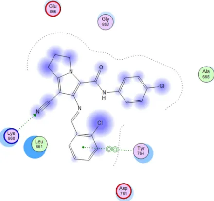
[image:6.595.195.395.227.557.2]All compounds were nicely and in a comparable manner of AEE 7 bound to the EGFR binding domain and form a hydrogen bond through the nitrogen atom of the cyano group with amino acid Lys860 which is an important binding site of EGFR inhibitor AEE 7as shown in Fig 4. As shown in Fig5-6, compounds18b and18c have a good interactions with EGFR, with the highest scores and less distance and this result was reliable with anticancer activity of these compounds which are the most active compounds against MCF-7 and HEPG2 cell lines.
Fig 4. 2D interactions of AEE ligand with EGFR
Fig 6. 2D interactions of comp 18c with EGFR
EXPERIMENTAL SECTION 4.1. Chemistry
Chemical reagents and solvents were obtained from commercial sources. Solvents are dried by standard methods when necessary. Melting points (m.p.) were uncorrected and were carried out by open capillary tube method using IA 9100MK-Digital Melting Point Apparatus. Microanalyses were carried out at the microanalytical Center, Faculty of Science, Cairo University. Infrared spectra were made on BRUKER Vector 22 (Japan), infrared spectrophotometer and were expressed in wavenumber (cm-1) using potassium bromide disc. The proton magnetic resonance1H NMR spectra were recorded on a Varian Mercury VX-300 NMR spectrometer at 400 MHz and
BRUKER APX400 spectrometer at 400 MHz in the specified solvent, chemical shifts were reported on the δ scale
and were related to that of the solvent and J values are given in Hz.13C NMR spectra were obtained on a Bruker APX400 at 100 MHz at the faculty of pharmacy, Beni-Suef University. Mass spectra were recorded on Fennigan MAT, SSQ 7000, Mass spectrometer, at 70 eV (EI) at themicroanalytical Center, Faculty of Science, Cairo University. All mass spectra were recorded in the EI mode. Thin layer chromatography, was done using Macherey Nagel Alugram Sil G/UV254 silica gel plates and benzene–ethanol (9.5:0.5) as the eluting system.
Compounds 14[26],16a-c[27],17a-c[28]were prepared according previously reported procedures.
4.2. General procedure for the preparation of compounds (18-20)
A mixture of the carboxamide derivatives 17(3.75 mmol) and the appropriate aldehyde (3.75 mmol) was refluxed in absolute ethanol (20 ml) in the presence of glacial acetic acid (0.5 ml) for 4 h. The reaction mixture was concentrated, set aside to cool, the formed crystals was collected and recrystallized from ethanol.
7-Cyano-6-[(2-chloro-benzylidene)-amino]-N-phenyl-2,3-dihydro-1H-pyrrolizine-5-carboxamide (18a):
Compound 18a was prepared by refluxing compound 17a with 2-chlorobenzaldehyde. The product obtained yellow crystals, m.p. 245-7 0C, yield 84%. IRʋmax/cm-1, 3230 (NH)3065 (C-H aromatic, 2907 (CH2), 2212 (CN),1665 (
7-Cyano-6-[(2-chloro-benzylidene)-amino]-N-p-tolyl-2,3-dihydro-1H-pyrrolizine-5-carboxamide (18b):
Compound 18b was obtained by refluxing compound 17b with 2-chlorobenzaldehyde. The product obtained yellow crystals, m.p. 265-6 0C, yield 87%. IRʋmax/cm-1, 3231 (NH ) 3093,3066 (C-H aromatic), 2856 (CH2), 2213 (CN
),1662 (C=O ) ,1595 (C=C ) ,1546 (C=N ) ,1462, 1403, 1257(C-N ) ,828,802 (C-Cl),1H-NMR (CDCl3-400 MHz):
δ2.25(s,3H,CH3Ph)2.50 (m, 2H, CH2-2), 2.99 (t, 2H, J = 7.2Hz, CH2-1), 4.55 (t, 2H, J = 6.8Hz, CH2-3), 7.12-7.38
(m, 8H, aromatic protons), 9.32 (s, 1H, N=CH), 10.14 (s,1H,NHC=O).13C-NMR (DMSOd6): 19.7, 20.9, 24.5, 40.58,
116.27, 118.19, 126.95, 129.67, 132.41, 133.06, 136.55, 148.39, 157.82, 158.07. Anal. Calcd. for C23H19ClN4O
(402.88). C, 68.57; H, 4.75; N, 13.91 Found: C, 68.71; H, 4.78; N, 14.08.
7-Cyano-6-[(2-chloro-benzylidene)-amino]-N-(4-chlorophenyl)-2,3-dihydro-1H-pyrrolizine-5-carboxamide (18c):
Compound 18c was obtained by refluxing compound 17c with 2-chlorobenzaldehyde. The product obtained yellow crystals, m.p. 273-6 0C, yield 81%. IRʋmax/cm-1, 3274 (NH ) 3097,3056 (C-H aromatic),2986, 2801 (CH2), 2217
(CN ) ,1667 (C=O ) ,1592 (C=C ) ,1544 (C=N ) ,1491, 1309, 1257(C-N ) ,829 (C-Cl),1H-NMR (CDCl3-400 MHz): δ
2.60 (m, 2H, CH2-2), 3.09 (t, 2H, J = 7.2Hz, CH2-1), 4.59 (t, 2H, J = 6.8Hz, CH2-3), 7.30-8.18 (m, 8H, aromatic
protons), 9.67 (s, 1H, N=CH), 10.71 (s,1H,NHC=O).13C-NMR (DMSOd6):24.60, 25.47, 50.18, 115.57, 118.11, 120.76,
127.24, 127.39, 128.87, 129.14, 130.76, 132.80, 133.18, 136.84, 137.02, 139.46, 148.56, 156.72, 158.34. Anal. Calcd. for C22H16Cl2N4O (423.29). C, 62.42; H, 3.81; N, 13.24 Found: C, 62.53; H, 3.79; N, 13.40.
7-Cyano-6-[(4-bromo-benzylidene)-amino]-N-phenyl-2,3-dihydro-1H-pyrrolizine-5-carboxamide (19a):
Compound19a was obtained by refluxing compound 17a with 4-bromobenzaldehyde. The product obtained yellow crystals, m.p. 223-5 0C, yield 82%. IRʋmax/cm-1, 3231 (NH ) 3062 (C-H aromatic, 2958,2848 (CH2), 2213 (CN
),1665 (C=O ) ,1596 (C=C ) ,1551 (C=N ) ,1474, 1413, 1315C-N ) ,770 (C-Br),1H-NMR (CDCl3-400 MHz): δ 2.51
(m, 2H, CH2-2), 2.92 (t, 2H, J = 7.2Hz, CH2-1), 4.74 (t, 2H, J = 6.8Hz, CH2-3), 7.16-7.60 (m, 9H, aromatic protons),
9.00 (s, 1H, N=CH), 10.41 (s,1H,NHC=O).13C-NMR (DMSOd6): 24.38, 25.29, 50.18, 116.20, 118.05, 119.32,
123.99, 127.06, 129.17, 129.78, 132.43, 134.12, 138.18, 138.50, 148.54, 157.09, 158.13, MS (EI): m/z (%): 434.85 (M+2,13.15), 433.85(M+1,49.06), 432.85 (M+,18.88), 431.9 (50.87), 341.85 (95.42), 340.85 (21.92), 339.85 (99.06),313.8 (5.69), 312.85 (4.09), 311.85 (7.03), 276.95(100), 92 (3.94), 91(4.9), 77(15.88), 65(19.83). Anal. Calcd. for C22H17BrN4O (433.3). C, 60.98; H, 3.95; N, 12.93. Found: C, 61.07; H, 3.99; N, 13.02.
7-Cyano-6-[(4-bromo-benzylidene)-amino]-N-p-tolyl-2,3-dihydro-1H-pyrrolizine-5-carboxamide (19b):
Compound19b was obtained by refluxing compound 17b with 4-bromobenzaldehyde. The product obtained yellow crystals, m.p. 240-2 0C, yield 80%. IRʋmax/cm-1, 3280 (NH ) 3070(C-H aromatic), 2968 (CH2), 2212 (CN ) ,1661
(C=O ) ,1611 (C=C ) ,1596 (C=N ) ,1418, 1315, 1292(C-N ) ,830,809 (C-Br),1H-NMR (CDCl3-400 MHz):
δ2.35(s,3H,CH3Ph)2.55 (m, 2H, CH2-2), 2.95 (t, 2H, J = 7.6Hz, CH2-1), 4.51 (t, 2H, J = 7.2Hz, CH2-3), 7.15-7.68
(m, 8H, aromatic protons), 9.01 (s, 1H, N=CH), 10.42 (s,1H,NHC=O).13C-NMR (DMSOd6):20.93, 24.43, 25.34,
50.17, 116.27, 118.19, 119.36, 126.99, 129.81, 132.41, 133.61, 134.21, 135.62, 138.38, 148.39 , 157.82, 158.07. Anal. Calcd. for C23H19BrN4O (447.33). C, 61.75; H, 4.28; N, 12.52 Found: C, 61.87; H, 4.32; N, 12.64.
7-Cyano-6-[(4-bromo-benzylidene)-amino]-N-(4-chlorophenyl)-2,3-dihydro-1H-pyrrolizine-5-carboxamide (19c):
Compound19c was obtained by refluxing compound 17c with 4-bromobenzaldehyde. The product obtained yellow crystals, m.p. 272-3 0C, yield 87%. IRʋmax/cm-1, 3282 (NH ) 3073(C-H aromatic),2968 (CH2), 2214 (CN ) ,1661
(C=O ) ,1611 (C=C ) ,1586 (C=N ) ,1418, 1315, 1298(C-N ) ,830,805 (C-Br),1H-NMR (CDCl3-400 MHz): δ 2.62 (m,
2H, CH2-2), 3.08 (t, 2H, J = 7.2Hz, CH2-1), 4.56 (t, 2H, J = 7Hz, CH2-3), 7.31-7.79 (m, 8H, aromatic protons), 9.16
(s, 1H, N=CH), 10.60 (s,1H,NHC=O).13C-NMR (DMSOd6):24.57, 25.44, 50.20, 116.08, 117.84, 120.70, 127.28,
128.93, 129.19, 129.85, 132.56, 134.22, 136.78, 138.95, 148.54, 158.31, 158.52. Anal. Calcd. for C22H16BrClN4O
(467.75). C, 56.49; H, 3.45; N, 11.98 Found: C, 56.64; H, 3.47; N, 12.13.
7-Cyano-6-[(4-dimethylamino-benzylidene)-amino]-N-phenyl-2,3-dihydro-1H-pyrrolizine-5-carboxamide (20a)
Compound20a was obtained by refluxing compound 17a with 4-dimethylaminobenzaldehyde. The product obtained yellow crystals, m.p. 250-1 0C, yield 79%. IRʋmax/cm-1, 3432 (NH ) 3065 (C-H aromatic, 2910 (CH2), 2209 (CN
),1665 (C=O ) ,1588 (C=C ) ,1537 (C=N ) ,1432, 1369, 1308(C-N ) ,1H-NMR (CDCl3-400 MHz): δ 2.52 (m, 2H,
CH2-2), 2.99 (t, 2H, J = 7.4Hz, CH2-1), 3.11(s,6H,N(CH3)2), 4.48 (t, 2H, J = 7Hz, CH2-3), 6.76-7.2 (m, 9H,
aromatic protons), 9.00 (s, 1H, N=CH), 10.91 (s,1H,NHC=O).13C-NMR (DMSOd6): 24.54, 25.36, 40.16,50.00,
158.96,159.58. MS (EI): m/z (%): 398 (M+1,28.46), 397 (M+,100), 306 (19.05), 305(92.42), 277.95(3.28), 277(13.6), 148.05(94.3), 92(2.9), 77(7.59), 65(5.55).Anal. Calcd. for C24H23N5O (397.47). C, 72.52; H, 5.83;
N, 17.62. Found: C, 72.68; H, 5.89; N, 17.84.
7-Cyano-6-[(4-dimethylamino-benzylidene)-amino]-N-p-tolyl-2,3-dihydro-1H-pyrrolizine-5-carboxamide (20b):
Compound20b was obtained by refluxing compound 17b with 4-dimethylaminobenzaldehyde. The product obtained yellow crystals, m.p. 258-9 0C, yield 81%. IRʋmax/cm-1, 3433 (NH ) 2914 (CH2), 2210 (CN ) ,1665 (C=O ) ,1581
(C=C ) ,1535 (C=N ) ,1475, 1370, 1307(C-N),1H-NMR (CDCl3-400 MHz): δ2.35(s,3H,CH3Ph)2.50(m, 2H, CH2-2),
3.01 (t, 2H, J = 7Hz, CH2-1), 3.11(s,6H,N(CH3)2) ,4.51 (t, 2H, J = 6.4Hz, CH2-3), 6.76-7.81 (m, 8H, aromatic
protons), 9.01 (s, 1H, N=CH), 10.86 (s,1H,NHC=O).13C-NMR (DMSOd6): 19.76, 20,91, 24.55,25.39, 40.16, 49.99,
111.66, 116.65, 16.81, 119.61, 120.19, 123.16, 129.53, 130.70, 133.07, 136.11, 140.95, 147.72, 153.12, 158.87, 159.45. Anal. Calcd. for C25H25N5O (411.5). C, 72.97; H, 6.12; N, 17.02 Found: C, 73.18; H, 6.19; N, 17.28.
7-Cyano-6-[(4-dimethylamino-benzylidene)-amino]-N-(4-chlorophenyl)-2,3-dihydro-1H-pyrrolizine-5-carboxamide (20c):
Compound20c was obtained by refluxing compound 17c with 4-dimethylaminobenzaldehyde. The product obtained yellow crystals, m.p. 262-5 0C, yield 85%. IRʋmax/cm-1, 3433 (NH ) 3045(C-H aromatic),2899 (CH2), 2208 (CN
),1666 (C=O ) ,1590 (C=C ) ,1536 (C=N ) ,1482, 1365, 1308(C-N ) ,1H-NMR (CDCl3-400 MHz): δ 2.58 (m, 2H,
CH2-2), 3.01 (t, 2H, J = 7.2Hz, CH2-1), 3.12(s,6H,N(CH3)2), 4.51 (t, 2H, J = 6.6Hz, CH2-3), 6.75-7.75 (m, 8H,
aromatic protons), 8.98 (s, 1H, N=CH), 11.00 (s,1H,NHC=O).13C-NMR (DMSOd6): 24.56, 25.33, 40.16, 49.97,
111.64, 116.28, 116.64, 120.63, 122.93, 128.21, 128.96, 130.70, 137.33, 141.24, 148.00, 153.19, 158.88, 159.59. Anal. Calcd. for C24H22ClN5O (431.92). C, 66.74; H, 5.13; N, 16.21 Found: C, 66.91; H, 5.16; N,
16.42.24,148.00,153.19,158.88,159.59. Anal. Calcd. for C24H22ClN5O (431.92).C, 66.74; H , 5.13 ; N, 16.21
Found C ,66.91 ; H, 5.16 ; N,16.42
4.2. Pharmacological screening
4.2.1. In vitro cytotoxic activity evaluation by SRB assay.
Cytotoxicity of the novel pyrrolizines (18-20) was evaluated against HEPG2 and MCF7 cancer cell lines using Sulforhodamine-B (SRB) assay method as previously reported by Skehan et al.[29]. Antitumor activity evaluation was completed at the Center for Genetic Engineering, Al-Azhar University, Cairo, Egypt. Reagents and chemicals were obtained from Sigma Aldrich Chemical Company (St. Louis, Mo, U.S.A.).The tested cell lines were obtained from the American Type Culture Collection (ATCC, Minnesota, USA) through the Tissue Culture Unit, The Egyptian Organization for Biological Products and Vaccines (Vacsera, Egypt).Cells were seeded for 24h in a 96 well microtiter plates at a concentration of 1000-2000 cells/well, 100 µl/well, then cells were incubated for 48 h with various concentrations ( 0, 6.25, 12.5, 25, 50, 100 µg/ml ) of the tested compounds, 3 wells were used for each concentration, after incubation for 48h the cells were fixed with 10% trichloroacetic acid 150 µl/well for 1 hr at 40C, washed by distilled water for 3 times. Wells were stained for 10-30 min at r.t. with 0.4% SRB, dissolved in 1% acetic acid 70 µl/well. Washed with acetic acid 1% to eliminate unbound dye till colorless drainage obtained. The plates were subjected to air drying, 24 hr not exposed to UV. The dye was solubilized with 150 µl/well of 10 mMTrise-EDTA (PH 7.4) for 5 min on a shaker at 1600 rpm. The optical density (OD) of each well was measured spectrophotometrically at 545 nm with an ELISA microplate reader. Survival curve was obtained by plotting the percent of surviving cells against different concentrations of the tested compounds. The IC50 values were calculated using sigmoidal concentration– response curve fitting models (Sigmaplot software).
4.3. Molecular docking
CONCLUSION
According to the results obtained during this work, we can conclude that:
(1) compounds with substituted phenyl ring are more active than the unsubstituted compounds. (2) Chlorosubstitued compounds are more active than methylsubstitued ones.
(3) compounds18band 18care the most active compounds against (HEPG2) and (MCF7) cancer cell lines respectively.
(4)Most of compounds are more active against (MCF7) than (HEPG2) cell lines.
Furthermore studies will be carried out to investigate the most probable mechanism of action of these compounds.
REFERENCES
[1] Jemal, F. Bray, M.M. Center, J. Ferlay, E. Ward, D. Forman, CA. Cancer J. Clin., 2011, 61, 69–90. [2] A.-M. Florea, D. Büsselberg, Cancers, 2011, 3, 1351–71.
[3] E.P. Simard, L.A. Torre, A. Jemal, Oral Oncol., 2014, 50, 387–403.
[4] P. Li, A. Znaor, I. Holcatova, E. Fabianova, D. Mates, M.B. Wozniak, et al., Eur. Urol., 2015, 67, 1134–1141. [5] M.M. Center, A. Jemal, J. Lortet-Tieulent, E. Ward, J. Ferlay, O. Brawley, et al., Eur. Urol., 2012, 61, 1079– 1092..
[6] S. Ghatak, A. Vyas, S. Misra, P. O’Brien, A. Zambre, V.M. Fresco, et al., Bioorganic. Med. Chem. Lett., 2014, 24, 317–324.
[7] W. Liu, J. Zhou, K. Bensdorf, H. Zhang, H. Liu, Y. Wang, et al., Eur. J. Med. Chem., 2011, 46, 907–913. [8] N.K. Narayanan, D. Nargi, M. Attur, S.B. Abramson, B. a. Narayanan, Anticancer Res., 2007, 27, 2393–2402. [9] S. Tavolari, M. Bonafè, M. Marini, C. Ferreri, G. Bartolini, E. Brighenti, et al., Carcinogenesis, 2008, 29, 371– 380.
[10] G. Kus, P. Oztopcu-Vatan, R. Uyar, S. Kabadere, Acta Biol. Hung., 2013, 64, 438–52.
[11] V. Lisowski, C. Enguehard, J.C. Lancelot, D.H. Caignard, S. Lambel, S. Leonce, et al., Bioorganic Med. Chem.
Lett., 2001, 11, 2205–2208.
[12] V. Lisowski, S. Léonce, L. Kraus-Berthier, J.S.D.O. Santos, A. Pierré, G. Atassi, et al., J. Med. Chem., 2004, 47, 1448–1464.
[13] C. Rochais, T. Cresteil, V. Perri, M. Jouanne, A. Lesnard, S. Rault, et al., Cancer Lett., 2013, 331, 92–98. [14] A.M. Gouda, A.H. Abdelazeem, E.-S. a Arafa, K.R. a Abdellatif, Bioorg. Chem., 2014, 53, 1–7.
[15] T. Ishikawa, M. Seto, H. Banno, Y. Kawakita, M. Oorui, T. Taniguchi, et al., J. Med. Chem., 2011, 54, 8030– 8050.
[16] N.E. Hynes, H.A. Lane, Nat Rev Cancer., 2005, 5, 341–354..
[17] P.Z. Gatzeva-Topalova, L.R. Warner, A. Pardi, M.C. Sousa, Structure,2010, 18, 1492–1501. [18] B.A. Chabner, Oncologist., 2004, 9, 245–246.
[19] C. Gridelli, F. De Marinis, M. Di Maio, D. Cortinovis, F. Cappuzzo, T. Mok, Lung Cancer, 2011, 71, 249–57. [20] L. Huang, L. Fu, Acta Pharm. Sin. B., 2015, 5, 390–401.
[21] M.J. Lavecchia, R. Puig de la Bellacasa, J.I. Borrell, C.N. Cavasotto, Bioorg. Med. Chem., 2015, 7, 768-778. [22] S. Mowafy, N.A. Farag, K.A.M. Abouzid, Eur. J. Med. Chem., 2013, 61, 132–145.
[23] A. Tarozzi, C. Marchetti, B. Nicolini, M. D’Amico, N. Ticchi, L. Pruccoli, et al., Eur. J. Med. Chem., 2016, (2016).
[24] S. Yin, L. Zhou, J. Lin, L. Xue, C. Zhang, Eur. J. Med. Chem., 2015, 101, 462–475.
[25] H.-Q. Zhang, F.-H. Gong, C.-G. Li, C. Zhang, Y.-J. Wang, Y.-G. Xu, et al., Eur. J. Med. Chem., 2016, 109, 371–379.
[26] A.Etienne, Y. Correia,Bull. Soc. Chem., 1969, 10, 3704-3712.
[27] W.A.Jacobs, M. Heidelberger, J. Am. Chem. Soc.1917, 39, 1435-1439.
[28] [1] A. Gouda, H. Ali, W. Almalki, M. Azim, M. Abourehab, A. Abdelazeem, Molecules, 2016, 21, 201. [29] P. Skehan, R. Storeng, D. Scudiero, A. Monks, J. McMahon, D. Vistica, et al., J. Natl. Cancer Inst., 1990, 82, 1107–1112.