INTRODUCTION
Intellectual disability (ID) is characterized by impairment of cognitive and adaptive functions, with onset before age 18 years. ID is usually identified during infancy or early child-hood because of developmental delay (DD). However, ID is formally diagnosed upon obtaining an intelligence quotient
(IQ) score of less than 70.1,2 ID occurs in approximately 1−3% of the population, and is largely caused by genetic abnormali-ties.1 However, it has extensive genetic heterogeneity, and 60% of the cases of ID still do not have a known etiology.3 ID is of-ten accompanied by clinical features of dysmorphism, congen-ital anomalies, or epilepsy. Chromosomal microarray (CMA), with a higher diagnostic yield (15−20%) than that of the G-banded karyotype (~3%), is now a first-titer clinical diagnostic test for individuals with unexplained DD/ID, autism, or mul-tiple congenital anomalies.4 CMA has a 100-fold higher reso-lution than G-banded karyotype, and detects submicroscopic copy number variants (CNVs) across the entire genome. In many cases, the detected CNVs are disease-causing genomic alterations, although benign variants or variants of uncertain significance (VOUS) are also frequent. Although CMA and clinical data have extended the spectrum of understanding the genetic causes of ID, interpretation thereof and appropriate management, including genetic counseling, remain problems Received: June 12, 2017 Revised: December 13, 2017
Accepted: January 19, 2018
Corresponding author: Dr. Hyon J. Kim, Department of Medical Genetics, Konyang University College of Medicine,158 Gwanjeodong-ro, Seo-gu, Daejeon 35365, Korea.
Tel: 82-2-523-9230, Fax: 82-2-581-9230, E-mail: [email protected] •The authors have no financial conflicts of interest.
© Copyright: Yonsei University College of Medicine 2018
This is an Open Access article distributed under the terms of the Creative Com-mons Attribution Non-Commercial License (http://creativecomCom-mons.org/licenses/ by-nc/4.0) which permits unrestricted non-commercial use, distribution, and repro-duction in any medium, provided the original work is properly cited.
Phenotypic Analysis of Korean Patients with Abnormal
Chromosomal Microarray in Patients with Unexplained
Developmental Delay/Intellectual Disability
Hyo Jeong Kim
1, Chang Il Park
2, Jae Woo Lim
3, Gyung Min Lee
3, Eunhae Cho
4, and Hyon J. Kim
5 1Department of Pediatrics, Gachon University Gil Medical Center, Incheon;Departments of 2Rehabilitation Medicine, 3Pediatrics, and 5Medical Genetics, Konyang University College of Medicine, Daejeon; 4Green Cross Genome, Yongin, Korea.
Purpose: The present study aimed to investigate chromosomal microarray (CMA) and clinical data in patients with unexplained developmental delay/intellectual disability (DD/ID) accompanying dysmorphism, congenital anomalies, or epilepsy. We also aimed to evaluate phenotypic clues in patients with pathogenic copy number variants (CNVs).
Materials and Methods: We collected clinical and CMA data from patients at Konyang University Hospital between September 2013 and October 2014. We included patients who had taken the CMA test to evaluate the etiology of unexplained DD/ID.
Results: All of the 50 patients identified had DD/ID. Thirty-nine patients had dysmorphism, 19 patients suffered from epilepsy, and 12 patients had congenital anomalies. Twenty-nine of the 50 patients (58%) showed abnormal results. Eighteen (36%) were considered to have pathogenic CNVs. Dysmorphism (p=0.028) was significantly higher in patients with pathogenic CNVs than in those with normal CMA. Two or more clinical features were presented by 61.9% (13/21) of the patients with normal CMA and by 83.3% (15/18) of the patients with pathogenic CMA.
Conclusion: Dysmorphism can be a phenotypic clue to pathogenic CNVs. Furthermore, pathogenic CNV might be more fre-quently found if patients have two or more clinical features in addition to DD/ID.
Key Words: Chromosomal microarray, developmental delay, intellectual disability, dysmorphism
pISSN: 0513-5796 · eISSN: 1976-2437 Yonsei Med J 2018 May;59(3):431-437
in a clinical setting.
Here, we present our CMA results with clinical data in pa-tients with unexplained DD/ID accompanying dysmorphism, congenital anomalies, failure to thrive (FTT), or epilepsy. We aimed to describe characteristics of clinical features in patients with pathogenic CNVs.
MATERIALS AND METHODS
Patients
We collected clinical and CMA data of the patients who visited Konyang University Hospital for evaluation of unexplained DD/ID in a period of one year. In total, 50 patients who had taken the CMA test to evaluate the etiology of unexplained DD/ ID between September 2013 and October 2014 were included. DD/ID was defined by IQ lower than 70 or developmental quotient lower than 85. All the patients had severe DD/ID with or without congenital anomalies, growth failure, or epilepsy. Exclusion criteria included 1) brain damage owing to hypoxic ischemic encephalopathy, periventricular leukomalacia, in-tracranial hemorrhage, infarction, or sequeale of encephalitis; 2) metabolic abnormalities, such as hypothyroidism, organic acidemia, amino acidopathy, peroxisomal disorder, etc.; and 3) recognizable chromosomal syndromes or single gene dis-orders, such as Down syndrome, Klinefelter syndrome, or Frag-ile-X syndrome. Phenotypes were described as follows. 1) Dys-morphism: anatomical structures or their measures are outside the normal range. 2) Major organ anomalies: CNS, heart, and uro-genital anomalies were included. 3) FTT: height and weight growth lie below the third percentile. 4) Microcephaly: head circumference is below third percentile. 5) Macrocephaly: head circumference is above the 97th percentile. 6) Epilepsy: recur-rent seizure disorder with abnormal EEG. 7) Autism: neuro-developmental disorder characterized by impaired social in-teraction, impaired verbal and non-verbal communication, and restricted and repetitive behavior.
Chromosomal microarray
DNA was extracted from peripheral blood leukocytes. CMA analysis was performed using CytoScan 750K (Affymetrix, Santa Clara, CA, USA). The array is characterized with >750436 CNV markers, including 200436 genotype-able SNP probes and >550000 non-polymorphism probes. The overall average marker space is 4127 base pairs. All data were visualized and analyzed with the Chromosome Analysis Suite (ChAS) soft-ware package (Affymetrix) using Human Genome build hg19. This software for CytoScan 750K was designed to detect a minimal size of 200 kb aberrations.
Interpretation
All detected CNVs were compared with known CNVs databas-es, such as the Database of Genomic Variants (http://dgv.tcag.
ca), University of California Santa Cruz Genome Browser (http://genome.ucsc.edu), and DECIPHER (http://decipher. sanger.ac.uk). In cases of potentially significant CNVs not list-ed in the above databases, literature searches in the PubMlist-ed database was performed. We classified CNVs as pathogenic, benign, or VOUS based on literature guidelines.3
Statistical analysis
Statistical analyses were performed using SPSS 19.0 (IBM Corp., Armonk, NY, USA). Clinical features of dysmorphism, FTT, microcephaly, and epilepsy in patients with pathogenic CNVs and normal CMA were compared using the chi-squared test. Fischer’s exact test was used in cases where expected num-bers of the patients were below 5, such as in the clinical features of CNS anomaly, heart anomaly, uro-genital anomaly, macro-cephaly, and autism. A p-value ≤0.05 was considered to indi-cate statistical significance.
Ethics statement
Approval was obtained from the Konyang University Hospital Institutional Review Board (2015-07-012-002). The title of the approved study is “Clinical utility of chromosomal microarray in patients with unexplained developmental delay/intellectu-al disability.” Written informed consent was obtained from the parents/legal guardians of all the participants. Written informed consent was about confirming that participants or parents/le-gal guardians understand CMA tests and agree for the genetic test and for their participation in human materials research. Konyang University Hospital Institutional Review Board ap-proved this consent procedure.
RESULTS
and 16 were deletions. The size of the CNVs ranged from 227 kb to 18 Mb.
Clinically known syndromes, such as the MECP2 duplica-tion syndrome, FOXG1 syndrome, DiGeorge syndrome, An-gelman syndrome, Nance-Horan syndrome, and other known microdeletion syndromes, were identified. DiGeorge syn-drome could be diagnosed without the CMA test because of its typical phenotype. However, the patient was suspected to have additional genomic alterations because of severe lan-guage impairment and autistic behavior. The results showed a 22q11.2 deletion and benign CNVs of 8p23.2 duplication. An-gelman syndrome in patient 11 was finally confirmed by meth-ylation PCR after CMA test. In this study, 14 patients present-ed reportpresent-ed rare CNVs (Table 2).
Among the nine familial patients, eight patients were found in VOUS and one patient (patient 16) was in pathogenic CNVs. In the eight patients in VOUS, all the parents were normal, whereas the father of patient 16 manifested a mild phenotype, such as mild atophic dermatitis and low body weight during childhood.
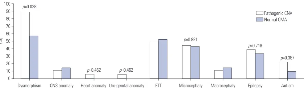
Further, phenotypes among the patients with pathogenic CNVs, VOUS, benign CNVs, and normal CMA were compared (Table 3). The clinical features most often seen in patients with pathogenic CNVs were dysmorphism (88.9%), FTT (50.0%), microcephaly (44.4%), epilepsy (38.9%), major organ anomaly (22.4%), and autism (22.2%), in the order of frequency. Patients with normal CNVs presented dysmorphism (57.1%), FTT (52.4%), microcephaly (42.9%), epilepsy (33.3%), major organ anomaly (14.3%), and autism (9.5%). Dysmorphism (p=0.028) was significantly more frequent in patients with pathogenic CNVs than in those with normal CMA (Fig. 1). Autism (p=0.387), epilepsy (p=0.718), and microcephaly (p=0.921) were more fre-quent in patients with pathogenic CNVs than in patients with normal CMA, although the difference was not significant (Fig. 1). Among the nine clinical features listed in Table 3, patients with normal CMA presented 2.2±1.3 (range: 1−4) manifesta-tions, and patients with pathogenic CMA presented 2.8±1.3 (range: 1−5) manifestations. Two or more symptoms were presented by 61.9% (13/21) of the patients with normal CMA and by 83.3% (15/18) of the patients with pathogenic CMA.
DISCUSSION
In this study, we identified chromosomal imbalances in 58.0% (29/50) of our patients. Of these, pathogenic CNVs were found in 62.1% (18/29), benign CNVs in 20.7% (6/29), and VOUS in 17.2% (5/29) of the patients. The overall diagnostic yield was, therefore, 36.0% (18/50). In recent reports, the diagnostic yield of CMA was shown to have increased up to 10−28% for genetic testing in patients with unexplained DD/ID, autism spectrum disorders, or multiple congenital anomalies, as compared to 3% for G-banded karyotype.4-11 In our study, the diagnostic yield
was higher than that found in previous reports with similar pa-tients. In Korea, the cost of CMA is not covered by Korean Health Insurance; therefore, patients for the CMA test were very carefully selected by a clinical geneticist, Hyon J. Kim. The correct diagnosis included the MECP2 duplication syn-drome, FOXG1 synsyn-drome, Angelman synsyn-drome, and other known microdeletion or microduplication syndromes. In these cases, the diagnosis could be useful for predicting the clinical progress, preparing for symptoms not yet presented, and de-ciding the plan for the evaluation and management of the dis-ease. Furthermore, using a familial study, we can estimate a recurrent risk in the family and can give more information about the disease. A previous large-scale study showed that 54% of the abnormal variants generated a recommendation for clini-cal action.12 The CMA test is an important diagnostic test that influences medical management. It also influences medical management in cases of VOUS, although clinicians should pe-riodically review updated information in order to provide ap-propriate medical management. As data from genetic studies increase, CNV interpretation and useful information can be updated. Palmer, et al.13 reported that interpretation of CMA could change over time. For example, patients 22 and 23 sis-ters showed CNV of VOUS until now; however, the interpreta-tion of the 1q44 trisomy in these patients can change over time. In our study, among the 21 patients who showed no abnor-malities in the CMA test, three patients were further tested us-ing whole exome sequencus-ing. Two familial patients with ID, severe short stature, and macrodontia were diagnosed with the KBG syndrome, with confirmed ANKRD11 mutation.14 The other patient was diagnosed with Pitt-Hopkins syndrome, with confirmed missense mutation in TCF4. In cases where the CMA test did not allow diagnosis, whole exome or whole ge-nome sequencing could identify the genetic causes of ID. Fi-nally, genome sequencing could be applied as a single genetic test, with a higher diagnostic yield, in the majority of patients with severe ID.15
[image:3.595.314.554.87.205.2]Some researchers have investigated the characteristics of CNVs in patients with DD/ID. Patients with ID and multiple congenital anomalies show higher burden of CNVs than those with ID alone.16 DD/ID risk is known to increase in relation to CNV size and craniofacial anomalies. Further, cardiovascular
Table 1. Clinical Characteristics of the 50 Patients
Characteristics n=50
Gender (male:female) 26:24 Age (yr) (mean±SD) 5.4±5.9 Developmental delay/intellectual disability 50 (100.0%) Craniofacial dysmorphism 39 (78.0%) Failure to thrive 27 (54.0%) Epilepsy 19 (38.0%) Major organ anomalies 12 (24.0%)
Autism 9 (18.0%)
Table 2.
Clinical and Genetic Features in 29 Patients with CNVs
Patient
Age
Sex
Microarray (genome build:hg19)
Size
Critical genes or region
Classification
Inheritance
Clinical features
Syndrome
Reasons of classification
Pathogenic CNVs 1
6 yr F 3p26.3p24.3(234763_18360238) × 3, 4q34.1q35.2(175696310_190957460) × 1
18 Mb/ 15 Mb 3p26.3 4q 34 Pathogenic/ Pathogenic
Unknown
Severe DD/ID, dysmorphism, hypotonia 3p26.3 duplication 4q 34 deletion
1/1
2
10 yr 3 m
M 5q33.3q35.1(156409412_172584708) × 1 16 Mb 5q33.3q35.1 Pathogenic De novo
Severe DD/ID, dysmorphism, epilepsy
, FTT
, microcephaly
5q33.3q35.1 deletion
1 3 8 m M Xq28(149987236_155233098) × 2 5 Mb MECP2 Pathogenic De novo
Severe DD, dysmorphism, macrodactyly
, hypotonia, intestinal
pseudoobstruction, chylothorax
MECP2
duplication syndrome
1 4 12 yr 1 0 m M 4q13.2(68598997_69155166) ×3, 6q12.3(47653195_48811902) × 3, Xq28(152916789_153421838) × 3
556 kb/ 1159 kb/ 505 kb GNRHR, TMPRSS11D/OPN5, GPR111, GPR115/SLC6A8, ABCD1, L1CAM, AVPR2, MECP2, NAA10, MRX3 VOUS/ VOUS/ Pathogenic
Unknown
Severe DD/ID no speech, dysmorphism, epilepsy
MECP2
duplication syndrome 3, 4/ 3, 4/1
5 7 m M 14q12q13.3(25363718_36872996) × 1 11.5 Mb FOXG1 Pathogenic De novo
Severe DD, dysmorphism, emangioma in midline forehead, simian crease, epilepsy
, FTT
,
microcephaly
, agenesis of corpus
callosum FOXG1 syndrome 1 6
9 yr 11 m
M 8p23.2(3685300_5935671) × 3, 18q11.2q12.2(24000546_34855 669) × 1
2.2 Mb/ 11 Mb CSMD1/KCTD1, DSC2, DSC3, ASXL3, DTNA, DSG1, DSG2, DSG4 Benign/ Pathogenic
De novo
DD/ID, dysmorphism
18q11.2 microdeletion
2/1
7
2 yr 11 m
M 2q36.3q37.3(228613832_242782258) × 3, 10p15.3(100047_2979784) × 1
14 Mb/ 2.8 Mb
COL6A3, TWIST2, NDUF
A10,
A
TG16L1, MLPH, HDAC4, AGXT
,
SP110/ DIP2C, ADARB2, IDI2AS1, IDI1, IDI2 Pathogenic/ Pathogenic
De novo
DD/ID, dysmorphism, FTT
, microcephaly , cryptochidism 1/1 8 8 yr F 8p23.2(3685300_5935671) ×3, 22q11.21(18648855_21800471) × 1
2.2 Mb/ 3 Mb
CSMD1
/22q11.21
Benign/ Pathogenic
Unknown
DD/ID, no speech, dysmorphism, FTT
, microcephaly
, tetralogy
of Fallot, autism
DiGeorge syndrome
2/1
9
8 yr 10 m
F
9q21.11q21.2(70966261_80322287)
×
1
9.4 Mb
FXN, TRPM6, VPS13A, TMC1, TJP2
Pathogenic
Unknown
DD/ID, dysmorphism, epilepsy
,
FTT
, microcephaly
9q21 microdeletion
1
10
2 yr 1 m
M 8p21.3(20328613_21241707) × 3, 10q21.3(68716001_68942534) × 1
913 kb/ 227 kb
LOC286114/CTNNA3
Pathogenic/ VOUS
Unknown
Severe DD/ID, no speech, dysmorphism, epilepsy
, FTT , microcephaly , hypotonia 8p duplication 1/3, 4 11 2 yr M 15q11.2q13.1(23290787_28540345) × 1 5.3 Mb
MKRN3, SNRPN, UBE3A, GABRB3, OCA2, MAGEL2, HERC2, NDN
Pathogenic
Unknown
DD, dysmorphism, epilepsy
,
FTT
, microcephaly
Angelman syndrome
1
12
1 yr 11 m
M Xp22.2p22.13(16985909_17728652) ×2 743 kb NHS Pathogenic Unknown
DD, no speech, FTT
, microcephaly
,
autistic behavior
R/O Nance- Horan syndrome
1 13 2.9 yr M 22q11.21(18648855_21800471) ×3 3.2 Mb
PRODH, GP1BB, COMT
, DCARF2, HCF2,
LZTR1, TBX1, RTN4R, SLC25A1
Pathogenic
Unknown
DD, autistic behavior
22q11.2 duplication
1 14 18 yr F 2q36.3(229975072_230647161) × 1, 16p11.2(29580020_30176508) × 1
672 kb/ 596 kb PID1, DNER, TRIP12/KIF22, PRRT2, ALDOA, TBX6 VOUS/ Pathogenic
De novo
Severe ID, dysmorphism, macrocephaly
, DM, obesity
,
abnormal behavior
, sister
of patient 15
16p 11.2 microdeletion syndrome
Table 2.
Clinical and Genetic Features in 29 Patients with CNVs (continued)
Patient
Age
Sex
Microarray (genome build:hg19)
Size
Critical genes or region
Classification
Inheritance
Clinical features
Syndrome
Reasons of classification
15 32 yr F 16p11.2(29580020_30177916) ×1 596 kb
KIF22, PRRT2, ALDOA, TBX6
Pathogenic
De novo
Severe ID, dysmorphism, macrocephaly
, obesity
, mood
disorder
, sister of patient 14
16p 11.2 microdeletion syndrome
1 16 1 yr M 21q22.2q22.3(41891664_42708105) × 3 816 kb
DSCAM, BACE2, PLAC4, F
AM3B
Pathogenic
Paternal
DD, dysmorphism, FTT
, atophic dermatitis 1 17 0.1 yr F 5q31.2q31.3(138934568_139624833) ×1 690 kb
UBE2D2, CXXC5, NRG2, PURA
Pathogenic
De novo
DD, dysmorphism, epilepsy
, hypotonia 1 18 2 yr M 5q14.3q21.3(89183371_105989481) × 1 17 Mb
GPR98, NR2F1, TTC37, PCSK1
Pathogenic
Unknown
DD, dysmorphism, FTT
1
CNVs of VOUS 19
9 m M 15q26.3(100738522_101136059) × 3 400 kb ADAMTS17,CERS3, SP AT A41, LINS, PRKXP1 VOUS Maternal
Mild DD, hypotonia, atrial septal defect
3, 4
20
2 yr 1 m
M
5q23.2q23.3(126540520_128636176)
×1
2 Mb
MEGF10, PRRC1, CTXN3, FLJ33630, SLC12A2, FBN2, SLC27A6, ISOC1
VOUS Paternal DD, epilepsy , FTT , macrocephaly , ATP1A2 mutation
Alternating hemiplegia
3, 4 21 1 m M Y(19585046_21028944) × 2 1.4 Mb VOUS Unknown
DD, dysmorphism, seizure, FTT
,
hemangioma in philtrum, severe VUR, thinning of corpus callosum
3, 4 22 15 yr F 1q44(247575767_248639486) × 3 1 Mb NLRP3, CIAS1 VOUS Paternal
Severe ID, dysmorphism, short stature, microcephaly
,
sister of patient 23
3, 4
23
13 yr 8 m
F 1q44(247584363_248660805) × 3 1 Mb NLRP3, CIAS1 VOUS Paternal
Severe ID, dysmorphism, short stature, microcephaly
,
autism, sister of patient 22
3, 4
Benign CNVs 24
9 yr 3 m
F 8p23.2(3685300_5935671) × 3 2.2 Mb CSMD1 Benign Maternal
Severe DD/ID, no speech, dysmorphism, cleft palate, epilepsy
, FTT
, microcephaly
2
25
11 yr 7 m
M Yq11.223q11.23(24660113_28464713) ×3 3.8 Mb DAZ, PRY , PRY2 Benign Unknown
Severe DD/ID, autism
2
26
1 yr 8 m
F 8p23.2(3688709_5950611) × 3 2.3 Mb CSMD1 Benign Paternal
Severe DD, dysmorphism, epilepsy
, FTT
, hypotonia, VUR
2 27 4.1 yr F 11q21(95577614_96054413) × 3 477 kb MTMR2, MAML2 Benign Paternal
Severe DD, dysmorphism, epilepsy
, FTT
, double ureter
, microcephaly , autism 2 28 0.2 yr M 17q21.32q21.33(47070357_47637376) × 1 567 kb PHB Benign Maternal
DD, dysmorphism, hyperpigmentation, FTT
, pulmonary stenosis 2 29 6.3 yr F 8p23.2(3685300-5935671) × 3 2.3 Mb CSMD1 Benign Unknown
DD, dysmorphism, hypotonia
2
CNVs, copy number variants; VOUS, variants of uncertain significance; CMA, chromosomal microarray; DD, developmental delay; ID,
intellectual disability; FTT
, failure to thrive; DM, diabetes millitus; VUR, vesicoure
-teral reflux. 1. Overlapping with a known imbalance syndrome; 2. In the category of genomic imbalance in healthy individuals as per public da
tabase; 3. CNV is not a common polymorphism; 4. Genes in the CNV are not associ
-ated with patient’
defects are enriched for large CNVs relative to epilepsy and autism spectrum disorder.17 Multiple large CNVs, including CNVs of unknown significance, are known to result in severe clinical presentation.18
We investigated the relation between other phenotypes and pathogenic CNV in patients with DD/ID. Dysmorphism (p= 0.028) alone was significantly more frequent in patients with pathogenic CNVs than in those patients with normal CMA. A previous study showed that congenital anomalies, microceph-aly, short stature, and FTT were more frequent in children with pathogenic CNVs.19 However, in our study, such pheno-types were not different between the two groups, because most of the patients with normal CNVs were also in the spec-trum of genetic variation that have yet to be identified by fur-ther testing, such as whole exome sequencing. Therefore, most of the phenotypes, such as congenital anomalies, micro-cephaly, and FTT were as frequent in patients with normal CMA as in patients with pathogenic CNVs. The number of phenotypes, however, was different. A greater number of pa-tients with pathogenic CMA, compared to those with normal CMA, presented two or more symptoms. We can, therefore, expect that in patients with unexplained DD/ID, the patho-genic CNV might be more frequently found if the patients have two or more phenotypes in addition to DD/ID.
Our results emphasize the importance of the CMA test in the clinical evaluation of patients with unexplained DD/ID. This study was limited to a small number of patients; thus, di-verse statistical analysis was not possible. However, our data involving detailed phenotype analysis and CNVs can add to databases for future genetic studies for discovering new can-didate genes and molecular pathways underlying unex-plained DD/ID.
ORCID
Hyo Jeong Kim https://orcid.org/0000-0003-2191-5341 Hyon J. Kim https://orcid.org/0000-0002-2945-8731
REFERENCES
1. Leonard H, Wen X. The epidemiology of mental retardation: chal-lenges and opportunities in the new millennium. Ment Retard Dev Disabil Res Rev 2002;8:117-34.
2. Vissers LE, Gilissen C, Veltman JA. Genetic studies in intellectual disability and related disorders. Nat Rev Genet 2016;17:9-18. 3. Rauch A, Hoyer J, Guth S, Zweier C, Kraus C, Becker C, et al.
Diag-nostic yield of various genetic approaches in patients with unex-plained developmental delay or mental retardation. Am J Med Genet A 2006;140:2063-74.
[image:6.595.49.540.85.214.2]4. Miller DT, Adam MP, Aradhya S, Biesecker LG, Brothman AR,
Table 3. Percentage of Presented Clinical Manifestations in Different CNVs Groups
Characteristics Pathogenic CNVs (n=18) VOUS (n=5) Benign CNVs (n=6) Normal CMA (n=21)
Dysmorphism (%) 16/18 (88.9) 5/5 (100.0) 6/6 (100.0) 12/21 (57.1) CNS anomaly (%) 2/18 (11.2) 1/5 (20.0) 0/6 (0.0) 3/21 (14.3) Heart anomaly (%) 1/18 (5.6) 1/5 (20.0) 1/6 (16.7) 0/21 (0.0) Uro-genital anomaly (%) 1/18 (5.6) 1/5 (20.0) 2/6 (33.3) 0/21 (0.0) FTT (%) 9/18 (50.0) 3/5 (60.0) 4/6 (66.7) 11/21 (52.4) Microcephaly (%) 8/18 (44.4) 2/5 (40.0) 2/6 (33.3) 9/21 (42.9) Macrocephaly (%) 2/18 (11.2) 1/5 (20.0) 0/6 (0.0) 3/21 (14.3) Epilepsy (%) 7/18 (38.9) 2/5 (40.0) 3/6 (50.0) 7/21 (33.3) Autism (%) 4/18 (22.2) 1/5 (20.0) 2/6 (33.3) 2/21 (9.5) CNVs, copy number variants; VOUS, variants of uncertain significance; CMA, chromosomal microarray; CNS, central nervous system; FTT, failure to thrive.
(%)
Dysmorphism CNS anomaly Heart anomaly Uro-genital anomaly FTT Microcephaly Macrocephaly Epilepsy Autism 100
90 80 70 60 50 40 30 20 10 0 − − − − − − − − − − −
p=0.028
p=0.462 p=0.462
p=0.921
p=0.718
p=0.387 Pathogenic CNV Normal CMA
Fig. 1. Comparison of phenotypes between the patients with pathogenic CNVs and normal CMA. Dysmorphism (p=0.028) was significantly more
[image:6.595.46.539.236.379.2]Carter NP, et al. Consensus statement: chromosomal microarray is a first-tier clinical diagnostic test for individuals with develop-mental disabilities or congenital anomalies. Am J Hum Genet 2010;86:749-64.
5. Battaglia A, Doccini V, Bernardini L, Novelli A, Loddo S, Capalbo A, et al. Confirmation of chromosomal microarray as a first-tier clinical diagnostic test for individuals with developmental delay, intellectual disability, autism spectrum disorders and dysmor-phic features. Eur J Paediatr Neurol 2013;17:589-99.
6. Zilina O, Teek R, Tammur P, Kuuse K, Yakoreva M, Vaidla E, et al. Chromosomal microarray analysis as a first-tier clinical diagnos-tic test: Estonian experience. Mol Genet Genomic Med 2014;2: 166-75.
7. Chong WW, Lo IF, Lam ST, Wang CC, Luk HM, Leung TY, et al. Performance of chromosomal microarray for patients with intel-lectual disabilities/developmental delay, autism, and multiple con-genital anomalies in a Chinese cohort. Mol Cytogenet 2014;7:34. 8. Howell KB, Kornberg AJ, Harvey AS, Ryan MM, Mackay MT,
Free-man JL, et al. High resolution chromosomal microarray in undi-agnosed neurological disorders. J Paediatr Child Health 2013; 49:716-24.
9. Byeon JH, Shin E, Kim GH, Lee K, Hong YS, Lee JW, et al. Applica-tion of array-based comparative genomic hybridizaApplica-tion to pediat-ric neurologic diseases. Yonsei Med J 2014;55:30-6.
10. Iourov IY, Vorsanova SG, Kurinnaia OS, Zelenova MA, Silvanovich AP, Yurov YB. Molecular karyotyping by array CGH in a Russian cohort of children with intellectual disability, autism, epilepsy and congenital anomalies. Mol Cytogenet 2012;5:46.
11. Uwineza A, Caberg JH, Hitayezu J, Hellin AC, Jamar M, Dideberg V, et al. Array-CGH analysis in Rwandan patients presenting
de-velopment delay/intellectual disability with multiple congenital anomalies. BMC Med Genet 2014;15:79.
12. Coulter ME, Miller DT, Harris DJ, Hawley P, Picker J, Roberts AE, et al. Chromosomal microarray testing influences medical man-agement. Genet Med 2011;13:770-6.
13. Palmer E, Speirs H, Taylor PJ, Mullan G, Turner G, Einfeld S, et al. Changing interpretation of chromosomal microarray over time in a community cohort with intellectual disability. Am J Med Genet A 2014;164A:377-85.
14. Kim HJ, Cho E, Park JB, Im WY, Kim HJ. A Korean family with KBG syndrome identified by ANKRD11 mutation, and phenotypic comparison of ANKRD11 mutation and 16q24.3 microdeletion. Eur J Med Genet 2015;58:86-94.
15. Gilissen C, Hehir-Kwa JY, Thung DT, van de Vorst M, van Bon BW, Willemsen MH, et al. Genome sequencing identifies major causes of severe intellectual disability. Nature 2014;511:344-7.
16. Girirajan S, Brkanac Z, Coe BP, Baker C, Vives L, Vu TH, et al. Rela-tive burden of large CNVs on a range of neurodevelopmental phe-notypes. PLoS Genet 2011;7:e1002334.
17. Cooper GM, Coe BP, Girirajan S, Rosenfeld JA, Vu TH, Baker C, et al. A copy number variation morbidity map of developmental de-lay. Nat Genet 2011;43:838-46.
18. Girirajan S, Rosenfeld JA, Coe BP, Parikh S, Friedman N, Gold-stein A, et al. Phenotypic heterogeneity of genomic disorders and rare copy-number variants. N Engl J Med 2012;367:1321-31. 19. Shoukier M, Klein N, Auber B, Wickert J, Schröder J, Zoll B, et al.