Vol. 46, No. 4, pp. 511 - 518, 2005
Because obesity is frequently complicated by other cardio-vascular risk factors, the impact of a reduction in visceral adiposity on vascular endothelial dysfunction (VED) in obese patients is difficult to determine. In the present study, we evaluated the impact of a reduction in visceral adiposity on VED in obese women. Thirty-six premenopausal obese women (BMI 25 kg/m2) without complications were enrolled in the study. VED was evaluated by determining the augmentation index (AIx) from radial artery pulse waves obtained by appla-nation tonometry. Changes in AIx in response to nitroglycerin-induced endothelium-independent vasodilatation ( AIx-NTG) and in response to salbutamol administration ( AIx-Salb) were determined before and after weight reduction. After a 12-week weight reduction program, the average weight loss was 7.96±3.47 kg, with losses of 21.88±20.39 cm2 in vis-ceral fat areas (p< 0.001). Pulse wave analysis combined with provocative pharmacological testing demonstrated preserved endothelium-independent vasodilation in healthy premenopaus-al obese women ( AIx-NTG: 31.36±9.80% before weight re-duction vs. 28.25 ± 11.21% after weight rere-duction, p> 0.1) and an improvement in endothelial-dependent vasodilation fol-lowing weight reduction ( AIx-Salb: 10.03±6.49% before weight reduction vs. 19.33 ± 9.28% after reduction,p< 0.001). A reduction in visceral adipose tissue was found to be most significantly related to an increase in AIx-Salb (β= -0.57, p< 0.001). A reduction in visceral adiposity was significantly related to an improvement in VED. This finding suggests that reduction of visceral adiposity may be as important as the control of other major risk factors in the prevention of atherosclerosis in obese women.
Key Words:Augmentation index, visceral adiposity, vascular endothelial dysfunction
INTRODUCTION
Obesity is an independent risk factor for cardio-vascular disease.1,2 In particular, abdominal
vis-ceral adipose tissue is believed to have harmful effects on vascular endothelial function through various interactions of coagulants and inflamma-tory molecules.3 The vascular endothelium serves
as an endocrine organ, secreting several vasoactive substances, such as nitric oxide. These substances prevent the development of atheroma. Obesity is independently associated with vascular endothe-lial dysfunction (VED,) and VED is a key event in the early development of atherosclerosis. Therefore, VED may be an important link between obesity and atherosclerosis. Weight reduction has been re-ported to improve VED.4 However, because obesity is frequently accompanied by other cardiovascu-lar risk factors and metabolic diseases, the ability of reduced visceral adiposity alone to improve VED in obese patients has been difficult to determine. A simple, non-invasive pulse wave analysis base technique was recently used to evaluate va-somotor endothelial function.5,6 After
demonstra-ting that albuterol,7 a β2 -agonist, reduces wave
reflection by activating the L-arginine-NO path-way, many studies have shown that an assess-ment of global endothelial function can be per-formed by examining changes in the peripheral
Reduction in Visceral Adiposity is Highly Related to
Improvement in Vascular Endothelial Dysfunction among
Obese Women: An Assessment of Endothelial Function by
Radial Artery Pulse Wave Analysis
Si-Hoon Park1 and Kyung Won Shim2
1Department of Cardiology and Ewha Medical Research Institute, College of Medicine, Ewha Womans University, Seoul, Korea;
2Department of Family Medicine, College of Medicine, Ewha Womans University, Seoul, Korea.
Received January 19, 2005 Accepted April 21, 2005
waveform in response to inhalation of β2
-ago-nists. Changes can be measured by tonometry and quantified according to the augmentation index (AIx).5,6 The present study utilized the
advanta-ges of pulse wave analysis, i.e., its simplicity, re-peatability, operator-independency and non-inva-siveness. We evaluated the impact of substantial weight reduction on VED in premenopausal obese women without complications or any other car-diac risk factors to determine whether weight re-duction alone was associated with improved VED.
MATERIALS AND METHODS
Subjects
The same healthy, premenopausal obese women included in a previous study8were enrolled in the
present study. These subjects were visiting a university hospital obesity clinic and were 20 to 45 years-old (mean age: 33.81 ± 7.13) with a body mass index (BMI) 25 kg/m2. Obesity was
de-fined according to the guidelines established by the 'Redefining Obesity and its Treatment'- a Joint Enterprise by the Regional Office for the Western Pacific of the World Health Organization and the International Association for the Study of Obesity and International Obesity Task Force. Subjects with any of the following co-morbidities were ex-cluded: hypertension (systolic blood pressure > 140 mmHg or diastolic blood pressure > 90 mm Hg), dyslipidemia {LDL (low density lipoprotein) cholesterol 160 mg/dL, HDL (high-density lipo-protein) cholesterol < 40 mg/dL, or triglyceride 200 mg/dL}, type 2 diabetes, coronary heart dis-ease, stroke, thyroid disdis-ease, and depression. In-formation was obtained by administering a self-report questionnaire to patients and by clinical examination and blood tests. Current smokers and those who had undergone a commercial diet pro-gram or received anti-obesity medication during the previous three months were also excluded. As in our previous study, obese subjects were matched with healthy, premenopausal non-obese women for clinical, metabolic, and lipid charac-teristics. Initially, 42 obese patients were enrolled, but six dropped out over the 12-week study period, leaving a total of 36 subjects. All subjects
were given informed consent, and the study pro-tocol was approved by the Institutional Review Board of Ewha Womans University, Mokdong Hospital.
Data collection
All examinations were performed during the follicular phase of the menstrual cycle, within 10 days of the onset of menstruation. Subjects re-ceived dietary and exercise consultations every two weeks for three months, during which BP, height, weight, and waist and hip circumferences were measured.
Anthropometric measurements
Body weights were measured to an accuracy of 0.1 kg, with subjects wearing light clothing. Height was measured to an accuracy of 0.1 cm. The waist circumference was measured at the mid-point between the upper iliac and lower costal level at the end of normal expiration, to an accuracy of 0.1 cm, and the hip circumference was defined as the largest measurement around the hip area. Body mass index (BMI) was calculated as the weight in kilograms divided by the square of the height in meters (kg/m2).
Body fat measurements
Percent body fat (%) was measured using bio-electrical impedance analysis while subjects were in a fasting state (Inbody 2.0, Biospace Co., Seoul, Korea), and body fat distribution was determined by computed tomography (GE High Speed Ad-vantage CT scanner, GE Co., Waukesha, USA). Single slices of abdominal subcutaneous adipose tissue and visceral adipose tissue (measured in cm2) were taken at the 4-5 level of the lumbar
spine, as described elsewhere.9 The attenuation
interval was -40 to -140 Hounsfield units.
Blood tests
Abbott, Abbott Park, Illinois, USA). Levels of high-density C-reactive protein (hsCRP) were measured by nephelometry using BNII (Dade Behring, Liederbach, Germany) with a mini-mum detection concentration of 0.0175 mg/dL. Homeostasis Model Assessment (HOMA) scores were calculated to evaluate insulin resistance.10
Evaluation of vascular endothelial function
Pulse wave analysis using the provocative phar-macological test was performed by administering salbutamol or nitroglycerin (NTG) to determine VED.
Pulse wave analysis
A pulse wave is composed of two waves, i.e., an aortic wave produced when the left ventricle contracts and a reflected wave produced when the aortic wave is reflected from peripheral vessels. The velocity of the reflected waves is increased when the arterial walls are stiff. Accordingly, in cases with stiffened arteries, systolic pressure aug-mentation occurs during the systolic phase. Based on this principle, radial artery pressure was measured using a high-fidelity micromanometer (SphygmoCor, Medical ECONET Co., Sydney, Australia). In practice, the artery is compressed between a sensor and the underlying structures, and, thus, the intra-arterial pressure is transmitted through the arterial wall to the sensor. Data were collected directly using a portable computer. After 20 sequential waveforms had been acquired, in-tegral software was used to generate an averaged peripheral waveform and a corresponding central waveform. Further analysis was conducted to determine AIx and the ascending aortic pressure, as described previously.7,11,12 AIx was defined as
the difference between the first (P1) and second (P2) peaks of the central arterial waveform, ex-pressed as a percentage of pulse pressure.13,14 In
this study, repeated pulse wave analyses at one-week intervals in ten non-obese volunteers showed a coefficient of variance (CV) of 1.4 ± 2.3 % for AIx.
Measurements of aix after salbutamol or ntg admini-stration
AIx was evaluated after administering
nitrogly-cerin or salbutamol, which induce endothelial-independent vasodilation and endothelial-depen-dent vasodilation, respectively. AIx was deter-mined during the peak response which occurred 3 minutes after administering 0.6 mg of nitrogly-cerin. The difference in AIx before and after this administration was measured and is depicted as AIx-NTG. Twenty-five minutes after admini-stering nitroglycerin, 2.5 mg of nebulized salbuta-mol (Ventolin nebules), which acts on vascular endothelial cells through the L-arginine-NO path-way, was administered by inhalation using a mask. Thiry minutes later, the pulse wave an-alysis was again carried out to measure AIx. The difference between AIx before and after salbuta-mol administration was then determined and is depicted as AIx-Salb.5,7,11 The dosage and
timing of the administration of these drugs were based on pilot studies and previous experience. Pilot studies confirmed that 15 minutes was sufficient to allow NTG-induced hemodynamic changes to return to baseline, but that a longer period was required for salbutamol. Therefore, NTG was always administered first, and followed by salbutamol 25 minutes later.
Diet and exercise program
For effective weight control, a diet and exercise program was utilized, and subjects were ed-ucated regarding living habits. To maintain a low calorie/low fat diet, subjects received 1,000 kcal per day during the program. Initially, subjects ex-ercised for half an hour a day, 3 times a week. The length and frequency of exercise were then in-creased every other week, reaching 40-60 minutes a day and 4-5 times a week. Subject compliance was evaluated at every visit by examining the dietary diary and weight loss log. Subjects were administered 20 mg of fluoxetine daily, as well as a thermogenic agent (pseudoephedrine 60 mg, theolan 100 mg) as a supplementary measure. To avoid side-effects, drugs were administered for the first 4 weeks only. Three months later, physi-cal examinations, obesity measurements, and pulse wave analysis were repeated.
All data are expressed as means±SD. Changes in AIx-NTG, AIx-Salb and all other param-eters versus weight reduction were analyzed using a paired t-test. To identify parameters af-fecting (AIx-Salb, a multiple linear regression model was constructed using the step-wise method, after adjusting for BMI with (AIx-Salb as the dependent variable. hsCRP was log trans-formed to achieve a normal distribution. The level of significance was set at p< 0.05 (two-tailed). Statistical calculations were performed using the SAS system for Windows (version 8.0, SAS Inc. Raleigh, NC, USA).
RESULTS
Anthropometric and clinico-metabolic responses to weight reduction
After 12 weeks of the diet and exercise
pro-gram, all subjects were re-evaluated. The average weight reduction was 7.96 ± 3.47 kg (11% of base-line), with losses of 5.29±2.59 kg (13% of baseline) in fat mass (p<0.001) and a marked reduction in BMI and waist circumferences. The visceral and subcutaneous fat areas were reduced by 21.88 ± 20.39 cm2 and 49.01 ± 53.76 cm2, respectively (p<
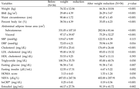
[image:4.595.60.536.413.715.2]0.001). Diastolic pressure was slightly reduced, without a significant change in systolic pressure. The levels of total cholesterol, LDL cholesterol, and triglycerides were all reduced after weight reduction, but the serum HDL cholesterol level did not show an increase following weight reduc-tion. Fasting glucose, fasting insulin, HOMA scores, and hsCRP levels were lower following weight reduction, while estradiol levels increased (Table 1).
Vascular reactivity and hemodynamic changes by weight reduction. Baseline heart rate, mean arterial pressure (MAP), and baseline AIx were not significantly changed by weight reduction.
Table 1. General Characteristics Before and After Weight Reduction
Variables Before(N=36) weight reduction After weight reduction (N=36) p-value
Weight (kg) 74.32 ± 12.04 66.36 ± 10.04 <0.001
BMI (kg/m2) 29.48 ± 4.39 26.29 ± 3.67 <0.001
Waist circumference (cm) 90.46 ± 1.72 83.47 ± 1.45 <0.001
Percent body fat (%) 38.54 ± 4.39 35.05 ± 4.23 <0.001
Abdominal adipose tissue area (cm2)
Subcutaneous 331.05 ± 107.10 282.04 ± 81.66 <0.001
Visceral 97.17 ± 39.87 75.29 ± 32.27 <0.001
SBP (mmHg) 119.67 ± 9.89 122.53 ± 8.38 0.115
DBP (mmHg) 72.03 ± 6.21 70.94 ± 6.59 0.016
Cholesterol (mg/dL) 197.03 ± 25.61 176.69 ± 26.44 <0.001
LDL cholesterol (mg/dL) 95.00 ± 18.32 85.03 ± 15.32 <0.001
HDL cholesterol (mg/dL) 53.53 ± 9.25 54.67 ± 11.11 0.393
Triglyceride (mg/dL) 100.78 ± 35.70 85.00 ± 40.76 0.030
Fasting glucose (mg/dL) 96.58 ± 7.41 91.58 ± 7.41 0.007
Fasting insulin (μIU/mL) 12.55 ± 17.31 6.67 ± 5.22 0.002
HOMA score 3.13 ± 4.63 1.53 ± 1.24 0.030
NEFA (μEq/L) 487.03 ± 247.56 402.00 ± 207.91 0.076
hsCRP* (mg/dL) 0.25 ± 0.41 0.19 ± 0.36 <0.001
Estradiol (pg/mL) 64.17 ± 27.76 91.19 ± 41.72 0.002
Changes in heart rate and MAP provoked by the administration of NTG or salbutamol before weight loss were similar to changes after weight reduction (Table 2). Pulse wave analysis combined with provocative pharmacological testing dem-onstrated preserved endothelium-independent vasodilation in healthy premenopausal obese women ( AIx-NTG: 31.36 ± 9.80% before weight reduction vs. 28.25 ± 11.21% after weight reduc-tion,p> 0.05) and an improvement in endothelial-dependent vasodilation following weight reduc-tion ( AIx-Salb: 10.03 ± 6.49% before weight re-duction vs. 19.33 ± 9.28% after rere-duction, p< 0.001) (Table 2).
Relationship between anthropometric and clinico-metabolic parameters and weight reduction
Improvements in endothelial-dependent vas-cular function following weight reduction (in-crease in AIx-Salb by weight reduction) were related to reductions in visceral adipose tissue area (r= -0.62,p<0.001), percent body fat (r= -0.35,
p< 0.05), BMI (r= -0.33, p<0.05), fasting insulin (r= -0.38, p<0.05), and HOMA scores (r= -0.375, p< 0.05). Moreover, step-wise multiple regression an-alysis identified visceral adipose tissue area (β= -0.57, p< 0.001) as the most significant indepen-dent parameter accounting for improved endo-thelial-dependent vascular function following weight reduction (Table 3).
DISCUSSION
Obesity is characterized by VED, and the degree of VED is predicted by body fat distribution, independent of metabolic and other hemodynamic parameters.15,16 This association between obesity
[image:5.595.58.537.117.266.2]and VED, an early feature of cardiovascular dis-ease, is of great importance because it may pro-vide clinicians with new preventive strategies against atherosclerosis in obese patients. Recently, a prospective study demonstrated VED improve-ment following substantial weight reduction in postmenopausal obese women with no additional
Table 3. Multiple Regression Analysis of Change in AIx-Salb in Obese Women
Obese women (N=36) β p-value
Visceral adipose tissue area (cm2) HOMA score
-0.566
-0.275 <0.0010.047
[image:5.595.58.530.354.396.2]AIx-Salb, change in augmentation index by administering salbutamol; Visceral adipose tissue area, change of visceral adipose tissue area; HOMA, change of homeostasis model assessment score.
Table 2. Pulse Wave Analysis Results Before and After Weight Reduction
Variables Before weight reduction (N=36) After weight reduction (N=36) p-value
Baseline AIx (%) 22.64 ± 11.25 22.92 ± 9.09 0.859
AIx NTG (%) 31.36 ± 9.80 28.25 ± 11.21 0.251
AIx Salb (%) 10.03 ± 6.49 19.33 ± 9.28 <0.001
Baseline HR (beat/min) 71.83 ± 7.95 70.31 ± 13.55 0.484
HR NTG (beat/min) 14.64 ± 11.15 6.25 ± 25.78 0.054
HR Salb (beat/min) 4.19 ± 5.98 0.81 ± 20.37 0.329
Baseline MAP (mmHg) 89.28 ± 1.14 86.14 ± 2.41 0.163
MAP NTG (mmHg) 3.56 ± 0.41 1.86 ± 2.27 0.574
MAP Salb (mmHg) 2.00 ± 0.52 0.36 ± 2.30 0.991
Values are mean ± SD.
risk factors.4 However, the study tested VED by
determining hemodynamic and rheologic re-sponses to L-arginine, a technique that may be inadequate for screening or clinical applications. A simple and noninvasive form of pulse wave analysis to determine VED was recently intro-duced.5,6 In the present study, we used pulse
wave analysis, combined with provocative phar-macological testing, to determine VED. To our knowledge, this is the first report to prospectively test the impact of weight reduction per se on VED by pulse wave analysis. The main finding of this study is that substantial weight reduction over a 12-week period can improve VED, and that visceral fat reduction is most strongly related to VED improvement in premenopausal obese women.
Relationship between visceral adiposity and VED
Adipose tissue acts as an endocrine organ and appears to secrete metabolic pro-inflammatory molecules. These cytokines are known to con-tribute to vascular inflammation and to regulate vascular endothelial function, an early indicator of atherosclerosis development. Moreover, visceral fat appears to produce these cytokines more ac-tively than other adipose tissues. Visceral fat cells show higher rates of catecholamine-induced lipolysis and express higher numbers of β1- and
β2-adrenergic receptors and levels of
glucocor-ticoid receptor mRNA, thus explaining the impor-tance of fat distribution.17 Aging and estrogen
deficiency can play a role in changes in vascular endothelium. After menopause, the possibility of obesity-related complications is likely to increase. Other cardiovascular risk factors, such as hyper-tension, diabetes mellitus, hypercholesterolemia, and smoking are also associated with VED. Thus, we confined our study to premenopausal obese women without additional cardiovascular risk factors in order to reduce the effect of such factors on VED.18 In a previous study, we found that
provocative pharmacological testing using NTG showed preserved endothelium-independent vaso-dilation in premenopausal, healthy, obese women. However, changes in AIx after the introduction of salbutamol nebulas, which induces vasodilation
through vascular endothelial cells, was less evident in the obese group than in the non-obese group. The results of the present study suggest that endothelial vasodilatory function is reduced in premenopausal healthy obese women despite a similar level of arterial stiffness, thus confirming that visceral fat per se is highly associated with VED.
Relationship between reduced visceral adiposity and improved VED
Vascular endothelial function is affected by car-diovascular risk factors, hormones, and medica-tions. Factors that affect VED include hyperten-sion,19 hypercholesterolemia,20 aging,21 estrogen,22
smoking, diabetes mellitus,23 cholesterol-lowering
agents,24,25 hormone replacement therapy,26,27 and physical inactivity.28 Weight reduction improves
many of the conditions frequently associated with or co-occur with obesity. This raises the question of whether the restoration of vascular endothelial function by weight reduction in obese people is due to visceral adiposity reduction, an improve-ment in cardiovascular risk factors, or increased physical activity. A recent study4 demonstrated
that a reduction of visceral adipose tissue in obese women is a more important factor than increased physical activity or improvement of cardiovas-cular risk factors in improving VED. The study4 showed that a reduction in adipose tissue by lipo-suction surgery improved VED and highlighted the importance of visceral adiposity reduction in and of itself in improving VED in obese women.
Pulse wave analysis for the evaluation of VED
Vascular endothelial cell function was measured using pulse wave analysis. The results of pulse wave analysis following the administration of salbutamol or NTG has been shown to correlate with the results of invasive methods based on acetylcholine infusion.5 The augmentation index
however, some questions have been raised about the validity and clinical utility of pulse wave analysis. Wide margins of error were found when comparing centrally measured and peripherally derived AIx.30 AIx was also found to be
depen-dent on heart rate and blood pressure during IV infusion of beta-adrenergic drugs, such as isopro-terenol.31However, the inhalation of albuterol was
not accompanied by significant alterations in heart rate and MAP.5 We also confirmed in a previous
study that a substantial change in heart rate or MAP was not induced by NTG or salbutamol. In the present study, changes in AIx-Salb by weight reduction were not related to a small change in heart rate or diastolic or systolic pres-sure (Table 3). Moreover, as the absolute value of AIx might not be of importance in follow-up studies,32 we used AIx with provocative pharma-cological testing in this study. Since all subjects were premenopausal women free of cardiovas-cular disease, it remains to be determined whether our findings can be applied to men, children, postmenopausal women, and cardiovascular patients.
In conclusion, we have demonstrated that a reduction in visceral adiposity is significantly re-lated to improvements in VED, as determined by radial artery pulse wave analysis, following weight reduction. In this context, clinicians may choose to focus on the reduction of visceral adi-posity, as much as on the control of other risk fac-tors, for the prevention of atherosclerosis in obese women.
REFERENCES
1. Eckel RH, Krauss RM. American Heart Association call to action: obesity as a major risk factor for coronary heart disease. Circulation 1998;37:2099-100.
2. Terry RB, Page WF, Haskell WL. Waist-hip ratio, body mass index and premature cardiovascular disease mortality in US Army veterans during a twenty-three year follow up study. Int J Obes Relat Metab Disord 1992;16:417-23.
3. Tracy RP. Inflammation in cardiovascular disease; cart, horse, or both? Circulation 1998;97:2000-2.
4. Ziccardi P, Nappo F, Giugliano G, Esposito K, Marfella R, Cioffi M, et al. Reduction of inflammatory cytokine concentrations and improvement of endothelial func-tions in obese women after weight loss over one year.
Circulation 2002;105:804-9.
5. Wilkinson IB, Hall IR, MacCallum H, Mackenzie IS, McEniery CM, van der Arend BJ, et al. Pulse-wave analysis: clinical evaluation of a noninvasive, widely applicable method for assessing endothelial function. Arterioscler Thromb Vasc Biol 2002;22:147-52. 6. Hayward CS, Kraidly M, Webb CM, Collins P.
Assess-ment of endothelial function using peripheral wave-form analysis. J Am Coll Cardiol 2002;40:521-8. 7. Chowienczyk PJ, Kelly RP, MacCallum H, Millasseau
SC, Andersson TL, Gosling RG, et al. Photoplethysmo-graphic assessment of pulse wave reflection: blunted response to endothelium-dependent beta 2-adrenergic vasodilation in type II diabetes mellitus. J Am Coll Cardiol 1999;34:2007-14.
8. Suh HS, Park YW, Kang JH, Lee SH, Lee HS, Shim KW. Vascular endothelial dysfunction tested by blunted response to endothelium-dependent vasodilation by salbutamol and its related factors in uncomplicated pre-menopausal obese women. Int J Obes Relat Metab Disord 2005;29:217-22.
9. Shuman WP, Morris LL, Leonetti DL, Wahl PW, Moceri VM, Moss AA, et al. Abdominal body fat distribution detected by computed tomography in diabetic men. Invest Radiol 1986;21:483-7.
10. Matthews DR, Hosker JP, Rudenski AS, Naylor BA, Treacher DF, Turner RC. Homeostasis model asses-ment: insulin resistance and beta-cell function from fasting plasma glucose and insulin concentration in man. Diabetologia 1985;28:412-9.
11. Dawes M, Chowienczyk PJ, Ritter JM. Effects of inhi-bition of the L-arginine/NO pathway on beta-adre-nergic mediated vasodilation in human forearm vas-culature. Circulation 1997;95:2293-7.
12. Kelly R, Hayward C, Avolio A, O'Rourke M. Nonin-vasive determination of age-related changes in the human arterial pulse. Circulation 1989;80:1652-9. 13. Seger P, Qasem A, DeBacker T, Carlier S, Verdonck P,
Avolio A. Peripheral 'oscillatory' compliance is asso-ciated with aortic augmentation index. Hypertension 2001;37:1434-9.
14. Mackenzie IS, Wilkinson IB, Cockcroft JR. Assessment of arterial stiffness in clinical practice. Q J Med 2002; 95:67-74.
15. Arcaro G, Zamboni M, Rossi L, Turcato E, Covi G, Armellini F, et al. Body fat distribution predicts the degree of endothelial dysfunction in uncomplicated obesity. Int J Obes Relat Metab Disord 1999;23:936-42. 16. Hashimoto M, Akishita M, Eto M, Kozaki K, Ako J. The impairment of flow-mediated vasodilatation in obese men with visceral fat accumulation. Int J Obes Relat Metab Disord 1998;22:477-84.
17. Montague CT, O'Rahilly S. The perils of portliness: causes and consequences of visceral adiposity. Diabetes 2000;49:883-8.
endo-thelial dysfunction. Circulation 2000;101:948-54. 19. Hayashi T, Boyko EJ, Leonetti DL, McNeely MJ,
Newell-Morris L, Kahn SE, et al. Visceral adiposity is an independent predictor of incident hypertension in Japanese Americans. Ann Intern Med 2004;140:992-1000.
20. Chowienczyk PJ, Watts GF, Cockcroft JR, Ritter JM. Impaired endothelium dependent vasodilation of fore-arm resistance vessels in hypercholesterolaemia. Lancet 1992;340:1430-2.
21. Celermajer DS, Sorensen KE, Spiegelhalter DJ, Georgakopoulos D, Robinson J, Deanfield JE. Aging is associated with endothelial dysfunction in healthy men years before the age-related decline in women. J Am Coll Cardiol 1994;24:471-6.
22. Lieberman EH, Gerhard MD, Uehata A, Walsh BW, Selwyn AP, Ganz P, et al. Estrogen improves endo-thelium-dependent, flow mediated vasodilation in post-menopausal women. Ann Intern Med 1994;121:936-41. 23. Clarkson P, Celermajer DS, Donald AE, Sampson M, Sorensen KE, Adams M, et al. Impaired vascular reactivity in insulin-dependent diabetes mellitus is related to disease duration and low density lipoprotein cholesterol levels. J Am Coll Cardiol 1996;28:573-9. 24. Vogel RA, Corretti MC, Plotnick GD. Changes in
flow-mediated brachial artery vasoactivity with lowering of desirable cholesterol levels in healthy middle-aged men. Am J Cardiol 1996;77:37-40.
25. O'Driscoll G, Green D, Taylor RR. Simvastatin, an HMG-coenzyme A reductase inhibitor, improves endo-thelial function within 1 month. Circulation 1997;95:
1126-31.
26. Gerhard M, Walsh BW, Tawakol A. Haley EA, Creager SJ, Seely EW, et al. Estradiol therapy combined with progesterone and endothelium-dependent vasodilation in postmenopausal women. Circulation 1998;98:1158-63. 27. Sorensen KE, Dorup I, Hermann AP, Mosekilde L. Combined hormone replacement therapy does not pro-tect women against the age-related decline in endothe-lium-dependent vasomotor function. Circulation 1998; 97:1234-8.
28. Gill JM, Al-Mamari A, Ferrell WR, Cleland SJ, Packard CJ, Sattar N, et al. Effects of prior moderate exercise on postprandial metabolism and vascular function in lean and centrally obese men. J Am Coll Cardiol 2004;21: 2375-82.
29. Anderson TJ, Uehata A, Gerhard MD, Meredith IT. Close relation of endothelial function in the human coronary and peripheral circulations. J Am Coll Cardiol 1995;26:1235-41.
30. Hope SA, Tay DB, Meredith IT, Cameron JD. Use of arterial transfer functions for the derivation of arterial wave form characteristics. J Hypertens 2003;21:1299-305.
31. Lemogoum D, Flores G, Van den Abeele W, Ciarka A, Leeman M, Degaute JP. Validity of pulse pressure and augmentation index as surrogate measures of arterial stiffness during beta-adrenergic stimulation. J Hyper-tens 2004;22:511-7.