Early Experience Using a Left Atrial Appendage Occlusion
Device in Patients with Atrial Fibrillation
Yung Ly Kim,
1Boyoung Joung,
1Young Keun On,
2Chi Young Shim,
1Moon Hyoung Lee,
1Young-Hoon Kim,
3and Hui-Nam Pak
11Department of Cardiology, Yonsei University Health System, Seoul; 2Department of Cardiology, Samsung Medical Center, Sungkyunkwan University, Seoul;
3Department of Cardiology, Korea University Cardiovascular Center, Seoul, Korea.
Received: February 8, 2011 Revised: March 25, 2011 Accepted: March 28, 2011
Corresponding author: Dr. Hui-Nam Pak, Department of Cardiology,
Yonsei University Health System, 50 Yonsei-ro, Seodaemun-gu, Seoul 120-752, Korea.
Tel: 82-2-2228-8459, Fax: 82-2-393-2041 E-mail: hnpak@yuhs.ac
∙ The authors have no financial conflicts of interest.
© Copyright:
Yonsei University College of Medicine 2012
This is an Open Access article distributed under the terms of the Creative Commons Attribution Non-Commercial License (http://creativecommons.org/ licenses/by-nc/3.0) which permits unrestricted non-commercial use, distribution, and reproduction in any medium, provided the original work is properly cited.
Purpose: Atrial fibrillation (AF) is one of the major risk factors for ischemic stroke, and 90% of thromboembolisms in these patients arise from the left atrial appendage (LAA). Recently, it has been documented that an LAA occlusion de-vice (OD) is not inferior to warfarin therapy, and that it reduces mortality and risk
of stroke in patients with AF. Materials and Methods: We implanted LAA-ODs
in 5 Korean patients (all male, 59.8±7.3 years old) with long-standing persistent
AF or permanent AF via a percutaneous trans-septal approach. Results: 1) The
major reasons for LAA-OD implantation were high risk of recurrent stroke (80%), labile international neutralizing ratio with hemorrhage (60%), and 3/5 (60%) pa-tients had a past history of failed cardioversion for rhythm control. 2) The mean LA size was 51.3±5.0 mm and LAA size was 25.1×30.1 mm. We implanted the LAA-OD (28.8±3.4 mm device) successfully in all 5 patients with no complica-tions. 3) After eight weeks of anticoagulation, all patients switched from warfarin to anti-platelet agent after confirmation of successful LAA occlusion by
trans-esophageal echocardiography. Conclusion: We report on our early experience
with LAA-OD deployment in patients with 1) persistent or permanent AF who cannot tolerate anticoagulation despite significant risk of ischemic stroke, or 2) re-current stroke in patients who are unable to maintain sinus rhythm.
Key Words: Atrial fibrillation, left atrial appendage, occlusion device, thrombo-embolism
INTRODUCTION
post-mor-ing. Here, we report our very early experiences with LAA occlusion devices in Korean patients with AF.
MATERIALS AND METHODS
Study population
This study included patients with persistent or permanent AF who had a significant risk of stroke or could not tolerate war-farin therapy. Proper informed consent was obtained from all patients. The inclusion criteria were as follows: 1) permanent AF refractory to the electrical cardioversion, 2) persistent AF with failed maintenance of sinus rhythm with anti-arrhythmic drugs, 3) persistent AF and recurrent ischemic stroke despite proper anticoagulation, and 4) inability to tolerate warfarin due to adverse effects, labile INR, or recurrent hemorrhagic complications. We excluded patients with AF who were opti-mal candidates for rhythm control strategy, anticoagulation, or who were at low risk for ischemic stroke.
Structure of the LAA occlusion device
We used a WATCHMAN LAA occlusion device (Atritech, Plymouth, MN, USA) for LAA closure. The WATCHMAN device is composed of three parts as displayed in Fig. 1: 1) a delivery catheter (Fig. 1A, B and C), 2) a trans-septal tem and echocardiographic studies.5-7 Therefore, it has been
established that appropriate anticoagulation is the best treat-ment for stroke prevention with mortality benefits in pa-tients with AF.8,9 However, anticoagulation with warfarin has many limitations, such as clinical under-utility,10,11 diffi-culties in achieving optimal international neutralizing ratio (INR) values (64% in Rely, 63.8% in ACTIVE W),12,13 phar-macokinetic interactions with other drugs, food, and a life-style that requires regular blood test monitoring.14 Warfarin has an annual 3-5% risk of major bleeding and still has a 1.4-1.6% risk of stroke during anticoagulation in patients with AF.12,13 The rate of intracerebral hemorrhage has been found to be between 0.1% and 0.6% during warfarin mono-therapy in contemporary reports, but the major bleeding risk increases dramatically to 7.4-10.3% when warfarin is combined with aspirin and clopidogrel.15 In contrast to the warfarin strategy, surgeons have been reducing the risk of stroke by excising the LAA during mitral valve surgery or coronary artery bypass surgery.16,17 Recently, a PROTECT-AF investigation revealed the percutaneous mechanical oc-clusion of LAA not to be inferior to that of warfarin therapy.18 Therefore, percutaneous closure of the LAA might provide an alternative strategy to chronic warfarin therapy for stroke prophylaxis in patients with AF, especially to those who can-not tolerate warfarin or who have high risk of major
bleed-Fig. 1. LAA occlusion device. (A, B and C) Delivery catheter (A) including folded WATCHMAN device inside the catheter lumen (B) con-nected to the deployment knob (C) and detachable by being unscrewed. (D) Trans-septal sheath has multiple radio-opaque marker bands that indicate the locations of delivery catheter and LAA ostium. (E and F) WATCHMAN device is unfolded by elastic recoil outside of de-livery sheath (E) and remains in LAA after being detached from the dede-livery catheter (F). LAA, left atrial appendage.
A B
C
D E F
Deployment knob
Core wire
Hemostasis valve
Radio-opaque marker bands
1*(21 mm) sheath marker band=20.2 mm
loaded length
30 mm
33 mm 27 mm 21 mm 24 mm
Constrained device
[image:2.595.84.496.433.689.2]by intra-procedural TEE at the angles of 0°, 45°, 90°, and 135° (Fig. 2A, B and C), and decided the optimal WATCH-MAN device diameter, which was 8-20% larger in size than the maximal LAA ostial size. We performed a trans-septal puncture with an 8 Fr Schwartz Left 1 sheath (St. Jude Medical Inc., Minnetonka, MN, USA) via the right femoral vein approach, and exchanged it with an 11 Fr trans-septal sheath (WATCHMAN Access system, Atritech, Plymouth, MN, USA) for the delivery catheter. Immediate-ly after the trans-septal puncture, 150 U/kg of unfractionat-ed heparin was administerunfractionat-ed intravenously, and activatunfractionat-ed clotting time (ACT) maintained at 300-350 sec. An LAA angiogram was taken at the right anterior oblique 45° with a pig-tail catheter (6Fr, A&A Medical Device Inc., Gyeong-gi-do, Korea) inside the trans-septal sheath. The delivery catheter replaced a pig-tail catheter and was introduced into the trans-septal sheath in LAA. We lined up a distal radio-opaque marker band on the trans-septal sheath with the de-livery catheter marker band, and a proximal marker band on the trans-septal sheath with ostium of LAA. After con-firming the correct position via injection of contrast media, we deployed the WATCHMAN device by withdrawing the whole system (trans-septal sheath and delivery catheter to-gether) slowly at the fixed position of the deployment knob. The WATCHMAN device is a self-expanding nickel titani-um (nitinol) frame structure with fixation barbs and a per-meable polyester fabric cover. After confirming secure cap-sheath (Fig. 1D), and 3) the WATCHMAN device (Fig. 1E
and F). The trans-septal sheath guides the delivery catheter safely to the target site, and its depth in the LAA can be es-timated under fluoroscopy by radio-opaque marker bands (Fig. 1D). The WATCHMAN device is folded inside the de-livery catheter (Fig. 1B) and is designed to open like an um-brella in the LAA via plastic recoil (Fig. 1E) when the opera-tor pulls back the delivery catheter, maintaining a fixed position of the deployment knob (Fig. 1C). The WATCH-MAN device can be detached from the delivery sheath by screwing out the deployment knob, and it remains in the LAA. The WATCHMAN device is a self-expandable nitinol frame covered with a polyethyl terephthalate fabric cap. The fabric cap works as a filter with 160 μm-sized micro-pores. Five different diameters of WATCHMAN devices are cur-rently available, depending on the size of the LAA (21, 24, 27, 30, and 33 mm).
Implantation procedure of the LAA occlusion device
[image:3.595.100.513.462.681.2]Before the procedure, LAA size and shape were evaluated by trans-esophageal echocardiography (TEE; iE33, Philips Medical System, Andover, MA, USA). Anticoagulation therapy was maintained on the date of procedure and con-tinued at least for 8 weeks after successful implantation of the LAA occlusion device. The procedure was performed under general anesthesia and using intra-procedural TEE guidance. The ostial size and depth of LAA were measured
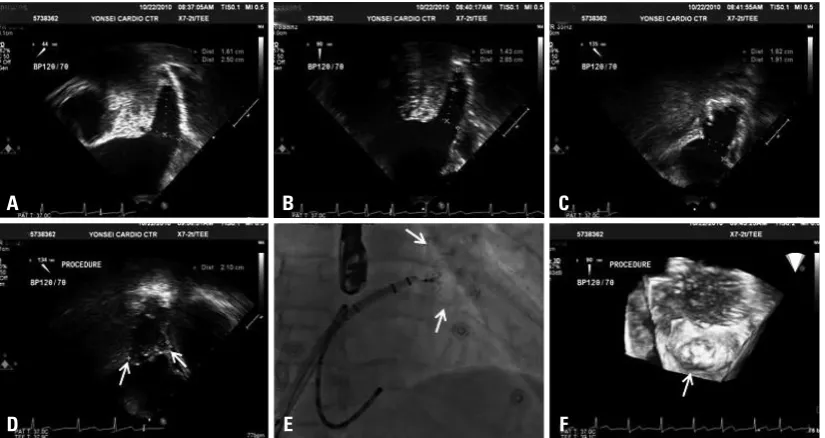
Fig. 2. Intra-procedural TEE images before (A, B and C) and after (D) deployment of LAA occlusion device. Diameter of LAA ostium and depth of LAA were measured from 4 different angles of TEE images to determine the appropriate size of WATCHMAN device. (D) Successful deployment of device should be confirmed using a tug test and color Doppler. (E) RAO 45° fluoroscopic view after deployment of WATCHMAN device. (F) Eight week follow-up 3-D TEE showed complete sealing off of LAA by WATCHMAN device. TEE, trans-esoph-ageal echocardiography; LAA, left atrial appendage; RAO, right anterior oblique.
A
D
B
E
C
ture of the device inside LAA via tug test and contrast injection, the device was disconnected from the deployment knob by being screwed out (Fig. 2C-F).
Post-procedural follow-up
After deployment of the WATCHMAN device, we stopped heparin and removed the sheath when ACT <250 sec. The patients were discharged the next day and maintained aspi-rin 100 mg and an optimal dose of warfaaspi-rin (INR 2.0-3.0) for 8 weeks. TEE was repeated 8 weeks after the procedure, and we stopped warfarin and added clopidogrel 75 mg after confirming that there was no flow leakage between the WATCHMAN device and LAA.
Data analysis
We reviewed the reasons for deploying the LAA occlusion
device, degrees of LA remodeling, CHADS2 score, shape
and size of LAA, procedure time, adverse effects, and clini-cal outcome.
RESULTS
[image:4.595.68.263.68.739.2]We implanted LAA occlusion devices in 5 patients with AF, and the characteristics of these patients are summarized in Table 1. The mean age of the patients was 59.8±7.3 years old, and all of them were male. The major reasons for LAA occlusion device implantation were high risk of recurrent stroke (80%) and labile INR with hemorrhage (60%); 3/5 (60%) patients had a history of failed cardioversion for rhythm control. The mean LA anterior posterior diameter was 51.3±5.0 mm and LAA size was 25.1×30.1 mm. We implanted LAA occlusion devices (28.8±3.4 mm device) successfully in all 5 patients without complications. The mean procedure time was 72.8±10.1 min. After 8 weeks of anticoagulation, all patients switched from warfarin to anti-platelet agent after confirmation of successful LAA occlu-sion by trans-esophageal echocardiography.
Case 1
A 53-year-old male taxi driver came to the emergency room after suddenly developing right-side motor weakness. His electrocardiography showed low voltage QRS with AF, and brain computed tomography revealed an embolic cerebral infarction in the region of the left side middle cerebral ar-tery. He had a history of persistent AF lasting longer than 4 years and initially presented with symptomatic pericardial
Ta bl e 1. P at ie nt C ha ra ct er ist ic s Pa tie nt nu m be r Se x A ge Re as on fo r L A A oc cl us io n Ty pe of A F CH A D S 2 sc or e LA A P di am et er (m m ) Pr ev io us hi sto ry o f rh yt hm co nt ro l t ria l Ty pe o f LA A Si ze o f LA A (m m ) D ev ic e siz e (m m ) Pr oc ed ur e tim e (m in ) Co m pl i-ca tio n 8 w ee ks TE E fo llo w up Cu rre nt an tit hr om bo tic str at eg y 1 M 53 St ro ke , l ab ile IN R, re cu rre nt bl ee di ng Pt A F 3 50 .0 Ye s Si ng le lo be 21 .5 ×2 9. 0 27 57 N on e N o le ak ag e A nt ip la te le t 2 M 67 Re cu rre nt st ro ke , w ar fa rin -in du ce d ce re br al h em or -rh ag e Pt A F 5 47 .6 N o M ul tip le lo be 27 .7 ×3 0. 7 30 72 N on e N o le ak ag e A nt ip la te le t 3 M 60 Re cu rre nt st ro ke Pe A F 4 49 .4 Ye s Si ng le lo be 19 .5 ×2 8. 5 24 75 N on e N o le ak ag e A nt ip la te le t 4 M 52 M CA in fa rc tio n, le ft Pt A F 3 49 .4 Ye s Br oc co li sh ap e 28 .2 ×2 9. 3 30 85 N on e N o le ak ag e A nt ip la te le t 5 M 67 La bi le IN R Pt A F 1 60 .0 N o Si ng le L ob e 28 .4 ×3 3. 2 33 75 N on e N o le ak ag e A nt ip la te le t Pt AF , p er m an en t A F; Pe AF , p er sis te nt A F; M CA , m id dl e ce re br al a rte ry ; I NR , i nt er na tio na l n or m al ize d ra tio cl op id og re
l; LA
complications (Fig. 2D and F). Because the device was well-fitted within the LAA (Fig. 2E) and there was no flow leakage between the LAA and the WATCHMAN device, we stopped warfarin and switched to clopidogrel.
Case 2
The second case was a 67-year-old male patient with hy-pertension and diabetes who had experienced an embolic stroke 3 years prior. Unfortunately, cerebral hemorrhage and optic nerve damage had occurred related to an adverse event of anticoagulation 2 years prior. Therefore, we switched from warfarin to aspirin and clopidogrel. However, the pa-tient had another ischemic stroke again 7 months after start-ing the antiplatelet agent in place of warfarin. We decided to deploy the LAA occlusion device due to his recurrent strokes and high risk of bleeding with anticoagulation. The WATCH-MAN device implantation was successful and we main-tained warfarin for 8 weeks with very careful INR monitor-ing, and finally stopped anticoagulation after confirmation of no leakage with TEE.
Cases 3 and 4
Cases 3 and 4 had recurrent ischemic strokes and a large middle cerebral artery infarction, respectively (Table 1). The patients did not want continuous warfarin therapy because they had frequent minor bleeding despite optimal INR. Rhythm control with electrical cardioversion was attempted in both patients, but AF recurred within a week despite anti-arrhythmic drugs. Therefore, we successfully deployed a effusion. At that time, we could not find any pathology for
[image:5.595.102.513.494.710.2]pericardial effusion except for mediastinal lymphadenopathy and high adenosine deaminase level in the pericardial fluid. The mediastinal lymph node biopsy results indicated reac-tive hyperplasia. Anti-tuberculous medication was pre-scribed for one year under the impression that the patient had tuberculous pericarditis. However, he still had a mild degree of pericardial effusion, despite medication. His LA anterior posterior diameter measured by trans-thoracic echo-cardiography was 50.0 mm and his left ventricular ejection fraction was within the normal limits. To prevent ischemic stroke, anti-coagulation was maintained, but it was very hard to keep INR at the optimal level, despite strict drug and diet control. INR levels fluctuated remarkably, and the patient came to the emergency room several times due to severe bruising (Fig. 3). Because of labile INR with warfarin, we attempted rhythm control using cardioversion, but AF re-curred very soon after cardioversion. We stopped warfarin and switched to clopidogrel; his CHADS2 score was 1 at that time. Unfortunately, this patient had an embolic stroke 9 months after switching to an anti-platelet agent. Fig. 3 dis-plays the warfarin dosage and INR values. When he started anti-tuberculous medication, his INR level was sub-optimal despite a high warfarin dosage, but it rose after stopping ri-fampin and increased rather drastically, even with 0.5 mg of warfarin. After an ischemic stroke, the neurologist pre-scribed low-dosage warfarin, but the INR value increased to higher than 8.0. Therefore, we decided to implant an LAA closing device, and the procedure was successful, with no
Fig. 3. Warfarin dosage and INR values of case 1. INR values were extremely labile, and ischemic stroke occurred 9 months after switch-ing to clopidogrel. INR, international neutralizswitch-ing ratio; MCA, middle cerebral artery.
0 1 2 3 4 5 6 7 8 9 10
mg/INR
Anti-TB medication
Stop W MCA infarction
Warfarin (mg) INR
percutaneous epicardial LAA suture ligation approach was also tested for pre-clinical feasibility.22 Recently, the PRO-TECT-AF trial, the largest-scale study to date, with 707 pa-tients, showed that hemorrhagic stroke occurred less often with the WATCHMAN device than with warfarin treatment; stroke and all-cause mortality outcomes were non-inferior.18 The inclusion criteria were: patients aged >18 years old with paroxysmal, persistent, or permanent non-valvular AF
and CHADS2 score ≥1. In the WATCHMAN group, the
procedure success rate was 88%; 85% of them had stopped warfarin after 45 days and 92% after six months, if TEE showed residual peri-device flow jet <5 mm in width. There-fore, the WATCHMAN device is ideally indicated for pa-tients with persistent or permanent AF who cannot tolerate anticoagulation despite a significant risk of ischemic or re-current stroke, and who are unable to maintain sinus rhythm. If anticoagulation is contraindicated, the patient is not can-didate for the WATCHMAN device, as the procedure requires that patients undergo anticoagulation for several weeks af-ter deployment. It is not clear whether an LAA occlusion device can replace warfarin or rhythm control strategy in patients with AF; further study with a longer follow-up pe-riod is warranted.
Potential complications of LAA occlusion device
Although the PROTECT-AF trial showed the non-inferiority of the LAA occlusion device compared to warfarin (>99.9% with regard to any kind of stroke, cardiovascular or unex-plained death, or systemic embolism within up to 3 years), procedure-related adverse events should be accounted for, such as cardiac tamponade (4.8%), device embolization or dislodgement (0.4%), procedure-related stroke (mostly air embolism 1.1%), or sepsis.18 However, Reddy, et al.23 recent-ly reported that such adverse events were dependent on the level of experience of the operator. The subsequent registry after the PROTECT AF trial showed a significantly im-proved success rate of implantation (91.3% to 95.0%), and reduced procedure time as well as the occurrence of proce-dure-related adverse events (6.5% to 3.7%). The procedure-related stroke rate was reduced to 0%.
Unsolved issue
Long-term safety data for the WATCHMAN device are still unavailable and need to be taken into account for clinical consensus. Complex aortic plaque on the descending aorta is an independent risk factor for stroke in AF24 and the de-gree of electroanatomical remodeling of LA may affect the WATCHMAN device in each patient. The shape of the LAA
in patient #4 was of the multi-lobulated Broccoli type, with a broad base and short length. In this type of LAA, the de-vice was easily shifted or tilted to the dominant lobe of the LAA. Therefore, we repeated deployment and recapture of the device several times, and finally implanted a 30.0 mm sized device at the optimal site without complication. The eight week follow-up TEE showed successful implantation of the device in both patients, and so warfarin was switched to anti-platelet agent.
Case 5
Case #5 was a 67-year-old patient with permanent AF.
Al-though his CHADS2 score was 1, his LA diameter was 60.0
mm and TEE showed severe spontaneous echo contrast. His INR value was very unstable and he experienced fre-quent purpura on his skin. Therefore, we successfully de-ployed the LAA occlusion device, and switched from war-farin to clopidogrel 8 weeks after implantation.
DISCUSSION
We report five cases of persistent or permanent AF that were successfully treated with LAA occlusion devices. None of the patients had been able to tolerate anticoagulation, de-spite significant risk of stroke because of labile INR or ad-verse hemorrhagic events. Three of them failed to maintain rhythm control after electrical cardioversion, and cardiover-sion was not attempted in two of them due to a huge atrium or to a high risk of bleeding with anticoagulation. Therefore, an LAA occlusion device might be considered for patients with persistent or permanent AF who cannot tolerate anti-coagulation and cannot maintain sinus rhythm.
Efficacy of LAA closure and ideal indications in patients with AF
It is well established that more than 90% of atrial thrombi
originate from the LAA,19 and the surgical MAZE
5. Stoddard MF, Dawkins PR, Prince CR, Ammash NM. Left atrial appendage thrombus is not uncommon in patients with acute atrial fibrillation and a recent embolic event: a transesophageal echocar-diographic study. J Am Coll Cardiol 1995;25:452-9.
6. Manning WJ, Weintraub RM, Waksmonski CA, Haering JM, Rooney PS, Maslow AD, et al. Accuracy of transesophageal echo-cardiography for identifying left atrial thrombi. A prospective, in-traoperative study. Ann Intern Med 1995;123:817-22.
7. Leung DY, Black IW, Cranney GB, Hopkins AP, Walsh WF. Prog-nostic implications of left atrial spontaneous echo contrast in non-valvular atrial fibrillation. J Am Coll Cardiol 1994;24:755-62. 8. Singer DE, Chang Y, Fang MC, Borowsky LH, Pomernacki NK,
Udaltsova N, et al. The net clinical benefit of warfarin anticoagu-lation in atrial fibrilanticoagu-lation. Ann Intern Med 2009;151:297-305. 9. Corley SD, Epstein AE, DiMarco JP, Domanski MJ, Geller N,
Greene HL, et al. Relationships between sinus rhythm, treatment, and survival in the Atrial Fibrillation Follow-Up Investigation of Rhythm Management (AFFIRM) Study. Circulation 2004;109: 1509-13.
10. Petersen P, Boysen G, Godtfredsen J, Andersen ED, Andersen B. Placebo-controlled, randomised trial of warfarin and aspirin for prevention of thromboembolic complications in chronic atrial fi-brillation. The Copenhagen AFASAK study. Lancet 1989;1:175-9. 11. Hylek EM, D’Antonio J, Evans-Molina C, Shea C, Henault LE,
Regan S. Translating the results of randomized trials into clinical practice: the challenge of warfarin candidacy among hospitalized elderly patients with atrial fibrillation. Stroke 2006;37:1075-80. 12. Connolly SJ, Ezekowitz MD, Yusuf S, Eikelboom J, Oldgren J,
Parekh A, et al. Dabigatran versus warfarin in patients with atrial fibrillation. N Engl J Med 2009;361:1139-51.
13. Connolly S, Pogue J, Hart R, Pfeffer M, Hohnloser S, Chrolavi-cius S, et al. Clopidogrel plus aspirin versus oral anticoagulation for atrial fibrillation in the Atrial fibrillation Clopidogrel Trial with Irbesartan for prevention of Vascular Events (ACTIVE W): a ran-domised controlled trial. Lancet 2006;367:1903-12.
14. Go AS, Hylek EM, Borowsky LH, Phillips KA, Selby JV, Singer DE. Warfarin use among ambulatory patients with nonvalvular atrial fibrillation: the anticoagulation and risk factors in atrial fi-brillation (ATRIA) study. Ann Intern Med 1999;131:927-34. 15. Camm AJ, Kirchhof P, Lip GY, Schotten U, Savelieva I, Ernst S,
et al. Guidelines for the management of atrial fibrillation: the Task Force for the Management of Atrial Fibrillation of the European Society of Cardiology (ESC). Europace 2010;12:1360-420. 16. Bonow RO, Carabello BA, Chatterjee K, de Leon AC Jr, Faxon
DP, Freed MD, et al. 2008 Focused update incorporated into the ACC/AHA 2006 guidelines for the management of patients with valvular heart disease: a report of the American College of Cardi-ology/American Heart Association Task Force on Practice Guide-lines (Writing Committee to Revise the 1998 GuideGuide-lines for the Management of Patients With Valvular Heart Disease): endorsed by the Society of Cardiovascular Anesthesiologists, Society for Cardiovascular Angiography and Interventions, and Society of Thoracic Surgeons. Circulation 2008;118:e523-661.
17. Healey JS, Crystal E, Lamy A, Teoh K, Semelhago L, Hohnloser SH, et al. Left Atrial Appendage Occlusion Study (LAAOS): re-sults of a randomized controlled pilot study of left atrial append-age occlusion during coronary bypass surgery in patients at risk for stroke. Am Heart J 2005;150:288-93.
18. Holmes DR, Reddy VY, Turi ZG, Doshi SK, Sievert H, Buch-binder M, et al. Percutaneous closure of the left atrial appendage
risk and event of stroke in patients with AF. The current guidelines recommend switching from warfarin to dural an-tiplatelet therapy 6-8 weeks after successful deployment of the LAA occlusion device.18 However, the risk of bleeding with dual anti-platelet therapy should be considered even after deployment of the LAA occlusion device. Theoretical-ly, complete rhythm control might be better than an LAA occlusion device in terms of etiology of ischemic stroke and hemodynamics. Therefore, LAA occlusion devices should be compared with catheter ablation of AF or newly devel-oped effective anti-arrhythmic drugs.25-27 Comparisons with direct thrombin or coagulation factor Xa inhibitors will be
required.28 The clinical efficacy of newly designed LAA
closure devices, such as the Amplatzer cardiac plug, need to be evaluated in a large randomized trial.29
In conclusion, here we report our early experience with LAA-OD deployment in patients with 1) persistent or per-manent AF who cannot tolerate anticoagulation despite sig-nificant risk of ischemic stroke or 2) recurrent stroke and inability to maintain sinus rhythm.
ACKNOWLEDGEMENTS
This work was supported by a grant (A085136) from the Korea Health 21 R&D Project, Ministry of Health and Wel-fare, and a grant (2010-0010537) from the Basic Science Research Program run by the National Research Founda-tion of Korea (NRF), which is funded by the Ministry of Ed-ucation, Science and Technology of the Republic of Korea. We also appreciate Mr. Soon Young Kwon’s (Heart Pace Control Inc. Seoul) technical and moderating support.
REFERENCES
1. Kannel WB, Abbott RD, Savage DD, McNamara PM. Epidemio-logic features of chronic atrial fibrillation: the Framingham study. N Engl J Med 1982;306:1018-22.
2. Go AS, Hylek EM, Phillips KA, Chang Y, Henault LE, Selby JV, et al. Prevalence of diagnosed atrial fibrillation in adults: national implications for rhythm management and stroke prevention: the AnTicoagulation and Risk Factors in Atrial Fibrillation (ATRIA) Study. JAMA 2001;285:2370-5.
3. Wolf PA, Abbott RD, Kannel WB. Atrial fibrillation as an inde-pendent risk factor for stroke: the Framingham Study. Stroke 1991;22:983-8.
tients with AF (PROTECT AF) clinical trial and the Continued Access Registry. Circulation 2011;123:417-24.
24. Lip GY, Lim HS. Atrial fibrillation and stroke prevention. Lancet Neurol 2007;6:981-93.
25. Hohnloser SH, Crijns HJ, van Eickels M, Gaudin C, Page RL, Torp-Pedersen C, et al. Effect of dronedarone on cardiovascular events in atrial fibrillation. N Engl J Med 2009;360:668-78. 26. Joung B, Chen PS, Lin SF. The role of the calcium and the voltage
clocks in sinoatrial node dysfunction. Yonsei Med J 2011;52:211-9. 27. Kim WH, Joung B, Shim J, Park JS, Hwang ES, Pak HN, et al. Long-term outcome of single-chamber atrial pacing compared with dual-chamber pacing in patients with sinus-node dysfunction and intact atrioventricular node conduction. Yonsei Med J 2010;51:832-7.
28. ROCKET AF Study Investigators. Rivaroxaban-once daily, oral, direct factor Xa inhibition compared with vitamin K antagonism for prevention of stroke and Embolism Trial in Atrial Fibrillation: rationale and design of the ROCKET AF study. Am Heart J 2010;159:340-7.e1.
29. Park JW, Bethencourt A, Sievert H, Santoro G, Meier B, Walsh K, et al. Left atrial appendage closure with Amplatzer cardiac plug in atrial fibrillation: initial European experience. Catheter Cardiovasc Interv 2011;77:700-6.
versus warfarin therapy for prevention of stroke in patients with atrial fibrillation: a randomised non-inferiority trial. Lancet 2009; 374:534-42.
19. Hwang JJ, Ko FN, Li YH, Ma HM, Wu GJ, Chang H, et al. Clini-cal implications and factors related to left atrial spontaneous echo contrast in chronic nonvalvular atrial fibrillation. Cardiology 1994;85:69-75.
20. Blackshear JL, Johnson WD, Odell JA, Baker VS, Howard M, Pearce L, et al. Thoracoscopic extracardiac obliteration of the left atrial appendage for stroke risk reduction in atrial fibrillation. J Am Coll Cardiol 2003;42:1249-52.
21. Block PC, Burstein S, Casale PN, Kramer PH, Teirstein P, Wil-liams DO, et al. Percutaneous left atrial appendage occlusion for patients in atrial fibrillation suboptimal for warfarin therapy: 5-year results of the PLAATO (Percutaneous Left Atrial Append-age Transcatheter Occlusion) Study. JACC Cardiovasc Interv 2009;2:594-600.
22. Singh SM, Dukkipati SR, d’Avila A, Doshi SK, Reddy VY. Per-cutaneous left atrial appendage closure with an epicardial suture ligation approach: a prospective randomized pre-clinical feasibili-ty study. Heart Rhythm 2010;7:370-6.