Department of Medical Physics & Biomedical Engineering
Proton and Advanced RadioTherapy GroupPhD Thesis
submitted for the degree of Doctor of Philosophy from University College London
Toward adaptive radiotherapy
Catarina Isabel Correia Veloso da Veiga
Supervisors: Gary Royle – Proton and Advanced Radiotherapy Group, UCL
Jamie McClelland – Centre for Medical Image Computing, UCL
Kate Ricketts – Division of Surgery and Interventional Sciences, UCL
I, Catarina Isabel Correia Veloso da Veiga, confirm that the work presented in this thesis is my own. Where information has been derived from other sources, I confirm this has been indicated in the thesis.
Intensity Modulated Radiotherapy (IMRT) and proton therapy are the state-of-art external radiotherapy modalities. To make the most of such precise delivery, accurate knowledge of the patient anatomy and biology during treatment is necessary, as unac-counted variations can compromise the outcome of the treatment. Treatment modification to account for deviations from the planning stage is a framework known as adaptive ra-diotherapy (ART).
To fully utilise the information extracted from different modalities and/or at different time-points it is required to accurately align the imaging data. In this work the feasibility of cone-beam computed tomography (CBCT) and deformable image registration (DIR) for ART was evaluated in the context of head and neck (HN) and lung malignancies, and for IMRT and proton therapy applications. This included the geometric validation of de-formations for multiple DIR algorithms, estimating the uncertainty in dose recalculation of a CBCT-based deformed CT (dCT), and the uncertainty in dose summation resulting from the properties of the underlying deformations. The dCT method was shown to be a good interim solution to repeat CT and a superior alternative to simpler direct usage of CBCT for dose calculation; proton therapy treatments were more sensitive to registration errors than IMRT. The ability to co-register multimodal and multitemporal data of the HN was also explored; the results found were promising and the limitations of current algorithms and data acquisition protocols were identified.
The use of novel artificial cancer masses as a novel platform for the study of imag-ing durimag-ing radiotherapy was explored in this study. The artificial cancer mass model was extended to generate magnetic resonance imaging (MRI)-friendly samples. The tu-moroids were imageable in standard T1 and T2 MRI acquisitions, and the relaxometric
properties were measured. The main limitation of the current tumour model was the poor reproducibility and controllability of the properties of the samples.
That sad look shake it off, the road has been too long. We’re all just
passengers, in time and space, an end we never chase.
Tom Barman
I would sincerely like to thank my supervisors, without whom this work would not have been possible. First, to Professor Gary Royle for giving me this unique opportunity along with his constant support, guidance, time and great sense of humour over the course of these long four years. To Dr Jamie McClelland, for all those long coffee breaks discussing papers and code, for all his patience when something went wrong, for all the kindness and trust; thank you for being my rock during the difficult moments of this PhD. And finally, to Dr Kate Ricketts, who always knew how to lift my spirits with her enthusiasm, support and encouragement.
During the duration of my PhD, I was extremely lucky to collaborate with fantastic people from different research groups and backgrounds. From the Centre for Medical Image Computing, I would like to acknowledge Marc Modat, Pankaj Daga, Gergely Zombori, Matt Clarkson, Sebastien Ourselin, Dave Hawkes, and Marcel van Herk for the support received with image registration in general, NifTK in particular and overall di-rection of this project. From the Departments of Radiotherapy and Radiotherapy Physics at University College London Hospital, I am indebted to Derek D’Souza, Ivan Rosenberg and Richard Amos for always so kindly motivating, encouraging, and guiding me in this journey; and to Rachel Bodey, Syed Moinuddin, Paul Doolan, Jailan Alshaiki, Phil Davies, Chris Stacey, Maria Kilkenny, Dr Dhanasekaran Kittappa, Dr Swee-Ling Wong, and Dr Ruheena Mendes for always finding the time and patience to help me with all the clinical needs of this project. From the Centre for Medical Imaging, I would like to show my appreciation for Dr Shonit Punwani and Heather Fitzke for the helpful discussions on the usability of MRI in radiotherapy and access to clinical trial data. From the Division of Surgery, I would like to thank Dr Marilena Loizidou, Tarig Magdeldin, Tong Long, Victor Lopez-Davila, and Bala Ramesh for all the patience required to guide a physicist through the complex world of biology. I am also very grateful to Bernard Siow, for all the long
Moss and George Randall for their amazing workshop skills, Dan O’Flynn for data ac-quisition on a benchtop CT scanner, and Reem Al-Samarraie for support in locating the materials of the box for transporting the tumoroids. Finally, my appreciation to Amber Cuming (Beekley Corporation) and Samuel Naslund (Naslund Medical AB) for kindly providing samples of CT/MR markers.
From my time at the University of Pennsylvania, I would first like to thank Dr Kevin Teo for receiving me so warmly in Philadelphia and at the Roberts Proton Therapy Center; I always felt very welcome and a valuable member of the team. A very special thanks goes to Guillaume Janssens for his 24/7 support, professionalism and sympathy; I was really lucky to always have the best collaborators. To Dr Ching-Ling Teng for her enthusiasm in the final phase of my stay in Philadelphia; without your excellent writing skills, full-time availability, girly lunch breaks and great attitude I do not believe this project could have been as successful. I am also grateful to Thomas Baudier and Lucian Hotoiu for their kindness and technical contributions to the project, and to Lingshu Yin, Sebastien Brousmiche, Liyong Lin, James Metz, Timothy Solberg, Zelig Tochner, and James McDonough for their contributions to the CBCT and adaptive lung proton therapy project. To David Weiss, Evan Meekins and Marcus Fager, for addingsweet, pickleand meatto mysaltyness. To my favouriteguapitas for cheering me up when I needed it the most. I was really my happiest during my time at UPenn, and I am so grateful to have been given the opportunity to do this project. Looking back I realise how much I grew as a woman and a scientist in the United States. Philly, you changed me for better and for good.
To everyone in the Department of Medical Physics & Biomedical Engineering at UCL, particularly, Alessandro Proverbio, Anna Zamir, Christiana Christodolou, Dan O’Flynn, Edgar Gelover Reyes, Emma Biondetti, Esther Bär, Vanessa La Rosa, George Randall, Ireneos Drakos, and Paul Burke. Thank you for all the good times, either at the office, common room, pub or outdoors! And coffee, thank you for all the coffee!
A very emotional thanks goes to my important friends, my lifetime friends: Abi-gail Moreira, Ana Luísa Castro Lopes, Ana Mónica Lourenço, Bruno Gomes, Consuelo Guardiola, Diana Barros, João Koch, João Nuno Mota, João Tavares, Juliana Narciso, Lucia Tejo, Mariana Dantas, Nuno Alcobia, Raphael Lopes, Raquel Koch, Raquel Leão, Reem Al-Samarraie, Sara Campos, Sofia C. Ribeiro, and Sofia J. Ribeiro. Distance can take quite a toll on relationships; nevertheless, you were always there for me. Knowing you always waited for my return, making me feel as if I had never left, meant more than words can convey. I am truly blessed to have such amasing people in my life. I would also like to express my genuine appreciation to Paulo Castro and Stavros Vorrias; while our paths may have split somewhere along this long road, your companionship was really important to me throughout our happy times.
apart: my parents Carlos and Isabel; my brother Nuno; my grandparents, Eurico, Maria Augusta and Ulisses; my amasing and favourite aunts, Patricia, Nocas and Sissi; and my little cousin, Eva. I know the moments we lost will never be recovered, but allow me to be selfish this one time and convince myself that we still have all the time in the world to recapture them all.
Finally, I would like to gratefully acknowledge the financial support received from Fundação para a Ciência e a Tecnologia (FCT) grant SFRH/BD/76169/2011, co-financed by ESF, POPH/QREN and EU; IOP, IPEM and UCL Graduate School for funding conference trips.
1 Introduction 27
1.1 Contextualisation of the research project . . . 27
1.2 Research question and aims . . . 28
1.3 My contribution to this work . . . 29
1.4 Novelty of this work . . . 31
1.5 Impact of this work . . . 32
1.6 Structure of this thesis . . . 34
2 The role of deformable image registration in adaptive radiotherapy 37 2.1 An introduction to cancer radiobiology . . . 38
2.2 An introduction to image registration . . . 41
2.2.1 Transformation Model . . . 41
2.2.2 Similarity metric . . . 42
2.2.3 Optimisation . . . 44
2.2.4 Symmetry, inverse-consistency and diffeomorphisms . . . 44
2.2.5 Evaluation and validation of deformable image registration . . . . 46
2.2.6 In-house software: NifTK . . . 46
2.2.6.1 Data transfer between NifTK and clinical systems . . . . 47
2.2.7 Other registration algorithms . . . 49
2.3 The role of image registration in image guidance and adaptive radiotherapy 49 2.3.1 The clinical problem: head and neck . . . 50
2.3.2 The clinical problem: lung. . . 51
2.4 Initial studies: optimisation of NiftyReg . . . 52
2.4.1 Choice of registration parameters and algorithms . . . 52
2.4.2 Image pre-processing to improve registration quality . . . 55
2.5 Optimisation of NiftyReg for CT to cone-beam CT deformable image reg-istration. . . 58
2.5.1 Methods and Materials . . . 59
2.5.1.1 Patients data acquisition . . . 59
2.5.1.2 Registration settings . . . 59
2.5.1.3 Contours comparison . . . 59
2.5.2 Results . . . 62
2.5.3 Discussion . . . 65
2.6 Conclusions . . . 65
3 Cone-beam CT and deformable image registration for “dose of the day” calcu-lations 67 3.1 Rationale . . . 67
3.2 Methods and Materials . . . 68
3.2.1 Patient data acquisition . . . 68
3.2.2 Image registration settings . . . 69
3.2.3 Evaluation of the suitability of deformable image registration for “dose of the day” calculations. . . 70
3.2.3.1 Geometric evaluation . . . 71
3.2.3.2 Dose comparison . . . 72
3.2.3.3 Propagation of structures and “dose of the day”. . . 74
3.3 Results . . . 75
3.3.1 Geometric evaluation . . . 75
3.3.2 Dose comparison . . . 77
3.3.3 Propagation of structures and “dose of the day” . . . 77
3.4 Discussion . . . 80
3.5 Conclusions . . . 83
4 Dose warping and summation applications 85 4.1 Rationale . . . 85
4.2 Methods and Materials . . . 87
4.2.1 Patient data acquisition . . . 87
4.2.2 Image registration settings . . . 87
4.2.3 Dose warping and summation in an adaptive radiotherapy workflow 87 4.2.4 Evaluation scheme . . . 89
4.2.4.1 Geometric matching. . . 89
4.2.4.2 Characteristics and similarity of the deformation fields. . 89
4.2.4.3 Computation times . . . 90
4.2.4.4 Dose warping comparison . . . 90
4.3 Results . . . 91
4.3.1 Geometric matching . . . 91
4.3.2 Deformation field analysis. . . 92
4.3.3 Computation times . . . 93
4.3.4 Dose warping comparison. . . 94
4.4 Discussion . . . 97
4.5 Conclusions . . . 100
5.1 An introduction to proton therapy . . . 101
5.2 Rationale . . . 103
5.3 Methods . . . 106
5.3.1 Patient data acquisition . . . 106
5.3.2 Treatment planning. . . 106
5.3.3 Image registration settings . . . 107
5.3.3.1 Geometric matching and properties of the deformation fields108 5.3.4 Dose comparison . . . 108 5.4 Results . . . 109 5.4.1 Geometric validation . . . 109 5.4.2 Dose comparison . . . 109 5.5 Discussion . . . 114 5.6 Conclusions . . . 116
6 Lung adaptive proton therapy 117 6.1 Rationale . . . 118
6.2 Methods and Materials . . . 119
6.2.1 Patient selection and data acquisition . . . 119
6.2.2 Overview of an adaptive lung proton therapy workflow . . . 122
6.2.2.1 Deformable registration. . . 123
6.2.2.2 Deformed CT correction . . . 124
6.2.2.3 Water equivalent thickness . . . 125
6.2.2.4 Range-corrected dose . . . 125
6.2.2.5 Clinical indicators . . . 125
6.2.3 Implementation details . . . 126
6.2.4 Evaluation of the adaptive proton therapy workflow . . . 127
6.2.5 Accuracy of cone-beam CT and deformable image registration for adaptive lung therapy . . . 127
6.2.5.1 Deformable registration. . . 127
6.2.5.2 Cone-beam CT dataset definition . . . 128
6.2.5.3 Validation workflow of the deformed CT method . . . 131
6.2.5.4 Comparison of the deformed CT method to simpler methods131 6.2.6 Clinical indicators of replanning . . . 132
6.3 Results . . . 133
6.3.1 Accuracy of cone-beam CT and deformable image registration for adaptive lung therapy . . . 133
6.3.1.1 Overall uncertainty of the deformed CT on water equiva-lent thickness and dose estimation . . . 133
6.3.1.2 Effect of different cone-beam CT datasets . . . 135
6.3.1.3 Effect of different registration algorithms . . . 137
6.3.1.4 Effect of deformed CT correction . . . 137
6.3.1.6 Comparison of the deformed CT method to simpler methods139
6.3.2 Clinical indicators of replanning . . . 139
6.3.2.1 Lung changes . . . 140
6.3.2.2 Tumour changes . . . 144
6.3.2.3 General considerations . . . 147
6.3.3 Discussion . . . 148
6.3.4 Conclusions . . . 152
7 Multimodal and multitemporal imaging in radiotherapy 155 7.1 The role of multimodal and multiparametric imaging in radiotherapy. . . 155
7.2 Rationale . . . 158
7.3 Methods and materials . . . 159
7.3.1 Patient data acquisition . . . 159
7.3.2 Multimodal and multiparametric imaging in a radiotherapy workflow160 7.3.3 Image registration settings . . . 162
7.3.4 Quantitative analysis. . . 163
7.4 Results . . . 164
7.5 Discussion . . . 166
7.6 Current status and future work . . . 166
7.7 Conclusion . . . 168
8 A novel artificial cancer mass model for imaging applications 169 8.1 Introduction to tissue engineering . . . 169
8.2 Engineering of a tridimensional cancer model. . . 172
8.2.1 Cell culture . . . 173
8.2.2 Collagen matrix . . . 174
8.3 Physics of magnetic resonance imaging . . . 175
8.3.1 Contrast mechanisms of conventional magnetic resonance imaging 175 8.3.2 Pulse sequences. . . 176
8.3.3 Measurement of T1and T2relaxation times . . . 177
8.4 Design of an artificial cancer mass for magnetic resonance imaging . . . . 178
8.4.1 Design specifications . . . 178
8.4.2 Biological properties of the samples . . . 178
8.4.2.1 Cell density. . . 178
8.4.2.2 Sample fixation. . . 181
8.4.3 Design of the imaging experiments. . . 182
8.4.3.1 Magnetic resonance system specifications . . . 182
8.4.3.2 Experimental setup and sample holder . . . 182
8.4.3.3 Fiducial markers . . . 183
8.4.3.4 Sample transportation and storage . . . 185
8.4.3.5 Timeline for imaging sessions . . . 186
8.5.1 Methods and materials. . . 187
8.5.1.1 Samples description . . . 187
8.5.1.2 Data acquisition . . . 189
8.5.1.3 Measurement of T1and T2relaxation times . . . 189
8.5.2 Results and discussion . . . 190
8.5.2.1 Study I . . . 190
8.5.2.2 Study II . . . 193
8.5.2.3 Study III. . . 195
8.5.2.4 Measurement of T1and T2relaxation times . . . 198
8.6 Current status and future work . . . 200
8.6.1 Sample production: design, reproducibility and engineering. . . . 203
8.6.2 Characterisation and biological properties of the samples. . . 204
8.6.3 Magnetic resonance imaging setup. . . 205
8.7 Conclusions . . . 207
9 Final remarks 209
A Clinical indicators of replanning 211
B Cell maintenance protocol 219
C Cell subculture protocol 221
D Cell counting protocol 223
E Collagen matrix preparation protocol 225
F Mould re-design 229
2.1 Direct and indirect actions of radiation. . . 38
2.2 Cell cycle and radiosensitivity. . . 40
2.3 Reoxygenation. . . 40
2.4 Key items of any image registration algorithm. . . 41
2.5 Importance of well constrained registrations. . . 53
2.6 Deformable vs rigid registration. . . 53
2.7 Similarity as function of the weight of penalty terms . . . 56
2.8 Computation time using different initial rigid alignments. . . 56
2.9 Transformation applied by gamma correction. . . 57
2.10 Structure set manually delineated for CT-to-CBCT DIR validation.. . . 60
2.11 DSC vs OI. . . 63
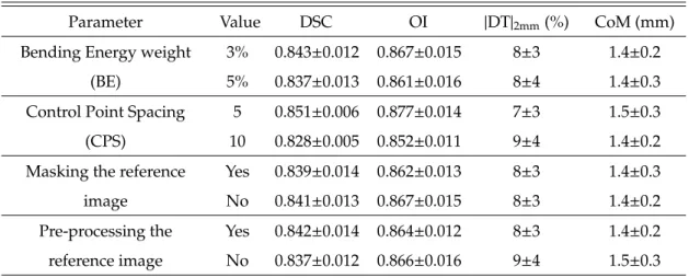
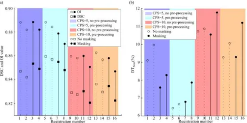
2.12 Variation of DSC, OI and|DT|2mmfor different combinations of parameters. 64 2.13 Dose similarity between choices of DIR parameters . . . 64
3.1 B-spline control point grid placement. . . 70
3.2 FN and FP versus DSC and OI . . . 72
3.3 Catphan 504. . . 73
3.4 CBCT-CT HU and RED calibration curves. . . 73
3.5 Diagram of the data and registrations used in the dosimetric evaluation. . 74
3.6 Geometric matching of manual and warped features. . . 75
3.7 Distribution of distance transform values. . . 77
3.8 Qualitative dose similarity results. . . 78
3.9 Dose differences inside organs at risk. . . 78
3.10 Dose volume histograms using different doses and structures. . . 80
4.1 Dose warping and summation in an adaptive radiotherapy workflow . . . 88
4.2 Distance to dose difference flow diagram. . . 91
4.3 L2-norm between deformation vector fields.. . . 93
4.4 Inverse-consistency error. . . 94
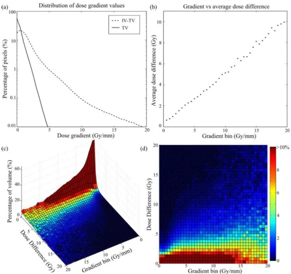
4.5 Dose uncertainty versus dose gradient. . . 96
4.6 Dose volume histogram using different DIR algorithms.. . . 98
5.2 Proton delivery systems. . . 104
5.3 Dose volume histogram comparing proton and photon plans. . . 107
5.4 Difference in dose between replan and deformed CT. . . 112
5.5 Dose volume histogram comparing dose in replan and deformed CT. . . . 113
6.1 Relative stopping power calibration curve. . . 122
6.2 Workflow for clinical lung adaptive proton therapy. . . 123
6.3 Pipeline for dCT correction. . . 124
6.4 Diagram of online adaptive proton therapy workflow. . . 127
6.5 Photos of the RANDO phantom setup. . . 129
6.6 Regular and simulated CBCT of RANDO phantom. . . 129
6.7 CBCT datasets. . . 130
6.8 Diagram of data and registrations. . . 130
6.9 Virtual CT versus other other methods. . . 132
6.10 Color overlay between rCT and pCT/dCTs. . . 134
6.11 Dose colorwash overlay on rCT using planned, range-corrected, and recal-culated on rCT doses. . . 135
6.12 Boxplot of the DTRMSvalues. . . 136
6.13 Examples of the dCT correction.. . . 138
6.14 Images used and generated by the lung adaptive proton therapy workflow (Example 1). . . 141
6.15 Color overlay of the CTs and corresponding dose distributions and DVHs. 142 6.16 Images used and generated by the lung adaptive proton therapy workflow (Example 2). . . 143
6.17 WET and WET difference maps.. . . 145
6.18 Images used and generated by the lung adaptive proton therapy workflow (Example 3). . . 146
7.1 Inclusion criteria. . . 160
7.2 MR data limitations. . . 161
7.3 Schematic diagram of registration pathways. . . 161
7.4 Structure set manually delineated for CT-MR and MR-MR DIR validation. 163 8.1 Platforms to study cancer and therapies. . . 170
8.2 Elements of tissue engineering . . . 171
8.3 Mould. . . 174
8.4 Original tumoroid model. . . 174
8.5 MR relaxation to equilibrium. . . 175
8.6 Components of the magnetisation due to the 90◦RF pulse. . . 175
8.7 Cell density measured over 14 days of ACMs with varying cell seeding value.179 8.8 Collagen density. . . 180
8.10 Microscopy images of the tumour model over 21 days. . . 181
8.11 MRI system and coil-holder. . . 182
8.12 Sample holder for MR imaging. . . 183
8.13 MR-compatible markers. . . 184
8.14 CT of Gold AnchorTM. . . 186
8.15 Tumoroid transport. . . 186
8.16 Timeline. . . 187
8.17 Samples used for the imaging studies. . . 188
8.18 Acellular and 30M tumoroid samples. . . 188
8.19 T1and T2theoretical and experimental fitting curves. . . 190
8.20 T1IR-RARE images of an acellular tumoroid. . . 191
8.21 T2MSME images of an acellular tumoroid. . . 191
8.22 T∗ 2FLASH images of an acellular tumoroid. . . 192
8.23 T1IR-RARE images of a 30M tumoroid. . . 192
8.24 T2MSME images of a 30M tumoroid.. . . 193
8.25 Intensity profile on T1and T2images. . . 194
8.26 T1IR-RARE images of an acellular tumoroid. . . 194
8.27 T2MSME images of an acellular tumoroid. . . 195
8.28 T1IR-RARE images of 0M, 20M and 40M tumoroids. . . 196
8.29 T2MSME images of 0M, 20M and 40M tumoroids. . . 197
8.30 T1and T2fitting.. . . 199
8.31 T1and T2colormaps. . . 200
8.32 T1histograms. . . 201
8.33 T2histograms. . . 201
8.34 Optimal acquisition for T1and T2contrast. . . 202
8.35 3D printer cap add-on . . . 205
8.36 Heath deflection for selected polymers. . . 206
8.37 Improvement to the sample holder.. . . 206
D.1 Squares of the haemocytometer used in cell counting. . . 224
E.1 Mould preparation for tumoroid production. . . 226
E.2 Preparation of the meshes for tumoroid production. . . 227
F.1 Mould re-design. . . 231
2.1 Functionalities implemented in NiftyReg. . . 47
2.2 DIR parameters in NiftyReg.. . . 48
2.3 Similarity between manual and registered contours for different registra-tion settings. . . 63
3.1 Characteristics of the patients. . . 69
3.2 Similarity between manual and registered contours . . . 76
3.3 Similarity between dose distributions . . . 79
3.4 Similarity between the isodose volumes . . . 79
4.1 Theoretical properties of DIR algorithms. . . 89
4.2 Geometric matching of manual and warped structures for different DIR algorithms. . . 92
4.3 Properties of the deformation vector fields for different DIR algorithms. . 93
4.4 Computation times for different DIR algorithms. . . 94
4.5 Dose warping similarity for different DIR algorithms. . . 95
4.6 Dose differences at organs at risk for different DIR algorithms. . . 97
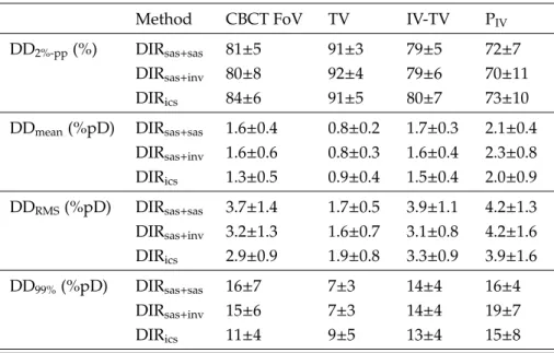
5.1 Dose statistics and properties of proton and photon plans. . . 108
5.2 Quantitative assessment of NMI vs LNCC registrations. . . 110
5.3 Qualitative dose similarity results for different methods and treatments (DD2%−pp). . . 111
5.4 Qualitative dose similarity results for different methods and treatments (DDRMS) . . . 112
5.5 Qualitative dose similarity results at organs-at-risk. . . 113
6.1 Patient characteristics. . . 120
6.2 Overall uncertainty in WET within the PTV, and on the distal and proximal surfaces. . . 134
6.3 Overall uncertainty in dose estimation. . . 136
6.4 Uncertainty in WET and dose for each CBCT dataset. . . 136
6.5 Uncertainty in WET and dose for each DIR algorithm. . . 137
6.7 Uncertainty in WET and dose for CT-to-CBCT and pCT-to-rCT registrations.139
6.8 Overall uncertainty for the virtual CT and simpler methods . . . 140
7.1 Quantitative assessment of the CT-MR1and MR1-MR2registrations. . . . 165
8.1 Parameters for SE and GE acquisitions. . . 177
8.2 T1and T2relaxation times for stroma and ACMs.. . . 199
A.1 Changes in WET between planning and verification scans. . . 212
A.2 Variation in DVH statistics from planning to verification doses . . . 214
1D unidimensional. 2D bidimensional. 3D tridimensional. 4D four-dimensional.
AAA analytical isotropic algorithm. ACM artificial cancer mass.
ADC apparent diffusion coefficient. AP anterior-posterior.
ART adaptive radiation therapy.
BE bending energy penalty term. BOLD blood oxygen level dependent.
CABI Centre for Advanced Biomedical Imaging.
CBCT cone-beam CT.
CMIC Centre of Medical Image Computing. CoM center-of-mass (centroid) position error. CP control point.
CPS control point spacing. CT computed tomography. CTV clinical tumour volume.
dCBCT deformed CBCT.
dCT deformed CT. DD dose-difference.
DICOM Digital imaging in communications in medicine. DIR deformable image registration.
DMEM Dulbecco’s Modified Eagle Medium. DNA deoxyribonucleic acid.
DSC dice similarity coefficient. DT distance transformation. DTA distance to agreement. DTD distance to dose-difference. DVF deformation vector field. DVH dose-volume histogram. DW diffusion-weighted.
ECM extracellular matrix.
EDTA ethylenediaminetetraacetic acid.
FBS foetal bovine serum. FFD free-form deformation. FID free induction decay. FLASH fast low angle shot. FN false negatives.
FoV field-of-view. FP false positives.
GE gradient echo.
GPU graphics processing unit. GTV gross tumour volume.
HE harmonic energy. HN head and neck. HU Hounsfield unit.
HUP Hospital of the University Pennsylvania.
IBA Ion Beam Applications, SA. IC inverse-consistency penalty term. ICE inverse-consistency error.
iCTV internal clinical tumour volume. IGRT image-guided radiation therapy. IMPT intensity modulated proton therapy. IMRT intensity modulated radiation therapy. IR inversion-recovery.
IV irradiated volume.
JL logarithm of the Jacobian determinant penalty term.
LINAC linear accelerator.
LNCC localised normalised cross correlation. LSCM left sternocleidomastoid muscle.
MEM minimum essential medium.
MFO multiple-field optimisation. MIP maximum intensity projection. MR magnetic resonance.
MRI magnetic resonance imaging. MRS magnetic resonance spectroscopy. MSME multi-slice multi-echo.
NCC normalised cross correlation. NMI normalised mutual information.
OAR organ-at-risk. OI overlap index.
P/S penicillin/streptomycin. PA posterior-anterior.
PBS phosphate buffed saline.
pCT planning computed tomography. pD prescribed dose.
PET positron emission tomography. PLA polylactic acid.
PSPT passive scattering proton therapy. PTV planning target volume.
RARE rapid acquisition refocused echoes. rCBCT regular CBCT.
rCT replan/rescan CT.
RED relative electron density. RF radio-frequency.
RL right-left.
RMS root mean square. ROI region of interest.
RSCM right sternocleidomastoid muscle. RTK Reconstruction Toolbox.
sCBCT simulated CBCT. SE spin-echo.
SFUD single-field uniform dose. SI superior-inferior.
SOBP spread-out Bragg peak.
SPECT single-photon emission computed tomography. SSD sum of the squared differences.
TE echo time. TI inversion time.
TPS treatment planning system. TR repetition time.
TRE target registration error. TV treated volume.
UCL University College London.
UCLH University College London Hospital.
UK United Kingdom.
USA United States of America.
VOI volume of interest.
WET water equivalent thickness. WHO World Health Organization.
Peer-reviewed journal papers
(In preparation) N. Hobson, X. Weng-Jiang, C. Veiga, B. Siow, M. Ashford, N. T. K. Thanh, A. Schätzlein, and I. Uchegbu, “Design and synthesis of self-assembling polymeric iron oxide nanoparticle theranostics for applications in cancer diagnostics and cancer therapy,” ACS Nano (2016).
(In preparation) X. Weng-Jiang, N. Hobson, C. Veiga, B. Siow, N. T. K. Thanh, A. Schätzlein, and I. Uchegbu, “Aqueous in-flow synthesis of superparamagnetic iron oxide nanoparticles for dual T1/T2-weighted magnetic resonance imaging,” ACS Nano (2016).
(In preparation)C. Veiga, G. Janssens, T. Baudier, L. Hotoiu, S. Brousmiche, J. R. Mc-Clelland, C.-L. Teng, L. Yin, G. Royle, and B.-K. K. Teo, “The accuracy of CBCT and deformable registration for adaptive lung proton therapy” (2016).
C. Veiga, G. Janssens, C.-L. Teng, T. Baudier, L. Hotoiu, J. R. McClelland, G. Royle, L. Lin, L. Yin, J. Metz, T. D. Solberg, Z. Tochner, C. B. Simone II, J. McDonough, and B.-K. K. Teo, “First clinical investigation of CBCT and deformable registration for adaptive proton therapy of lung cancer,” Int. J. Radiat. Oncol. Biol. Phys. 95(1) 549-559 (2016).
C. Veiga, J. Alshaikhi, R. Amos, A. M. Lourenço, M. Modat, S. Ourselin, G. Royle, and J. R. McClelland, “CBCT and deformable registration based “dose of the day” calculations for adaptive proton therapy,” Int. J. Particle Ther. 2(2) 404-414 (2015).
A. K. Hoang Duc, G. Eminowicz, J. McClelland, M. Modat, M. J. Cardoso, A. F. Mendel-son,C. Veiga, T. Kadir, D. D’Souza, and S. Ourselin, “Validation of clinical acceptability of an atlas-based segmentation algorithm for the delineation of organs at risk in head and neck cancer,” Med. Phys. 42(9) 5027-5034 (2015).
C. Veiga, A. Lourenço, S. Moinuddin, M. van Herk, M. Modat, S. Ourselin, D. D’Souza, G. Royle, and J. R. McClelland, “Toward adaptive radiotherapy for head and neck patients: uncertainties in dose warping due to the choice of deformable registration algorithm,” Med. Phys. 42(2) 760-769 (2015).
C. Veiga, J. McClelland, S. Moinuddin, A. Lourenço, K. Ricketts, J. Annkah, M. Modat, S. Ourselin, D. D’Souza, and G. Royle, “Toward adaptive radiotherapy for head and neck patients: feasibility study on using CT-to-CBCT deformable registration for “dose of the day” calculations,” Med. Phys. 41 031703 (2014).
Peer-reviewed conference papers
C. Veiga, R. Mendes, D. Kittapa, S.-L. Wong, R. Bodey, M. Modat, S. Ourselin, G. Royle, and J. McClelland, “Optimization of Multimodal and Multitemporal Deformable Image Registration for Head and Neck Cancer”, Imaging and Computer Assistance in Radiation Therapy Workshop of the 18th International Conference on Medical Image Computing and Computer Assisted Intervention (Munich, Germany, 2015).
N. Burgos, M. J. Cardoso, F. Guerreiro,C. Veiga, M. Modat, S. Ourselin, J. McClelland, A.-C. Knopf, S. Punwani, D. Atkinson, S. R. Arridge, B. F. Hutton, and S. Ourselin, “Robust CT Synthesis for Radiotherapy Planning: Application to the Head & Neck Region”, Proceedings of the 18th International Conference on Medical Image Computing and Computer Assisted Intervention (Munich, Germany, 2015).
C. Veiga, J. McClelland, S. Moinuddin, K. Ricketts, M. Modat, S. Ourselin, D. D’ Souza, and G. Royle, “Towards adaptive radiotherapy for head and neck patients: validation of an in-house deformable registration algorithm,” J. Phys.: Conf. Ser. 489 012083 (2014).
C. Veiga, J. McClelland, S. Moinuddin, K. Ricketts, D. D’Souza, and G. Royle, “De-formable registrations for head and neck cancer adaptive radiotherapy”, Image Guidance and Multimodal Dose Planning in Radiation Therapy Workshop of the 15th International Conference on Medical Image Computing and Computer Assisted Intervention (Nice, France, 2012).
Conference abstracts
A. J. Cole, J. R. McClelland,C. Veiga, U. Johnson, D. D’Souza, and M. Bidmead, “To-ward adaptive radiotherapy for lung patients: Feasibility study on deforming planning
Kingdom, 2016).
C. Veiga, G. Janssens, C.-L. Teng, T. Baudier, L. Hotoiu, Lingshu Yin, J. R. McClelland, G. Royle, C.B. Simone II, and B.-K. K. Teo, “Quantitative assessment of proton range de-viations using lung CBCT,” Proceedings of the 55th Annual Conference Particle Therapy Co-Operative Group (Prague, Czech Republic) (2016).
C. Veiga, T. Long, B. Siow, M. Loizidou, G. Royle, and K. Ricketts, “MO-F-CAMPUS-I-04: Magnetic resonance imaging of anin vitro3D tumor model,” Med. Phys. 42(6):3579 (2015).
C. Veiga, J. Alshaikhi, M. Modat, S. Ourselin, G. Royle, R. Amos, and J. R. McClel-land, “CBCT and deformable registration based dose calculations for adaptive proton radiotherapy,” 4D Treatment Planning Workshop (London, United Kingdom, 2014).
A. M. Lourenço,C. Veiga, G. Royle, and J. McClelland, “Dose remapping and summa-tion for head and neck adaptive radiotherapy applicasumma-tions”, NPL PPRIG Proton Therapy Physics Workshop (London, United Kingdom, 2014).
C. Veiga, J. McClelland, S. Moinuddin, K. Ricketts, M. Modat, S. Ourselin, D. D’ Souza, and G. Royle, “Towards adaptive radiotherapy for head and neck patients: validation of an in-house deformable registration algorithm,” Proceedings of the 17th International Conference on the Use of Computers in Radiotherapy (Melbourne, Australia, 2013).
C. Veiga, J. McClelland, S. Moinuddin, K. Ricketts, D. D’ Souza, and G. Royle, “Calcula-tion of the dose of the day using an in-house validated deformable registra“Calcula-tion algorithm,” Radiother. Oncol. 106(S2), S478 (2013) (Geneva, Switzerland, 2013).
S. Moinuddin, P. Davies, R. Bodey,C. Veiga, R. Mendes, D. D’Souza, G. Royle and I. Rosenberg, “Adaptive re-planning for H/N IMRT: How to choose when to do it!” IPEM: Adaptive radiotherapy (Leeds, United Kingdom, 2013).
Prizes
Runner-up: UCL Graduate School Poster Competition 2013/14 (Built Environment, Engineering Sciences, Mathematical & Physical Sciences), “Image-guided and adaptive
radiation therapy for head and neck cancer” (2014).
Grants
PTCOG Travel Fellowship (2016).
IPEM bursary (2014).
IOP Research Student Conference Fund (2013).
UCL Graduate School Student Conference Fund (2013).
Introduction
Living is worthwhile if one can contribute in some small way to this endless chain of progress.
Paul Dirac
1.1
Contextualisation of the research project
Radiation therapy stands for the medical use of ionising radiation as part of cancer treatment. Radiotherapy works by damaging the genetic material of cancerous cells [1]. The treatment is devised such that the prescribed dose is delivered to the tumour while minimising the dose to the surrounding healthy tissues, and delivered over multiple and smaller doses over a period time (fractionation) to minimise the negative side effects of the treatments. Intensity modulated radiation therapy (IMRT)[2, 3] and proton therapy [4] are the state-of-art external radiotherapy modalities.IMRTand proton therapy deliver very precise dose maps that minimise the dose to healthy tissues, and therefore the risks of secondary effects. To make the most of such precise delivery it becomes crucial to have accurate knowledge of the patient anatomy, biology, and tumour response during treatment, as unaccounted variations can compromise the outcome of the treatment. A typical radiotherapy treatment starts with the acquisition of a computed tomography (CT)scan, which is used to plan an individualised treatment for the patient. CTis the universal imaging modality in radiotherapy due to its good image quality, volumetric information, and how it correlates with the dose deposited during the actual treatment. Thus, a radiotherapy treatment is planned on a “snapshot” of the patient, but is actually delivered daily over several weeks, based on the (not always correct) premise that the anatomy is unchanged since the planning stage. During treatment delivery the patient positioning is verified with image guidance techniques. Image-guided radiation therapy (IGRT)is a useful tool that can detect and correct random and systematic change errors
that occur during treatment delivery [5, 6]. Several imaging techniques can be used, such as in-roomCT, cone-beam CT (CBCT),magnetic resonance imaging (MRI), ultrasound or planar X-rays. Each technique is associated with costs in terms of machine time and patient imaging dose [7]. By combining different and complementary medical imaging modalities it becomes possible to closely monitor the patient’s physical and biological responses throughout the treatment course. This information can then be used to rapidly modify the treatment to take in consideration any changes that could impact the final outcome. This framework is known asadaptive radiation therapy (ART)[8].
1.2
Research question and aims
Even though different imaging modalities provide additional and complementary information of the patient, their further introduction in the radiotherapy pathway is still limited by several reasons. Two of the major challenges in modern radiotherapy are how to combine the information that different imaging modalities at different time points provide in a comprehensive way, and how to use all this information in the best possible way to improve patient outcome.
The answer these questions, this project focuses on two main aims:
1. Development of in-house tools to facilitate the integration of different imaging modalities intoARTworkflows for clinical investigation. This aim is broken down into the following technical objectives:
• Investigate and optimise the use ofdeformable image registration (DIR) for the alignment ofCT,CBCTandMRIimages, in the context ofhead and neck (HN)and/or lung malignancies.
• Proposing and applying methodologies to validate the use ofDIRfor the clin-ical applications of contour propagation, dose recalculation and summation.
• Implementation of theARTworkflows as prototype in a research platform tool. 2. Development anin vitrotumour model (tumoroid) that can be used for multimodal and sequential imaging studies, and therefore act as a test subject that provides pre-clinical evidence of the benefits of incorporating additional imaging information in radiotherapy. This objective is broken down into the following technical objectives:
• Design specifications and procedure development to achieve MRI-friendly samples.
• Design of the experimental setup of pre-clinicalMRIsessions of the tumoroids.
1.3
My contribution to this work
This research project is in the field of Medical Physics, particularlyIGRTandART, and is part of a collaboration effort across various and interdisciplinary research groups. The following institutions and departments were involved in this collaboration:
• Proton and Advanced RadioTherapy Group, Department of Medical Physics & Biomedical Engineering, London,United Kingdom (UK)
• Centre of Medical Image Computing (CMIC), Department of Medical Physics & Biomedical Engineering and Department of Computer Science,University College London (UCL), London,UK
• Radiotherapy Physics Department, University College London Hospital (UCLH), London,UK
• Radiotherapy Department,UCLH, London,UK
• Centre for Advanced Biomedical Imaging (CABI),UCL, London,UK
• Division of Surgery and Interventional Science,UCL, London,UK
• Ion Beam Applications, SA (IBA), Louvain-la-Neuve, Belgium
• Department of Radiation Oncology,Hospital of the University Pennsylvania (HUP),
United States of America (USA)
The different areas of research are listed below, along with my particular contribution to each one:
Clinical needs in radiotherapy: I was part-time based at the Radiotherapy Physics and Radiotherapy departments atUCLHfrom the beginning of this project to understand the clinical needs ofHNand lung patients. This included observing the clinicalIMRT treat-ment pathway, learning about how to use two treattreat-ment planning systems (Eclipse and RayStation) forARTapplications, and practical sessions on proton treatment planning.
During the time I completed my research project in UCLHmultimodal imaging was not used inARTapplications, so to fill gaps in knowledge due to lack of in-house expertise, I attended the “Multimodal imaging towards individualized RT treatments” SUMMER consortium summer school in Delft, The Netherlands (July 2014).
Proton beam therapy centres are currently being developed atUCLHand The Christie (Manchester), and will start treating patients from 2018. Therefore, this is still a growing area in theUKand the expertise is still very limited. To further specialise in this area, I attended the NPL PPRIG Proton Workshop in London,UK(March 2014). This workshop was very valuable to learn about the different areas of research in proton therapy, and
was an unique opportunity to understand the need of image guidance in proton therapy directly from experts in the field.
I was also responsible for organising a visit to the Centre of Protonthérapie d’Orsay (Orsay, France) to establish research links with this institution (December 2014).
Finally, I have spent 6 months at Roberts Proton Center at the HUP (Philadelphia,
USA) in collaboration withIBA(April to September 2015). I had a privileged role in the development and evaluation of the world’s firstCBCTsystem for adaptive lung proton therapy. During my stay I also participated in several clinical activities, including proton therapy patient specific and machine quality assurance.
Computational medical imaging tools: NiftK was the main research tool used in this project, and it was developed by computer scientists atCMIC; NiftyReg is the open-source
DIRtool available as part of the NifTK project. My work is in the interface between theo-retical/technical developments and clinical usage, and my contributions include applying technologies developed by computer scientists to clinical applications, modifying outputs to a language that can be interpreted by thetreatment planning system (TPS)available clinically, implementing an accessible framework that will spark the clinicians’ interest, providing validation protocols that answer their concerns, and highlighting the benefits of translating this new technology to the clinic. As part of this process, I was also closely involved in testing cutting-edge improvements of the software tools, identifying and reporting malfunctioning of the code, and assessing its performance and relevance for different applications.
In the context of my placement at the University of Pennsylvania, I was invited toIBA
headquarters (Louvain-la-Neuve, Belgium) where I stayed for a week (March 2015) and was introduced to the iMagX project. I was trained to be competent in the in-houseDIR
tools developed atIBA (REGGUI), which I independently used and further developed during the whole placement.
Tissue Engineering: I was part-time based at theUCLDivision of Surgery and Inter-ventional Sciences from September to November (2013) to be trained on the basics of cell culture techniques and tissue engineering. I learnt the protocol for the production of the artificial tumour model, and identified the limitations of the model for multimodal and sequential imaging. I worked very closely with Tong Long (Division of Surgery and Interventional Sciences, UCL) to design atridimensional (3D) phantom more adequate for such applications. The samples used in the experimental work of this thesis were manufactured by my colleague, but the sample optimisation and imaging experiments were designed by me.
Pre-clinical imaging: I was responsible for setting a collaboration link with CABI to access their pre-clinicalMRIscanner for experimental sessions. Dr. Bernard Siow was the responsible for the scanner and for the optimisation of the acquisitions during the experiments. I was responsible for designing and preparing the experimental setup, was trained to independently operate theMRIscanner, and analysed the resulting imaging data.
1.4
Novelty of this work
Research efforts are being focused in developing reliable workflows to incorporate additional imaging as part of the clinical radiotherapy pathway. Several aspects of the work presented in this thesis are novel:
• While the concept of usingCBCTandDIRforARTforHNpatients itself was first investigated by Yang et al. [9] and Peroni et al. [10], this was the first time the uncertainties associated with dose calculations and dose warping were reported. Particularly, in the context of proton therapy this was, simultaneously with the work of Landry et al. [11, 12], one of the first studies assessing the clinical implications of
CBCTandDIRbased dose calculations.
• This thesis reports the first clinical use of on-boardCBCTfor adaptive proton therapy for lung cancer. This included the proposition of a novel adaptive therapy workflow, based on a fast decision online followed by a more careful offline review. This workflow was benchmarked both in terms of clinical indicators generated, and on the uncertainties associated with the approximations used.
• On a more technical aspect, several points of this work were novel. Different gold-standards for the validation of CT-to-CBCT registration were proposed. Addi-tionally, NiftyReg had not been previously validated in theHNregion for different image modalities (CT, CBCT, and MRI), or validated specifically for ART appli-cations. A new method to deal with missing image information for CBCT dose calculations was proposed, which was adequate for theHNregion. Finally, a cor-rection method was employed to deal with the limitations of deformable registration regarding non-deformable changes the thoracic region.
• The idea of developing a tumour model tailored for MRI is novel, and so is the tumour model engineered toward this application. This was the first attempt to acquire images of the artificial cancer masses on pre-clinical MRI system and to quantify its relaxometric properties.
The work presented in this thesis resulted in the following peer-reviewed journal papers:
McClelland, C.-L. Teng, L. Yin, G. Royle, and B.-K. K. Teo, “The accuracy of CBCT and deformable registration for adaptive lung proton therapy” (2016).
• C. Veiga, G. Janssens, C.-L. Teng, T. Baudier, L. Hotoiu, J. R. McClelland, G. Royle, L. Lin, L. Yin, J. Metz, T. D. Solberg, Z. Tochner, C. B. Simone II, J. McDonough, and B.-K. K. Teo, “First clinical investigation of CBCT and deformable registration for adaptive proton therapy of lung cancer,” Int. J. Radiat. Oncol. Biol. Phys. 95(1) 549-559 (2016).
• C. Veiga, J. Alshaikhi, R. Amos, A. M. Lourenço, M. Modat, S. Ourselin, G. Royle, and J. R. McClelland, “CBCT and deformable registration based “dose of the day” calculations for adaptive proton therapy,” Int. J. Particle Ther. 2(2) 404-414 (2015).
• C. Veiga, A. Lourenço, S. Moinuddin, M. van Herk, M. Modat, S. Ourselin, D. D’Souza, G. Royle, and J. R. McClelland, “Toward adaptive radiotherapy for head and neck patients: uncertainties in dose warping due to the choice of deformable registration algorithm,” Med. Phys. 42(2) 760-769 (2015).
• C. Veiga, J. McClelland, S. Moinuddin, A. Lourenço, K. Ricketts, J. Annkah, M. Modat, S. Ourselin, D. D’Souza, and G. Royle, “Toward adaptive radiotherapy for head and neck patients: feasibility study on using CT-to-CBCT deformable registration for “dose of the day” calculations,” Med. Phys. 41 031703 (2014).
1.5
Impact of this work
The work conducted was truly collaborative and multidisciplinary; thus it had impact beyond the content explicitly exhibited in this thesis:
Clinical tools forDIR: The tools developed throughout this project were implemented in a friendly way in clinical research settings, and are currently being used at the De-partment of Radiotherapy Physics (UCLH) and Radiation Oncology (HUP) to monitor patients that may benefit from treatment adaptation.
Adaptive lung therapy for photon therapy: Following the studies performed on the context ofHNmalignancies for photon/proton therapy atUCLHand for lung atHUP, I collaborated with Alison Cole (Department of Radiotherapy Physics, UCLH) to extend the work performed on HN to lung malignancies in the context of photon therapy at
UCLH. This work resulted in the following output:
• A. J. Cole, J. R. McClelland, C. Veiga, U. Johnson, D. D’Souza, and M. Bidmead, “Toward adaptive radiotherapy for lung patients: Feasibility study on deforming planning CT to CBCT to assess the impact of anatomical changes on dosimetry,”
Proceedings of the 18th International Conference on the Use of Computers in Ra-diotherapy (London, UK, 2016).
Range verification for eye proton therapy based on proton-induced x-ray emissions from implanted metal markers: Due to my computational skills and expertise of proton therapy, I was part of a team consisting of members fromUCLand Centro de Adroterapia e Applicazioni Nucleari Avanzate (CATANA) proton source at the Istituto Nazionale Fisica Nuclear - Laboratori Nazionali del Sud (INFN-LNS) (Catania, Italy), which operated to collect experimental data in April 2013 [13].
Validation of clinical acceptability of an atlas-based segmentation algorithm for the delineation of organs at risk in head and neck cancer: I collaborated with Albert Duanc (CMIC, UCL) to facilitate the transfer of data between NifTK and the clinical systems for validation of automatic segmentation in theHN, and provided expertise into the validation of the application for clinical use. This work resulted in the following output:
• A. K. Hoang Duc, G. Eminowicz, J. McClelland, M. Modat, M. J. Cardoso, A. F. Mendelson, C. Veiga, T. Kadir, D. D’Souza, and S. Ourselin, “Validation of clinical acceptability of an atlas-based segmentation algorithm for the delineation of organs at risk in head and neck cancer,” Med. Phys. 42(9) 5027-5034 (2015).
SynthesisingCTfromMRIdata: I collaborated closely with Dr. Albert Duanc (CMIC,
UCL) on a syntheticCTproject based on atlas registration, and ran preliminary analysis on the clinical performance of the method [14]. The preliminary results obtained were promising, and therefore a collaboration group was formed with the Institute of Cancer Research, that within theMRI-linear accelerator (LINAC) project was developing treat-ment planning onMRI. I provided expertise in the optimisation ofDIRfor registrations of images of the HN region, and how to clinically validate the dose calculations. This work resulted in the following output:
• N. Burgos, M. J. Cardoso, F. Guerreiro, C. Veiga, M. Modat, S. Ourselin, J. Mc-Clelland, A.-C. Knopf, S. Punwani, D. Atkinson, S. R. Arridge, B. F. Hutton, and S. Ourselin, “Robust CT Synthesis for Radiotherapy Planning: Application to the Head & Neck Region”, Proceedings of the 18th International Conference on Medical Image Computing and Computer Assisted Intervention (Munich, Germany, 2015).
Six degrees-of-freedom couch quality assurance in clinical settings: I provided exper-tise in image registration and research/commercial system integration to generate syn-thetic rotated phantoms for quality assurance of the new six degrees-of-freedom couch at the Radiotherapy department,UCLH. This is now being used clinically.
Development of oxide contrast agents: The tools developed to quantify the relaxomet-ric properties of the tumour models from the data extracted from the pre-clinicalmagnetic resonance (MR)system atCABI were modified for other application. This project was conducted by Nicholas Hobson and Xian Weng Jiang (School of Pharmacy, UCL) and consisted of developing oxide contrast-agents for cancer detection and drug delivery. This work resulted in the following outputs:
• (In preparation) N. Hobson, X. Weng-Jiang, C. Veiga, B. Siow, M. Ashford, N. T. K. Thanh, A. Schätzlein, and I. Uchegbu, “Design and synthesis of self-assembling polymeric iron oxide nanoparticle theranostics for applications in cancer diagnostics and cancer therapy,” ACS Nano (2016).
• (In preparation) X. Weng-Jiang, N. Hobson, C. Veiga, B. Siow, N. T. K. Thanh, A. Schätzlein, and I. Uchegbu, “Aqueous in-flow synthesis of superparamagnetic iron oxide nanoparticles for dual T1/T2-weighted magnetic resonance imaging,” ACS
Nano (2016).
1.6
Structure of this thesis
The current chapter consisted of a brief introduction to the research context where this thesis is inserted, detailing the research questions, novelty and personal contribution to the work. The structure of the remaining of the thesis resulted from grouping its contents in two major lines of research, described and justified in the following paragraphs.
The first line of research of this thesis, consisting of chapters 2 to 7, presents very focused and coherent studies into the same common global topic of the use and validation ofDIRinARTapplications. This research line was motivated by clinical needs from the Radiotherapy Department at UCLH. Chapter 2 introduces the theoretical concepts of cancer radiobiology, image registration, and the clinical problems tackled in this context. It also includes all the preliminary work conducted regarding the optimisation of DIR, and the identification of strategies for its validation for clinical applications. Building on this introduction, the following chapters evaluate the use of image registration for different and sequential applications. Chapter3is focused on the use ofDIRfor contour propagation and “dose of the day” calculations forHNmalignancies and in the context of IMRT treatments. Then, this work was extended to the study of dose warping and summation in chapter 4, and to proton therapy in chapter5. The work of these three chapters builds up the expertise that culminates in chapter6, where a clinical adaptive therapy workflow based on CBCT and DIR was implemented, thoroughly validated and clinically investigated in the context of lung proton therapy. Finally, in chapter 7
preliminary work on co-registration of multimodal (CTandMR) imaging was reported. The focus onMRIinstead ofCBCTon this chapter creates a bridge with the work presented in the following chapter.
The second line of research consists of the final chapter (chapter 8), in which the development of anin vitroartificial cancer mass (ACM)for multimodal and multitemporal imaging experiments was investigated. The methods and materials used in this chapter differ substantially from those used in the previous chapters (i.e., computational methods and patient data versus experimental methods andin vitrodata). In the previous chapters, two of the major technical difficulties found while conducting the studies presented was the limitations of the readily available clinical data (both in acquisition protocols and size of the cohorts), and the difficulties in defining ideal gold-standards for DIRvalidation based on patient models. Those two points are the reason why this study is presented at the end of this thesis, and in conjunction with excellent collaboration links with theUCL
The role of deformable image
registration in adaptive radiotherapy
I learned very early the difference between knowing the name of something and knowing something.
Richard Feynman
This chapter introduces the role of image registration in ART and NifTK, the main software tool used in the project. The word described here allowed to identify strategies for the validation ofDIRfor radiotherapy applications, as well as developing of the tools needed for its in-house clinical translation.
The work in this chapter resulted in the following outputs:
• C. Veiga, J. McClelland, S. Moinuddin, K. Ricketts, M. Modat, S. Ourselin, D. D’ Souza, and G. Royle, “Towards adaptive radiotherapy for head and neck patients: validation of an in-house deformable registration algorithm,” J. Phys.: Conf. Ser. 489 012083 (2014).
• S. Moinuddin, P. Davies, R. Bodey, C. Veiga, R. Mendes, D. D’Souza, G. Royle and I. Rosenberg, “Adaptive re-planning for H/N IMRT: How to choose when to do it!” IPEM: Adaptive radiotherapy (Leeds, United Kingdom, 2013).
• C. Veiga, J. McClelland, S. Moinuddin, K. Ricketts, D. D’Souza, and G. Royle, “Deformable registrations for head and neck cancer adaptive radiotherapy”, Im-age Guidance and Multimodal Dose Planning in Radiation Therapy Workshop of the 15th International Conference on Medical Image Computing and Computer Assisted Intervention (Nice, France, 2012).
Figure 2.1: Direct and indirect actions of radiation [1].
2.1
An introduction to cancer radiobiology
Cancer begins when a cell breaks free from the normal restraints on cell division and begins to follow its own agenda for proliferation [15]. In order to fully understand cancer treatment using radiotherapy, an understanding of the biology of the tumour microenvironment and biological effects of radiation is necessary.
The biological effects of radiation result principally from damage to the deoxyribonu-cleic acid (DNA), which is the critical target in radiotherapy. The radiation is known to interact in two distinct pathways: (1) direct action, i.e., the radiation interacts directly with theDNAmolecule, and (2) indirect action, i.e., the radiation interacts with the water inside the cell producing free radicals that interact with theDNA(Figure2.1). About two-thirds of the biological damage caused by x-rays results from indirect action. The timeline of physical/chemical and biological effects are of very different orders of magnitude. The physics of the absorption process is 10−15s; the chemistry takes 10−5s for the reactions betweenDNAand free radicals; the biology takes hours, days or months for cell killing [1]. In radiobiology, cell death is defined as the process that leads to permanent loss of reproductive capacity which includes several mechanisms. The most common process in radiotherapy is mitotic death, but other mechanisms such as apoptosis, autophagy, necro-sis, and senescence are also possible responses [16]. The prevalence of each mechanism differs between different types of normal and tumour cells.
support given by theextracellular matrix (ECM)and secreted soluble factors that regulate the growth and signalling between cells. Hence, the complexity of the in vivosystem causes a non linear relationship between physical dose and biological effect. The biological consequences of DNA damage are complex and influenced by pathways within the DNA damage response system, which determines the likelihood of the cells dying after irradiation and the type of cell death that occurs. Depending on the severity of the damage caused by a single irradiation, this damage may be irreversible and irreparable, leading to cell death (i.e., lethal damage). However, if the damage is not lethal the cells have mechanisms of DNArepair and may be able to recover for sublethal damage. Tumour cells are known to have lost the ability of repair damage, and thus are in general more sensitive to irradiation than normal tissue. Moreover, biological effects that are not related with direct dose delivery also occur, as irradiated cells signal nearby unirradiated cells that also exhibit response to radiation (bystander effect) [17].
Several factors are known to influence the radiosensitivity of human cells (i.e., the biological outcome will differ when the same physical dose is delivered):
• Cell cycle: cells are more sensitive to radiation depending on the phase of their cell cycle (Figure2.2) [18]. Between cell cycle phases checkpoints exist, such that dam-aged normal cells stop progressing through the cycle to attempt to repair damage. Abnormalities in the genetic material of cancer cells interfere with the repair mech-anisms, and the cells progress to mitosis anyway leading to mitotic catastrophe. In general, cells are more sensitive to radiation during the mitotic phase as repair is not possible at this point.
• Oxygenation: aerobic cells are generally more radiosensitive than hypoxic cells;
• Proliferation: the higher the rate of proliferation, the greater the radiosensitivity;
• Differentiation: undifferentiated cells are more radiosensitive than differentiated cells.
Therefore, and considering the biological mechanisms of dose response, two param-eters are of utmost importance when devising a radiotherapy treatment: dose rate and fractionation. Radiation-induced cell death is directly proportional to dose rate. How-ever, both normal and tumour cells show this increased radiosensitivity, hence high dose rates are rarely used to improve radiotherapy outcome. Dose is then delivered in fractions for the following reasons: (1) it allows for re-oxygenation of previously hypoxic tumour areas (Figure 2.3), (2) permits the redistribution of cells in the cell cycle, increasing the proportion of cancer cells in more radiosensitive phases of the cell cycle on the next radio-therapy fraction; and (3) normal cells exhibited higher rates of repair than tumour cells, and hence are given time to recover from radiation damage and repopulate. The schemes of fractionation used in the clinic are based on empirical data and convenience [1, 19].
Figure 2.2: (a) Cell cycle phases: gap 0 (G0), gap I (G1), synthesis (S), gap II (G2) and mitosis (M). (b)
Variation of radiosensitivity with the phase of the cells in the cell cycle [18].
Figure 2.3: Tumours contain a mixture of aerated and hypoxic cells. Irradiation kills a greater fraction of aerated than hypoxic cells, leaving mostly hypoxic cells surviving. Given time reoxygenation occurs, and the distribution of aerated/hypoxic cells returns to pre-irradiation state. This allows to successfully target previously radioresistant hypoxic cells. Adapted from [1].
Figure 2.4: Key items of any image registration algorithm: transformation model, similarity metric, optimisation method and validation protocol [20].
2.2
An introduction to image registration
The ability to fully utilise the information extracted from images acquired with different modalities and/or at different time-points relies on the accuracy to align the multiple sources of information. Image registration is the process of aligning different sets of data into a single coordinate system. It can be used to find the corresponding anatomical, biological or functional locations between two or more images. Depending on the specific application, it can be divided based on if the subject is the same or not for the different images (intra-subject or inter-subject registration), or by taking in consideration if the images being registered are of the same modality or not (monomodal or multimodal registration).
The key items of an image registration algorithm are summarised in Figure2.4, and will be discussed in further detail in the following sections.
2.2.1 Transformation Model
Image registration results in a mathematical transformationTthat maps every point in a source (or floating) image to the corresponding point in a target (or reference) image.
T: (x,y,z)→(x0,y0,z0) (2.1)
There are several transformation models, ranging from quite simple, like rigid and affine transformations, to more complex, like deformable transformations.
A rigid transformation in three dimensions involves the rotation and translation in the three different Cartesian axes. An affine transformation combines rigid alignment with scaling and shearing. Rigid and affine transformations are usually applied in the registration of anatomical structures like the brain and bones. However, when significant deformation is expected, like in soft tissue, such simple transformations do not properly
characterise the deformation of the tissue. In situations like this non-rigid (or deformable) transformations are used. There are a plethora ofDIR algorithms available in the liter-ature, which can be divided into parametric methods (B-splines [21], linear elastic finite element method [22], etc.) and nonparametric methods (viscous fluid [23], Demons [24], etc.).
Free-form deformations (FFDs) are a popular type ofDIRalgorithm. The basic idea is to deform an object by deforming the space around it, that is, by manipulating a
3D parallelepiped lattice containing the object. This manipulated lattice determines a deformation function that specifies a new position for each point in the object [25]. The originalFFDscheme was based on trivariate Bernstein polynomials [26], but tri-variate B-splines tensor products are used nowadays [25, 27]. The use ofFFDbased on B-splines was first proposed by Rueckert et al. for the registration of contrast-enhanced breastMRI[21]. Spline-based transformations are based on the assumption that a set of corresponding
control points (CPs)can be identified in the source and target images. At theCPposition the spline-based transformations interpolate or approximate the displacements, which are necessary to map the location of theCPin the target image to its corresponding counterpart in the source image. BetweenCPs, they provide a smoothly varying displacement field. The local control properties of B-splines make them computationally efficient even for a large number ofCPs, and the continuity of the transformation is guaranteed when any
CPsare moved [28], as only the local neighbourhood of thatCPis affected [21].
To define a B-spline based FFD, the domain of the image volume is defined asΩ = {(x,y,z)|0≤x<X,0≤y<Y,0≤z<Z}, andΦis anx×ny×nzmesh of control pointsφi,j,k with spacingδ. Then, theFFDcan be written as a3Dtensor of theunidimensional (1D)
cubic B-splines. Tlocal = 3 X l=0 3 X m=0 3 X n=0 Bl(u)Bm(v)Bn(w)φi+l,j+m,k+n (2.2) wherei= bx/δxc −1, j= by/δyc −1,k = bz/δzc −1,u =x/δx− bx/δxc,v = y/δy− by/δyc, w=z/δz− bz/δzcandBlis thelth basis function of the B-spline [25, 27].
B0(u)=(1−u)3/6 (2.3a)
B1(u)=(3u3−6u2+4)/6 (2.3b)
B2(u)=(−3u3+3u2+3u+1)/6 (2.3c)
B3(u)=u3/6 (2.3d)
2.2.2 Similarity metric
The registration looks to find correspondences of voxel intensities in thefield-of-view (FoV)of the images, and the algorithm will maximise some measure of similarity. The similarity metric measures globally or locally the degree of alignment between the images
registered (i.e., how well the images are matched to each other). For monomodal regis-trations, images of similar histogram content are registered by establishing a relationship between pixel intensities, while for multimodal registrations the assessment of pixel sim-ilarities is replaced by the likelihood of a pixel position being occupied [29]. Some of the most popular measures of similarity are described below:
• Sum of the squared differences (SSD)
SSD= 1 N N X i (A(i)−B(i))2 (2.4)
whereNis the number of voxels in the region of overlap.
• Normalised cross correlation (NCC)
NCC= P (A(i)−A¯)(B(i)−B¯) pP (A(i)−A¯)2P (B(i)−B¯)2 (2.5)
where ¯Aand ¯Bare the average intensities of the two images.
• Normalised mutual information (NMI), which is based on the information content, or entropy, of the images:
NMI(A,B)= H(A)+H(B)
H(A,B) (2.6)
The entropyH(A) of an imageAis: H(A)=−X
a∈A
p(a) logp(a) (2.7)
wherep(a) is the probability that a voxel in imageAhas intensitya. The joint entropy H(A,B) of the overlapping region of imagesAandBis
H(A,B)=−X
a∈A
X
b∈B
p(a,b) logp(a,b) (2.8)
wherep(a,b) is the joint probability that a voxel in the overlapping region ofAand Bhas valuesaandb.
BothSSDandNCCassume that both image modalities have the same intensity char-acteristics. If the images are correctly aligned, the different between them should be zero except for the noise produced. Such measures can be calculated globally (i.e., over the whole commonFoV) or localised (i.e., over a specified neighbourhood of the pixel). An example of such a measure is thelocalised normalised cross correlation (LNCC).
NMIis based on the notion of the marginal and joint probability distributions of the two images, and therefore is adequate for multimodal applications. It can be estimated by using histograms whose bins count the frequency of occurrence (or co-occurrence) of intensities. Dividing these frequencies by the total number of voxels yields the estimate of the probability of that intensity.
2.2.3 Optimisation
Optimisation in image registration aims at (1) maximising the similarity of the images and (2) minimising the cost associated with particular transforms. Thus, a cost function is defined as the sum of the measure of similarity and constrains, added to stop the registrations from being ill-posed (i.e., having no unique or stable of solution). The constraints act as a regularisation of the transformation.
In clinical applications, it is commonly accepted that the local deformation of soft tissue should be characterised by a smooth transformation. A B-splineFFDcan be constrained to be smooth by introducing a 3Dpenalty term that regularises the transformation. A popular constrain is thebending energy penalty term (BE)[21]:
BE= Z Z Z Ω ∂2T ∂x2 !2 + ∂2T ∂y2 !2 + ∂2T ∂z2 !2 +2 ∂2T ∂xy !2 + ∂∂2T xz !2 + ∂∂2T yz !2
Other penalty terms can be used to constrain the registrations, such as thelogarithm of the Jacobian determinant penalty term (JL):
JL= 1 n
X
|log (det(∇T)| (2.9)
The Jacobian determinant has an important physical meaning: det(Jac)=1 means that there is no volume change, while det(Jac)<1 is a compression and det(Jac)>1 an expansion. Negative det(Jac) is in general unwanted since it means that the pixel disappears (a process also known as folding). JLpenalises the regions where the algorithm tries to do extreme contractions or expansions, and enforces one-to-one mapping in the resulting transformation.
The cost function is maximised using an optimisation algorithm. Popular choices of algorithms are the gradient descend and conjugate gradient methods.
![Figure 2.4: Key items of any image registration algorithm: transformation model, similarity metric, optimisation method and validation protocol [20].](https://thumb-us.123doks.com/thumbv2/123dok_us/445729.2551635/49.892.260.714.110.322/figure-registration-algorithm-transformation-similarity-optimisation-validation-protocol.webp)

![Figure 3.3: Capthan 504 (a) position and composition of the inserts (adapted from [106]) and (b) image acquired with a CT scanner](https://thumb-us.123doks.com/thumbv2/123dok_us/445729.2551635/81.892.231.744.106.361/figure-capthan-position-composition-inserts-adapted-acquired-scanner.webp)