Copyright © 2013 Asian Pacific Prostate Society (APPS)
This is an Open Access article distributed under the terms of the Creative Commons Attribution Non-Commercial License (http://creativecommons.org/licenses/by-nc/3.0/) which permits unrestricted non-commercial use, distribution, and reproduction in any medium, provided the original work is properly cited.
http://p-international.org/
pISSN: 2287-8882 • eISSN: 2287-903X
Can microfocal prostate cancer be regarded as low-risk
prostate cancer?
Seung Hwan Lee, Kyu Hyun Kim, Jae Hyuk Choi, Kyo Chul Koo, Dong Hoon Lee, Byung Ha Chung
Department of Urology, Yonsei University Health System, Seoul, KoreaPurpose: Prostate specific antigen (PSA) screening for prostate cancer has become widespread, the prostate biopsy technique has evolved, and the occurrence of low-risk prostate cancer has been increasing. Even low-risk patients may demonstrate disease upgrading or upstaging. We aimed to evaluate the clinical importance of a single microfocal prostate cancer at biopsy in patients subsequently treated with radical prostatectomy.
Methods: A total of 337 cases of patients who underwent radical prostatectomy after prostate biopsies were retrospectively reviewed. Microfocal prostate cancer was defined as Gleason score 6 and a single positive core with ≤5% cancer involvement after the standard 12-core extended biopsy.
Results: Of the 337 prostatectomy specimens, 22 (6.5%) were microfocal prostate cancer based on prostate biopsy. On final pathology, microfocal patients were found to have significant 45% Gleason score upgrading (P=0.02) and 27% positive surgical margins (P=0.04) despite low PSA, compared with the nonmicrofocal prostate cancer group. Gleason upgrading was significantly higher in the microfocal prostate cancer group (P=0.02), whereas Gleason downgrading was significantly higher in the nonmicrofocal prostate cancer group (P<0.01). Furthermore, biochemical recurrence rate was no different between microfocal and nonmicrofocal prostate cancer at mean 31 months (P=0.18). Overall, 13 of 22 cases (53.1%) in the microfocal prostate cancer group showed Gleason upgrading or stage upgrading.
Conclusions: Based on higher rates of Gleason score upgrading or stage upgrading cases in microfocal prostate cancer group, compared with nonmicrofocal prostate cancer group, active surveillance should be cautiously applied to these patients.
Keywords: Prostate neoplasms, Biopsy, Low-risk prostate cancer, Prostatectomy
Prostate Int 2013;1(4):158-162 • http://dx.doi.org/10.12954/PI.13028
Corresponding author:Byung Ha Chung
Department of Urology, Gangnam Severance Hospital, Yonsei University Health System, 211 Eonju-ro, Gangnam-gu, Seoul 135-720, Korea E-mail: chung646@yuhs.ac / Tel: +82-2-2019-3474 / Fax: +82-2-3462-8887
Submitted: 8 October 2013/Accepted after revision: 26 November 2013
INTRODUCTION
Prostate specific antigen (PSA) screening for prostate cancer has become widespread, the prostate biopsy technique has evolved, and the detection of low-risk prostate cancer has been increasing [1]. Concerns have been expressed that the increased detection of indolent prostate cancer leads to pa-tients receiving unnecessary treatment and dealing with un-necessary side effects [2].
Patients diagnosed with Gleason score (GS) 6 microfocal prostate cancer are often considered to have low-risk disease
during initial counseling [3]. However, according to the Epstein criteria [4], the preoperative diagnosis of low-risk prostate cancer is a difficult decision to make since prostate cancer is a multifocal, heterogeneous disease. Some studies have re-ported that even low-risk patients may demonstrate disease upgrading or upstaging [5].
A strong connection between microfocal prostate cancer at biopsy and clinically insignificant disease would be a strong argument against treating these patients [6]. We aimed to evaluate the clinical importance of single microfocal prostate cancer (GS≤6) at biopsy in patients subsequently treated with
radical prostatectomy (RP). We characterized pathological stage, surgical margin, tumor volume, and PSA density in men with low-risk cancer and identified pretreatment clinical pa-rameters that may predict pathological outcomes.
MATERIALS AND METHODS
1. Patients and procedure
The study was approved by the Institutional Review Board of our institution. From January 2002 to September 2012, 337 cases that underwent RP after 12-core extended prostate bi-opsies were retrospectively reviewed. Microfocal prostate can-cer was defined as GS 6 and a single positive core with ≤5% cancer involvement after the 12-core biopsy. We excluded patients who had undergone prostate biopsy at another insti-tution, hormone therapy, or radiation therapy before the RP. In all patients, serum PSA levels were obtained before digital rectal examination and transrectal ultrasonography. Clinical staging was performed according to the TNM staging system, and the ellipsoid formula was used to derive prostate volume via transrectal ultrasonography. All biopsy and RP specimens were reviewed by a single genitourinary patholo-gist. All biopsy cores were individually labeled. For each bi-opsy protocol, the number of cores involved by cancer, total length of tissue sampled, total length of cancer detected, and GS were determined.
Patient age, preoperative PSA level, and clinical stage were recorded in all patients. The RP was performed by a single surgeon (B.H.C.). Lymph node dissection was selectively performed in patients with clinical stage T3 or greater. Patho-logical grade and stage were defined, and surgical margin status was noted following light microscopy examination of the specimen slides. The prostatectomy specimens were fixed
overnight in 10% neutral buffered formaldehyde and coated with India ink. Transverse whole mount step section speci-mens were obtained with 4-mm intervals on a plane paral-lel to that in which transverse T2-weighted sequences were performed. Upstaging was defined as pathological stage T3a, T3b, and T4. Patients were followed postoperatively at every 3 months for the first year and every 6 months afterward with serum PSA measurement. We define biochemical recurrence as PSA greater than 0.2 ng/mL.
2. Statistical analysis
Statistical analyses were performed using Student t-test to evaluate the demographic and clinical differences between microfocal prostate cancer and nonmicrofocal prostate cancer groups. The Mann-Whitney U test was used to compare the microfocal tumor characteristics, including biopsy location, as well as pathologic findings between the disease upgrading or upstaging group and the other group. All P-values less than 0.05 were considered statistically significant. The Kaplan-Mei-er method was used to compare biochemical recurrence-free survival between microfocal prostate cancer and nonmicro-focal prostate cancer. All statistical analyses were performed using IBM SPSS ver. 18.0 (IBM Co., Armonk, NY, USA).
RESULTS
Of the total 337 RP cases, 22 patients were diagnosed with microfocal prostate cancer upon biopsy. Mean age was com-parable between both groups, and mean PSA and GS were 5.6 ng/mL and 5.8, respectively, in the microfocal prostate cancer group and 13.2 ng/mL and 7.1, respectively (Table 1). PSA density in the microfocal prostate cancer group was sig-nificantly lower than in nonmicrofocal prostate cancer group Table 1. Patient characteristics and pathological outcome
Characteristic Microfocal PCa Nonmicrofocal PCa P-value
Number 22 315
Age (yr) 63.6±7.0 (49–71) 63.5±5.8 (48–74) 0.49
PSA (ng/mL) 5.6±2.6 (2.5–11.3) 13.2±3.8 (3.2–21.7) 0.02
PSA density (ng/mL) 0.18±0.09 (0.07–0.37) 0.36±0.07 (0.10–0.78) 0.01 Prostate volume (mL) 30.2±10.5 (16.4–64.5) 36.7±11.4 (14.8–121.3) 0.48
Gleason score, mean (range) 5.8 (4–6) 7.1 (5–9) <0.01
Pathology, n (%) PSM 6 (27.2) 45 (14.3) 0.04 GS upgrading 10 (45.4) 69 (21.9) 0.02 GS downgrading 1 (4.5) 101 (32.1) <0.01 Stage upgrading 11 (50.0) 152 (48.3) 0.55 Biochemical recurrence 3 (13.6) 56 (17.6) 0.18
Values are presented as mean±standard deviation (range) unless otherwise indicated.
(P=0.01) (Table 1). Among RP specimens, there were higher margin positive rates in the microfocal prostate cancer group (27.2%) than in the nonmicrofocal prostate cancer group (14.3%, P=0.03). On the final pathology, microfocal patients were found to have 45% Gleason upgrading, 50% staging up-grading, and 27% positive surgical margins despite low PSA. In addition, the rate of GS upgrading in the microfocal pros-tate group (45.4%) was significantly higher than in the non-microfocal prostate cancer group (21.9%, P=0.02), whereas Gleason downgrading was significantly higher in the non-microfocal prostate cancer group (P<0.01). The biochemical recurrence rate was no different between microfocal and non microfocal prostate cancer (Table 1). However, after a mean postoperative follow-up of 31 months, a log-rank test of the Kaplan-Meier survival curves demonstrated that overall bio-chemical recurrence-free survival rate is significant higher in the microfocal group compared with non microfocal group (Fig. 1) (P=0.004).
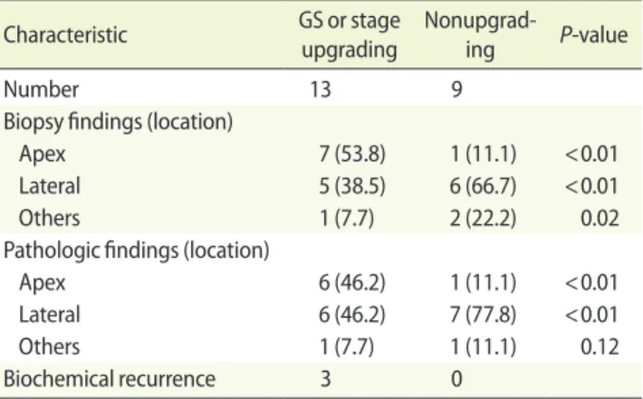
Of the 22 cases of microfocal prostate cancer upon biopsy, 13 cases (59.09%) showed GS upgrading or staging upgrading. Seven out of 13 patients with prostate cancer (53.8%) were detected at the foci of the apex lesion upon biopsy. Six out of 13 GS (46.2%) or stage upgrading cases were detected with prostate cancer located at the apex portion of the prostate. However, only one case out of 9 nonupgrading cases (11.1%) was detected at the apex (Table 2).
DISCUSSION
PSA screening for prostate cancer has become widespread, and the occurrence of low-risk prostate cancer has been dra-matically increasing [5]. Using definite therapy such as RP, clinically localized prostate cancer might be curatively treat-ed, especially in low-risk prostate cancer patients. However,
for low-risk prostate cancer patients with insignificant pros-tate cancer, RP is obviously an overtreatment considering the morbidities, postoperative complications, and oncologic fea-tures of these cases [7]. Despite the variation of the terminol-ogy and definitions used for insignificant prostate cancer in the literature, the intellectual concept of insignificant prostate cancer is well established: a low-grade, small-volume, and organ-confined prostate cancer that is unlikely to be clinically or biologically significant without treatment [8]. There have been many attempts to establish criteria to predict insignifi-cant prostate cancer before surgery, using biopsy results, PSA density, and PSA/free PSA ratio [9].
The high rates of GS or staging upgrading (59.1%) in mi-crofocal prostate cancer in this study might result from can-cer foci (apical portion of the prostate) which were hard to detect lesions at taking biopsies. At the apex portion of the prostate gland, the peripheral zone extends anteriorly to the distal prostatic urethra. It may be difficult to palpate by digital rectal examination cancers that arise in this apico-anterior peripheral zone [10]. Furthermore, an apical biopsy may not be performed in the initial biopsy because it is widely recog-nized as being more painful than a biopsy of the remainder of the prostate and difficulty in palpating by digital rectal examination [11]. The zonal origin of prostate cancer affects the pathological findings and biochemical recurrence rate after RP [12]. Anterior prostate cancer including apical le-sion were not only of lower clinical stage, but they also had lower GS on preoperative prostate biopsy compared with peripheral zone tumor [12]. However, data from whole mount specimens showed that anterior tumors are not insignificant cancers [13]. Patients with anterior prostate cancers had a higher tumor volume and a higher rate of positive surgical margins than patients with peripheral prostate cancers [12]. Table 2. Microfocal tumor characteristics
Characteristic GS or stage upgrading Nonupgrad-ing P-value
Number 13 9
Biopsy findings (location)
Apex 7 (53.8) 1 (11.1) <0.01 Lateral 5 (38.5) 6 (66.7) <0.01 Others 1 (7.7) 2 (22.2) 0.02 Pathologic findings (location)
Apex 6 (46.2) 1 (11.1) <0.01 Lateral 6 (46.2) 7 (77.8) <0.01 Others 1 (7.7) 1 (11.1) 0.12 Biochemical recurrence 3 0
Values are presented as number (%). GS, Gleason score.
Fig. 1. Comparison of Kaplan-Meier biochemical recurrence-free survival curves between two groups.
100 90 80 70 60 50 40 30 20 10 0 Biochemical r ecurr enc e-fr ee sur viv al (%) Months 0 10 20 30 40 50 60 70 Microfocal Nonmicrofocal Group
Furthermore, extraprostatic extension was more likely to be associated with positive surgical margins for anterior prostate cancers than peripheral prostate cancers, suggesting that an-terior positive margins might be clinically significant, and at greater risk of biochemical recurrence [14].
In our previous study [15], insignificant prostate cancer based on an Epstein criteria from a prostate biopsy underesti-mated the true nature of prostate cancer in as many as 42.1% of Koreans. This high inaccuracy rate of the Epstein criteria might result from more aggressive and poorly differentiated prostate cancer in Korean men, despite a low clinical stage or low serum PSA level [16]. Prostate cancer arising in Korean men that is of a predominantly high grade may be attributed to reduced testosterone metabolism. Hoffman et al. [17] dem-onstrated that patients with a low serum-free testosterone level have an increased mean percentage of biopsies revealing cancer with a GS of 8 or higher, suggesting that a low serum-free testosterone level may be a marker of more aggressive disease. However, in our study we do not know the exact rea-son why the high incidence of stage migration from insignifi-cant disease at biopsy to signifiinsignifi-cant disease at final pathology was occurred. Additional studies from a large data would be needed to confirm our results.
When counseling patients with low grade, microfocal pros-tate cancer on biopsy, final decision making regarding man-agement should be guided by the sampling technique, the potential risk of upgrading or upstaging, and contextual con-siderations, such as patient age and comorbidity [15]. Further improved biopsy sampling technique and imaging in patients who choose active surveillance may help minimize the risk of understaging and/or undergrading [18].
There are several limitations to our study. First, the pres-ent study consists of a relatively small number of patipres-ents; therefore, statistical results should be cautiously interpreted. Another limitation is a retrospective study design. Future pro-spective, large cohort study should be needed to confirm our current results.
In our study, microfocal prostate cancer showed higher rate of Gleason upgrading compared to nonmicrofocal pros-tate cancer. In GS or stage upgrading cases, prospros-tate cancer was usually located at the apical portion. Based on higher rates of GS upgrading or stage upgrading cases in microfocal prostate cancer group, compared with nonmicrofocal pros-tate cancer group, active surveillance should be cautiously applied to these patients.
CONFLICT OF INTEREST
No potential conflict of interest relevant to this article was re-ported.
REFERENCES
1. Catalona WJ, Smith DS, Ratliff TL, Basler JW. Detection of or-gan-confined prostate cancer is increased through prostate-specific antigen-based screening. JAMA 1993;270:948-54. 2. Ploussard G, Epstein JI, Montironi R, Carroll PR, Wirth M,
Grimm MO, et al. The contemporary concept of significant versus insignificant prostate cancer. Eur Urol 2011;60:291-303.
3. D’Amico AV, Wu Y, Chen MH, Nash M, Renshaw AA, Richie JP. Pathologic findings and prostate specific antigen outcome after radical prostatectomy for patients diagnosed on the ba-sis of a single microscopic focus of prostate carcinoma with a gleason score </= 7. Cancer 2000;89:1810-7.
4. Carter HB, Epstein JI. Prediction of significant cancer in men with stage T1c adenocarcinoma of the prostate. World J Urol 1997;15:359-63.
5. Pepe P, Fraggetta F, Galia A, Candiano G, Grasso G, Aragona F. Is a single focus of low-grade prostate cancer diagnosed on saturation biopsy predictive of clinically insignificant cancer? Urol Int 2010;84:440-4.
6. Hong SK, Na W, Park JM, Byun SS, Oh JJ, Nam JS, et al. Pre-diction of pathological outcomes for a single microfocal (≤3 mm) Gleason 6 prostate cancer detected via contemporary multicore (≥12) biopsy in men with prostate-specific antigen ≤10 ng/mL. BJU Int 2011;108:1101-5.
7. Boccon-Gibod LM, Dumonceau O, Toublanc M, Ravery V, Boccon-Gibod LA. Micro-focal prostate cancer: a compari-son of biopsy and radical prostatectomy specimen features. Eur Urol 2005;48:895-9.
8. Trpkov K, Yilmaz A, Bismar TA, Montironi R. ‘Insignificant’ prostate cancer on prostatectomy and cystoprostatectomy: variation on a theme ‘low-volume/low-grade’ prostate can-cer? BJU Int 2010;106:304-15.
9. Thong AE, Shikanov S, Katz MH, Gofrit ON, Eggener S, Zagaja GP, et al. A single microfocus (5% or less) of Gleason 6 pros-tate cancer at biopsy: can we predict adverse pathological outcomes? J Urol 2008;180:2436-40.
10. Presti JC Jr. Prostate biopsy strategies. Nat Clin Pract Urol 2007;4:505-11.
11. Jones JS, Zippe CD. Rectal sensation test helps avoid pain of apical prostate biopsy. J Urol 2003;170(6 Pt 1):2316-8. 12. Koppie TM, Bianco FJ Jr, Kuroiwa K, Reuter VE, Guillonneau
B, Eastham JA, et al. The clinical features of anterior prostate cancers. BJU Int 2006;98:1167-71.
13. Epstein JI, Chan DW, Sokoll LJ, Walsh PC, Cox JL, Rittenhouse H, et al. Nonpalpable stage T1c prostate cancer: prediction of insignificant disease using free/total prostate specific an-tigen levels and needle biopsy findings. J Urol 1998;160(6 Pt 2):2407-11.
14. Swindle P, Eastham JA, Ohori M, Kattan MW, Wheeler T, Maru N, et al. Do margins matter? The prognostic signifi-cance of positive surgical margins in radical prostatectomy specimens. J Urol 2005;174:903-7.
15. Yeom CD, Lee SH, Park KK, Park SU, Chung BH. Are clini-cally insignificant prostate cancers really insignificant among
Korean men? Yonsei Med J 2012;53:358-62.
16. Song C, Ro JY, Lee MS, Hong SJ, Chung BH, Choi HY, et al. Prostate cancer in Korean men exhibits poor differentiation and is adversely related to prognosis after radical prostatec-tomy. Urology 2006;68:820-4.
17. Hoffman MA, DeWolf WC, Morgentaler A. Is low serum free testosterone a marker for high grade prostate cancer? J Urol 2000;163:824-7.
18. Harnden P, Naylor B, Shelley MD, Clements H, Coles B, Ma-son MD. The clinical management of patients with a small volume of prostatic cancer on biopsy: what are the risks of progression? A systematic review and meta-analysis. Cancer 2008;112:971-81.
