Jefferson Digital Commons
Kimmel Cancer Center Papers, Presentations, and
Grand Rounds
Kimmel Cancer Center
7-22-2018
Role of HOX Genes in Stem Cell Differentiation
and Cancer.
Seema Bhatlekar
Helen F. Graham Cancer Center and Research Institute; University of Delaware
Jeremy Z Fields
CATX Inc.
Bruce M. Boman
Thomas Jefferson University; Helen F. Graham Cancer Center and Research Institute; University of Delaware; CATX Inc., [email protected]
Let us know how access to this document benefits you
Follow this and additional works at:
https://jdc.jefferson.edu/kimmelgrandrounds
Part of the
Oncology Commons
This Article is brought to you for free and open access by the Jefferson Digital Commons. The Jefferson Digital Commons is a service of Thomas
Jefferson University'sCenter for Teaching and Learning (CTL). The Commons is a showcase for Jefferson books and journals, peer-reviewed scholarly
publications, unique historical collections from the University archives, and teaching tools. The Jefferson Digital Commons allows researchers and interested readers anywhere in the world to learn about and keep up to date with Jefferson scholarship. This article has been accepted for inclusion in Kimmel Cancer Center Papers, Presentations, and Grand Rounds by an authorized administrator of the Jefferson Digital Commons. For more information, please contact: [email protected].
Recommended Citation
Bhatlekar, Seema; Fields, Jeremy Z; and Boman, Bruce M., "Role of HOX Genes in Stem Cell
Differentiation and Cancer." (2018).
Kimmel Cancer Center Papers, Presentations, and Grand Rounds.
Paper 62.
Review Article
Role of HOX Genes in Stem Cell Differentiation and Cancer
Seema Bhatlekar
,
1,2Jeremy Z. Fields,
3and Bruce M. Boman
1,2,3,41Center for Translational Cancer Research, Helen F. Graham Cancer Center and Research Institute, Newark, DE, USA 2Department of Biological Sciences, University of Delaware, Newark, DE, USA
3CATX Inc., Princeton, NJ, USA
4Kimmel Cancer Center, Thomas Jefferson University, Philadelphia, PA, USA
Correspondence should be addressed to Bruce M. Boman; [email protected] Received 1 March 2018; Revised 8 May 2018; Accepted 15 May 2018; Published 22 July 2018 Academic Editor: Jing Huang
Copyright © 2018 Seema Bhatlekar et al. This is an open access article distributed under the Creative Commons Attribution License, which permits unrestricted use, distribution, and reproduction in any medium, provided the original work is properly cited.
HOX genes encode an evolutionarily conserved set of transcription factors that control how the phenotype of an organism becomes organized during development based on its genetic makeup. For example, in bilaterian-type animals, HOX genes are organized in gene clusters that encode anatomic segment identity, that is, whether the embryo will form with bilateral symmetry with a head (anterior), tail (posterior), back (dorsal), and belly (ventral). Although HOX genes are known to regulate stem cell (SC) differentiation and HOX genes are dysregulated in cancer, the mechanisms by which dysregulation of HOX genes in SCs causes cancer development is not fully understood. Therefore, the purpose of this manuscript was (i) to review the role of HOX genes in SC differentiation, particularly in embryonic, adult tissue-specific, and induced pluripotent SC, and (ii) to investigate how dysregulated HOX genes in SCs are responsible for the development of colorectal cancer (CRC) and acute myeloid leukemia (AML). We analyzed HOX gene expression in CRC and AML using information from The Cancer Genome Atlas study. Finally, we reviewed the literature on HOX genes and related therapeutics that might help us understand ways to develop SC-specific therapies that target aberrant HOX gene expression that contributes to cancer development.
1. HOX Genes
HOX genes are master transcriptional regulators that have diverse roles from embryogenesis to carcinogenesis. The HOX genes are an evolutionary conserved family of genes that control anterior-posterior axis and dorsal-ventral anatomic development during embryogenesis. In humans there are a total of 39 HOX genes situated in clusters on four different chromosomes (7p15, 17q21.2, 12q13, and 2q31). These clusters are named as four HOX families: HOXA, HOXB, HOXC, and HOXD. Each family consists of 13 para-log groups with nine to eleven numbers assigned based on their sequence similarity and position within the cluster (Figure 1(a)). HOX genes contain two exons and a single intron. Exon 2 contains a 120-nucleotide sequence, known as homeobox. This homeobox encodes a 61 amino acid helix-turn-helix motif known as a homeodomain (Figure 1(b)).
The protein products of the HOX genes are transcription factors that are capable of binding to specific nucleotide sequences on the DNA.
1.1. HOX Genes and Stem Cell Differentiation.SCs are multi-potent cells that have the ability to self-renew or to diff eren-tiate along multiple lineages. HOX genes have been shown to play crucial roles during SC differentiation from embryonic stages of development to tissue-specific SC functions. Of the 39 HOX genes, mutations in 10 HOX genes (HOXA1, HOXA2, HOXA11, HOXA13, HOXB1, HOXB13, HOXC13, HOXD4,HOXD10, andHOXD13) have been found to cause human disorders with variations in their inheritance pat-terns, penetrance, expressivity, and mechanisms of patho-genesis [1]. Congenital defects caused by mutations in HOX genes support the concept that HOX gene function is crucial for SCs during development and differentiation. Volume 2018, Article ID 3569493, 15 pages
Therefore, we reviewed the published literature for the role of HOX genes during differentiation of three main types of SC, namely, (i) embryonic SCs, (ii) adult SCs (hematopoietic SCs, colonic SCs, and mesenchymal SCs), and (iii) induced pluripotent SCs.
1.1.1. HOX Genes and Embryonic Stem Cell Differentiation. Embryonic stem cells (ESCs) are obtained from the inner cell mass of the blastocyst. ESCs are pluripotent cells that can give rise to most cell types except the placenta and umbilical cord. Retinoic acid (RA) signaling regulates HOX gene expression in ESCs during embryonic development. In adult neurogen-esis, ESCs treated with RA almost exclusively differentiate into neurons and develop a HOX-related expression profile [2]. Retinoic acid response elements (RAREs) are found in regulatory regions of many HOX genes [3, 4]. In mice, RAREs are found in Hoxa1, Hoxa4, Hoxb1, Hoxb4, and Hoxd4 [5–14]. Several HOX genes have been found to be strongly upregulated during differentiation in the presence of RA [15]. RA receptorγ(RARγ) was found to be essential for RA-induced HOX gene transcriptional activation in ESCs. Deletion of its binding site in the Hoxa1 enhancer attenuates transcriptional and epigenomic activation of both Hoxa and Hoxb gene clusters. It was reported that RA/RARγ signaling is critical for the removal of histone methylation occurring on the amino terminal tail core of the core histone H3 (H3K27me3) from activated Hox genes during ESC dif-ferentiation [16]. The entire Hox cluster is actively repressed in ESCs by polycomb repressor complexes and plays key regulatory roles during their differentiation to multipotent progenitors in developing tissues [17]. As ESCs differentiate into different lineages, Hox gene clusters become activated in a controlled and sequence-specific manner [17, 18]. It has been shown that timely induction of Hoxb1 in ESCs results in the differentiation of neuronal SCs and neural pro-genitors of distinct posterior identities [19]. It is well-known thatHoxb4overexpression in ESCs confers long-term repo-pulating ability to ESC-derived hematopoietic stem cells (HSCs). Furthermore, Hoxb4 acts as a master regulator of
ESC differentiation into HSCs by directly targeting multiple essential hematopoietic transcription factors and epigenetic factors [20]. Overall, these results show that complex regula-tory mechanisms exist through which Hox genes are expressed in ESCs and function during differentiation. 1.1.2. HOX Genes and Adult Stem Cells. Adult SCs, also referred to as somatic or tissue-specific SCs, give rise to different cell types that are specific to each tissue type or organ in which they reside. HOX genes are crucial for the maintenance and functioning of adult SCs. Here, we focus on three types of adult SCs: hematopoietic SCs that generate entire blood cell lineages, colonic SCs that reside at the base of the normal crypt and are responsible for colonic tissue renewal and proper functioning of the colon, and mesenchymal SCs isolated from the stroma and that generate various differentiated cell types from bone, carti-lage, and fat cells.
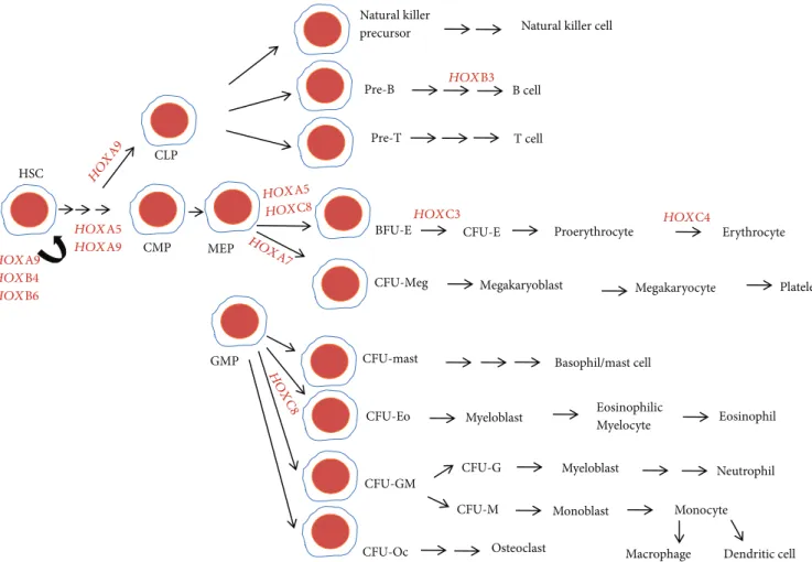
(1) HOX Genes and Hematopoietic Stem Cells. Hematopoietic stem cells (HSCs) are generally found in the bone marrow but can also be isolated from the peripheral blood, umbilical cord blood, and fetal liver. HOX genes are expressed in HSCs and progenitors in a lineage-specific and differentiation stage-restricted manner. For example, HOXB3, HOXB4, and HOXA9 are abundantly expressed in HSCs, whereas HOXB8 and HOXA10are expressed only in myeloid com-mitted cells. Recent studies showed that HOXA family genes are mostly expressed in the myeloid cells, HOXB family genes are mostly expressed in the erythroid cells, and HOXC family genes are commonly seen in the lymphoid cells. HOXD family genes are not expressed during hematopoiesis despite their similarities to other HOX gene clusters [21–24]. Since specific HOX genes are essential for SC differentiation into specific blood cell types, we present a diagram of all known HOX genes associated with hematopoiesis (Figure 2). 1.1.3. HOXA5. HOXA5 overexpression in bone marrow CD34+ SCs and cord blood CD34+ CD38−cells resulted in
A1 A2 A3 A4 A5 A6 A7 A9 A10 A11 A13
B1 B2 B3 B4 B5 B6 B7 B8 B9 B13 C4 C5 C6 C8 C9 C10 C11 C12 C13 D1 D3 D4 D8 D9 D10 D11 D12 D13 HOXA HOXB HOXC HOXD
Anterior Central Posterior
(a) Exon 2 5′ 3′ Exon 1 Intron Homeobox (b)
Figure1: HOX genes and genome organization. (a) In humans, there are a total of 39, clustered into four families, namely, HOXA, HOXB,
HOXC, and HOXD. Each family consists of 13 paralogous groups with nine to eleven numbers assigned based on their sequence similarity and position within the cluster. (b) HOX genes have two exons and 1 intron. Exon 2 has a 120-nucleotide sequence, called a Homeobox that encodes a 61 amino acid HOX protein.
a significant shift in myeloid differentiation [25]. When HOXA5 was overexpressed in HSCs, erythroid progenitors (burst-forming unit-erythroid BFU-E) were significantly decreased in frequency among all progenitors, with no reduc-tion in total colony-forming unit (CFU) numbers [25]. Sim-ilarly, the overexpression of HOXA5 inhibited erythroid differentiation of K562 cells [26]. In contrast, the knockdown of HOXA5 in human bone marrow cells resulted in the inhibition of granulocytic/monocytic hematopoiesis and increased erythroid progenitors [26]. Overall, these studies showed thatHOXA5is crucial for the balance between mye-loid and erythroid differentiation.
1.1.4. HOXA7. Hoxa7knockout mice showed reduced mega-karyocytic/erythroid progenitors (MEP) and exhibited reti-culocytosis and thrombocytopenia without anemia [27], suggesting thatHOXA7is required for MEP differentiation.
1.1.5. HOXA9. HOXA9 is one of the most abundant HOX genes in HSCs.HOXA9is downregulated during HSC diff er-entiation [24, 28]. Hoxa9knockout mice displayed marked lymphopenia (low levels of lymphocytes in the blood) and substantial reductions of common lymphoid progenitors (CLPs) and lymphoid precursors. In addition, significant reduction of common myeloid progenitors (CMPs) and granulocyte/monocyte progenitors (GMPs) was observed upon Hoxa9 knockout in vivo[27]. Hoxa9 knockout mice also showed a reduction in the number of total leukocytes [29], and the loss of expression ofHoxa9impaired the prolif-erating and repopulating ability of HSCs [30].HOXA9thus appears to regulate HSC proliferation, self-renewal, and mye-loid and lymphoid differentiation.
1.1.6. HOXA10. HOXA10 overexpression in human cord blood and fetal liver CD34+ cells resulted in a significant
CMP CLP HSC Pre-B Pre-T BFU-E CFU-Meg CFU-mast CFU-Eo CFU-GM CFU-Oc CFU-E CFU-G CFU-M T cell Proerythrocyte B cell Megakaryoblast Platelet Basophil/mast cell Eosinophilic Myelocyte Myeloblast Monocyte Osteoclast MEP GMP HOXA9 HOXB4 HOXB6 HOXA5 HOX A9 HOXA9 HOX A7 HOXC8 HO XC8 HOXA5 Natural killer
precursor Natural killer cell
HOXB3 HOXC3 Erythrocyte Megakaryocyte Myeloblast Eosinophil Neutrophil Monoblast
Macrophage Dendritic cell
HOXC4
Figure2: HOX gene expression during hematopoiesis. The hematopoietic stem cell (HSC) is a multipotent stem cell that has the ability to
give rise to common lymphoid progenitor (CLP) and common myeloid progenitor (CMP) cells.HOXA9,HOXB4, andHOXB6are known to be expressed in HSC and regulate HSC self-renewal.HOXA5andHOXA9are involved in the proliferation and differentiation of HSC to CMP, andHOXA9regulates the differentiation of HSC into CLP.HOXB3is expressed during the differentiation of pre-B cells into B cells. HOXA5 and HOXC8 are expressed during erythroid differentiation of megakaryocyte-erythrocyte progenitors (MEP) whereas HOXA7 is expressed during megakaryocyte differentiation. HOXC3 and HOXC4 are crucial during erythroid lineage differentiation. HOXC8 is shown to play a regulatory role during the differentiation of granulocyte-monocyte progenitor (GMP) cells. HSC: hematopoietic stem cells; CMP: common myeloid progenitor; CLP: common lymphoid progenitor; MEP: megakaryocyte-erythrocyte progenitor; GMP: granulocyte-monocyte progenitor; BFU-E: erythroid burst-forming units; CFU-E: erythroid colony-forming unit; CFU-Meg: megakaryocyte colony-forming unit; CFU-mast: mast colony-forming unit; CFU-Eo: eosinophil colony-forming unit; CFU-GM: granulocyte-monocyte colony-forming unit; CFU-Oc: osteoclasts colony-forming unit.
production of blast cells (undifferentiated blood cells, com-monly seen in acute leukemia) and myelopoiesis concomi-tant with a complete block of erythroid differentiation and a severe reduction in B cell development [31]. Thus, these
findings suggest that the regulation ofHOXA10expression is crucial for preventing abnormal development and diff er-entiation of HSCs.
1.1.7. HOXB3.In mice, the overexpression ofHoxb3blocked B and T cell differentiation and caused a delay in myeloid precursor proliferation [32], whereas the knockout ofHoxb3 in mice at 6 months of age caused significant impairment of B cell development in the bone marrow [33].
1.1.8. HOXB4. HOXB4is known to enhance primitive hema-topoietic cell growth by increasing self-renewal without affecting homeostatic control of HSC population size or of the rate of HSC production. The retention of full lympho-myeloid repopulating potential and enhancedin vivo regen-erative potential demonstrates the feasibility of achieving significant ex vivo expansion of HSCs without functional impairment [34–39]. Hoxb3 and Hoxb4 double-knockout mice showed defects in endogenous hematopoiesis with reduced cellularity of HSC regeneration. Hoxb3−/Hoxb4− mice showed reduction in hematopoietic progenitor num-bers without perturbing lineage commitment [40].
1.1.9. HOXB6. Hoxb6 overexpression in mice resulted in HSC and myeloid progenitor cell expansion but inhibited erythropoiesis and lymphopoiesis [41]. Upregulation of HOXB6 is often seen in acute myeloid leukemia (AML). Cytogenetic analysis of a subset of HOXB6-induced AMLs revealed recurrent deletions of chromosome band 2D-E4, a region frequently deleted in HOXA9-induced AMLs. The biologic effects ofHOXB6were seen to be largely dependent on DNA binding but they were independent of direct interac-tion of PBX1 [41]. The knockout of Hoxb6 resulted in an increase in early erythroid progenitors in murine bone mar-row and fetal liver cells [42]. Thus,HOXB6is critical not only for HSC self-renewal and maintenance but also for regulatory balance between lymphoid and myeloid differentiation. 1.1.10. HOXC3.An antisense oligonucleotide againstHoxc3 inhibited the formation of colony-forming units (CFUs) of erythroid-derived colonies without any changes in size or degree of hemoglobinization. Early erythroid burst-forming unit colonies or myeloid colonies were not affected, demonstrating that Hoxc3 is involved in an early step in proliferation of the erythroid colony-forming unit subset of progenitor cells [43].
1.1.11. HOXC4. The enforced expression of HOXC4 in human CD34+ cells induced a significant increase in the number of erythroid colonies compared with granulocyte/ macrophage colony-forming units (CFU-GM), without per-turbing, at least in vitro, the maturation program of these cells. On the other hand, HOXC4 overexpression did not induce any skewing in the colony types derived from the myeloid lineage [44].
1.1.12. HOXC8.A significant reduction in the number of ery-throid burst-forming units (BFU-E) and in CFU-GM occurred inHoxc8null mice, although the peripheral blood cell counts were normal [45] suggesting that HOXC8 plays a role during MEP (megakaryocyte-erythroid progenitor) differentiation into BFU-E and GMP differentiation into CFU-GM.
(1) HOX Genes and Colonic Stem Cells. We previously showed that normal colonic SCs are found at the base of the normal human colon crypt and can be isolated using SC markers such as aldehyde dehydrogenase (ALDH1), ALCAM (CD166), and LGR5 [46–48]. The overpopulation of colonic SCs drives colorectal cancer (CRC) development [47, 48]. We studied the expression of HOX genes in normal colonic SCs by microarray profiling. Our analysis showed thatHOXA4,HOXA9, andHOXD10are expressed more in colonic SCs than in proliferating cells or differentiating crypt cells [49]. Further studies showed thatHOXA4andHOXA9 are enriched in SCs during CRC development and that the dysregulation ofHOXA4and HOXA9expression promotes self-renewal and proliferation of colonic SCs [50] (Figure 3). The siRNA knockdown ofHOXA4andHOXA9 in colon cancer cell lines SW480 and HT29 reduced prolifer-ation and sphere-forming ability of colon SCs [50] thus sug-gesting regulatory roles of HOX genes during colon SC maintenance and differentiation.
(2) HOX Genes and Mesenchymal Stem Cells. Mesenchymal stem cells (MSCs) isolated from the umbilical cord blood express HOXA9, HOXB7, HOXC10, and HOXD6, whereas bone marrow-derived MSCs express HOXB7 and HOXD6 [51]. HOXC10 was found to be differentially expressed in amnion- and decidua-derived MSCs [51]. HOX genes, par-ticularlyHOXA9,HOXB7,HOXC10, andHOXD8, were used as biomarkers to distinguish between MSCs derived from unrestricted somatic stem cells and cord blood [51]. A study by Woo et al. [52] showed that the expression ofHOXC13 increased whereas HOXD13 expression decreased as bone marrow-derived MSCs differentiated into osteoblasts during osteogenesis. Taken together, thesefindings indicate that dis-tinct expression patterns of HOXA5, HOXA10, HOXB6, HOXB7, HOXC4, HOXC6, HOXC8, HOXC9, HOXC10, HOXD3, andHOXD8exist in MSCs derived from different human sources [52].Hoxb2,Hoxb5,Hoxb7, andHoxc4genes were found to regulate self-renewal and differentiation of murine MSCs [53].
HOX genes have been shown to play critical roles during osteogenesis of human MSCs. Histone demethylase KDM6B controlled HOX expression by removing histone 3K27 tri-methylation (H3K27me3) and reduced KDM6B significantly by reducing osteogenic differentiation and increasing adipo-genic differentiation [54]. The role of HOX genes during differentiation of human vascular wall-resident CD44+ multipotent stem cells (VW-MPSCs) was also studied [55]. VW-MPSCs are capable of differentiating into pericytes and smooth muscle cells. This study demonstrated that the expression ofHOXB7,HOXC6, andHOXC8is differentially expressed in VW-MPSCs as compared to terminally
differentiated human aortic smooth muscle cells, endothelial cells, and undifferentiated pluripotent ESCs. The knockdown of HOX genes in VW-MPSCs reduced their sprouting capac-ity and increased their levels of smooth muscle markers, transgelin and calponin, as well as histone H1. In addition, changes in methylation patterns of the TAGLN promoter were observed [55]. Overall, this study suggested a role for HOX genes in regulating differentiation of human VW-MPSC into smooth muscle cells via epigenetic mechanisms. The results of this study will help us understand VW-MPSC-dependent vascular disease processes such as neoin-tima formation and tumor vascularization [55].
1.1.13. HOX Genes and Induced Pluripotent Stem Cells. Induced pluripotent stem cells (iPSCs) are cells that are engineered in the lab by converting tissue-specific adult SCs into cells that possess ESC-like properties. iPSCs, like ESCs, did not express HOX genes [56]. Although suppres-sion of HOX gene expressuppres-sion was observed in iPSCs, tran-sient WNT/β-catenin signaling triggered the activation of the CDX/HOX pathway, which in turn conferred a hema-togenic posterior mesoderm phenotype to differentiating human iPSCs [57].
2. HOX Genes and Cancer
In recent years, it has been shown that HOX genes are not only responsible for proper embryonic development but they are also associated with cancer development [58]. In the next section of this review, we focus on the role of HOX genes in cancer development, particularly colorectal cancer (CRC) and acute myeloid leukemia (AML).
2.1. HOX Genes in CRC.Aberrant expression of HOX genes is seen in CRC [49, 50, 58, 59]. We previously reported that HOXA4, HOXA9, and HOXD10 are expressed in normal colonic SCs and dysregulation of HOX genes leads to aber-rant SC differentiation, contributing to CRC development and growth. Furthermore, we showed that HOXA4 and HOXA9 genes promote self-renewal and proliferation of colonic SCs, contributing to CRC development [49, 50].
In this review, we also evaluate the role of HOX genes during CRC development using The Cancer Genome Atlas (TCGA) database. Tumor samples were collected from newly diagnosed patients (i) with colon or rectum adenocarcinoma (ii) undergoing surgical resection, and (iii) having received no prior treatment for their disease, including no chemother-apy and no radiotherchemother-apy. All cases were collected regardless of surgical stage or histologic grade. Cases were staged according to the American Joint Committee on Cancer (AJCC) staging system. Each frozen tumor specimen had a companion normal tissue specimen which could be blood/ blood components, adjacent normal tissue taken from greater than 2 cm from the tumor, or previously extracted germline DNA from the blood [60]. Our analysis showed that HOXB9 was the most upregulated gene at all stages (Figure 4). HOXB6and HOXB8expression increased from stages I to IV but dramatically decreased at stage IVA. Inter-estingly, the expression ofHOXB6andHOXB8was increased during stages I and II and decreased at stage III but again increased at stage IV (Figure 4(f)).
HOXD family gene expression increased at stage IVA compared to all other stages of CRC (Figure 4). Our analysis showed that there is no difference in HOX gene expression based on gender (Figure 5). When we compared TCGA data-sets for HOX gene expression in CRC to overall survival,
Adenomatous crypt St em cells Tr an si t-am p lif yin g cell s Diff er en tia ted cell s HOXA4 HOXA9 HOXD10 HOXC8 HOXC9
Normal clonic crypt Colon carcinoma
Figure3: HOX gene expression during colonocyte differentiation. Normal colonic crypts consist of mainly three types of cells based on their
location in the crypt. Colon stem cells (SCs) reside at the base of the colonic crypt (shown in blue color).HOXA4,HOXA9, andHOXD10are expressed in colonic SCs and regulate colonic crypt SC differentiation [49, 50]. SCs generate transit-amplifying cells (shown in green color) that are actively proliferating and differentiating (shown in gold-bronze yellow color) as they move up the axis in the colonic crypt. Finally, fully differentiated or terminally differentiating cells are found at the top of the crypt (shown in brown color). Studies have shown that HOXA family genes are expressed mostly in proliferating colonic cells, and HOXC family genes are expressed in differentiating cells [68]. HOXB and HOXD family genes are expressed throughout the colonic crypts [68]. In colon tumors, the dysregulation ofHOXA4andHOXA9in colon SCs caused aberrant self-renewal and proliferation, contributing to colon carcinoma [50]. HOXC8 and HOXC9 are expressed in the differentiating cells in the colonic crypt [68].
Stage I HO X A 1 H O X A10 H O X A11 H O X A13 HO X A 2 HO X A 3 HO X A 4 HO X A 5 HO X A 6 HO X A 7 HO X A 9 H O XB1 H O XB13 H O XB2 H O XB3 H O XB4 H O XB5 H O XB6 H O XB7 H O XB8 H O XB9 H O X C10 H O X C11 H O X C12 H O X C13 HO X C 4 HO X C 5 HO X C 6 HO X C 8 HO X C 9 H O XD1 H O XD10 H O XD11 H O XD12 H O XD13 H O XD3 H O XD4 H O XD8 H O XD9 −10 0 10 20 30 Exp res si o n (a)
Stage IIA, IIB
HO X A 1 H O X A10 H O X A11 H O X A13 HO X A 2 HO X A 3 HO X A 4 HO X A 5 HO X A 6 HO X A 7 HO X A 9 H O XB1 H O XB13 H O XB2 H O XB3 H O XB4 H O XB5 H O XB6 H O XB7 H O XB8 H O XB9 H O X C10 H O X C11 H O X C12 H O X C13 HO X C 4 HO X C 5 HO X C 6 HO X C 8 HO X C 9 H O XD1 H O XD10 H O XD11 H O XD12 H O XD13 H O XD3 H O XD4 H O XD8 H O XD9 −10 0 10 20 30 Exp res si o n (b)
Stage IIIA, IIIB, IIIC
HO X A 1 H O X A10 H O X A11 H O X A13 HO X A 2 HO X A 3 HO X A 4 HO X A 5 HO X A 6 HO X A 7 HO X A 9 H O XB1 H O XB13 H O XB2 H O XB3 H O XB4 H O XB5 H O XB6 H O XB7 H O XB8 H O XB9 H O X C10 H O X C11 H O X C12 H O X C13 HO X C 4 HO X C 5 HO X C 6 HO X C 8 HO X C 9 H O XD1 H O XD10 H O XD11 H O XD12 H O XD13 H O XD3 H O XD4 H O XD8 H O XD9 −10 0 10 20 30 E xpre ss ion (c) Stage IVA HO X A 1 H O X A10 H O X A11 H O X A13 HO X A 2 HO X A 3 HO X A 4 HO X A 5 HO X A 6 HO X A 7 HO X A 9 H O XB1 H O XB13 H O XB2 H O XB3 H O XB4 H O XB5 H O XB6 H O XB7 H O XB8 H O XB9 H O X C10 H O X C11 H O X C12 H O X C13 HO X C 4 HO X C 5 HO X C 6 HO X C 8 HO X C 9 H O XD1 H O XD10 H O XD11 H O XD12 H O XD13 H O XD3 H O XD4 H O XD8 H O XD9 −5 0 5 10 15 20 E xpre ss ion (d) Stage IV HO X A 1 H O X A10 H O X A11 H O X A13 HO X A 2 HO X A 3 HO X A 4 HO X A 5 HO X A 6 HO X A 7 HO X A 9 H O XB1 H O XB13 H O XB2 H O XB3 H O XB4 H O XB5 H O XB6 H O XB7 H O XB8 H O XB9 H O X C10 H O X C11 H O X C12 H O X C13 HO X C 4 HO X C 5 HO X C 6 HO X C 8 HO X C 9 H O XD1 H O XD10 H O XD11 H O XD12 H O XD13 H O XD3 H O XD4 H O XD8 H O XD9 −10 0 10 20 30 E xpre ss ion (e) Figure4: Continued.
increasedHOXB13was found to be associated with decreased survival (data not shown). It was reported earlier that mis-sense germline HOXB13 mutations, most commonly in G84E (HOXB13 p. Gly84Glu), are associated with early-onset prostate cancer and possibly associated with breast can-cer and colorectal cancan-cer [61–63]. UnlikeHOXB13, increased HOXB8 expression was associated with increased survival. We observed similar trends in expression of other HOX genes, whereby changes in the level of expression was corre-lated with improved survival and greater in tumor than tumor-free CRC cases (discussed below).
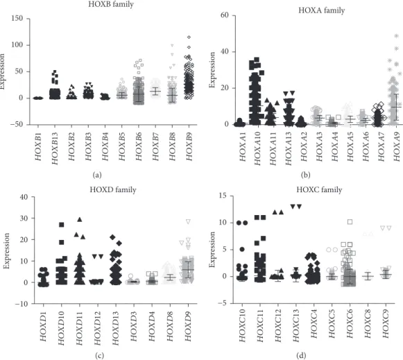
Next, we analyzed HOX expression based on their family gene clusters. Among all HOX families, the HOXB family showed the highest expression in CRC. In the HOXB family, HOXB9, HOXB8, HOXB6, HOXB13, HOXB, and HOXB7 were all overexpressed in CRC (Figure 6(a)). The HOXA family showed the next highest expression. In the HOXA
family, HOXA9 and HOXA10 showed higher expression than the remaining HOXA family genes (Figure 6(b)). Among HOXD family members, HOXD10, HOXD11, HOXD13, and HOXD9showed increased expression com-pared to the others (Figure 6(c)). The HOXC family of genes showed the least expression compared to HOXA, HOXB, and HOXD (Figure 6(d)).
We further assessed HOX genes as a function of overall patient survival. Because we previously found thatHOXA4 and HOXD10 are expressed in crypt SCs, we measured HOXA4and HOXD10levels in CRC cases (n= 220) based on high versus low expression (cutoff at 25th percentile). HOXA4 and HOXD10 high-expressing cases (n= 110) of CRC patients showed overall low survival rates. Increased HOXD10expression was found to be significantly associated with poor overall survival in CRC (Figure 7(a)). HOXD10 high expressers showed only a 15% survival rate versus 55% Stage I
Stage IIA and IIB Stage IIIA, IIIB, IIIC
Stage IV Stage IVA HOXB8 HOXB6 −2 −1 0 1 2 3 F o ld c h an ge co m p ar ed t o st ag e I (f)
Figure4: HOX gene expression during different stages of CRC. RNA sequencing data for CRC patients obtained from The Cancer Genome
Atlas (TCGA) for HOX gene expression (normalized FPKM) and analyzed based on different stages of CRC. We studied (a) 55 cases for stage I, (b) 102 cases for stage IIA and IIB combined, (c) 66 cases reporting stage IIIA, IIIB, and IIIC, (d) 39 samples for stage IV, and (e) 4 cases for stage IVA. (f) Fold changes in the expression ofHOXB6andHOXB8are shown for stages II, III, IV, and IVA compared to stage I.
HO X A 1 H O X A10 H O X A11 H O X A13 HO X A 2 HO X A 3 HO X A 4 HO X A 5 HO X A 6 HO X A 7 HO X A 9 H O XB1 H O XB13 H O XB2 H O XB3 H O XB4 H O XB5 H O XB6 H O XB7 H O XB8 H O XB9 H O X C10 H O X C11 H O X C12 H O X C13 HO X C 4 HO X C 5 HO X C 6 HO X C 8 HO X C 9 H O XD1 H O XD10 H O XD11 H O XD12 H O XD13 H O XD3 H O XD4 H O XD8 H O XD9 Females Males −10 0 10 20 30 Exp ressio n
Figure5: Gender-based HOX gene expression in CRC. The Cancer Genome Atlas (TCGA) was used to analyze gender-based differences in
H O XB1 H O XB13 H O XB2 H O XB3 H O XB4 HOXB family H O XB5 H O XB6 H O XB7 H O XB8 H O XB9 −50 0 50 100 150 Exp ressio n (a) HO X A 1 H O X A10 H O X A11 H O X A13 HO X A 2 HO X A 3 HO X A 4 HO X A 5 HO X A 6 HO X A 7 HO X A 9 HOXA family 0 20 40 60 Exp ressio n (b) HOXD family H O XD1 H O XD10 H O XD11 H O XD12 H O XD13 H O XD3 H O XD4 H O XD8 H O XD9 −10 0 10 20 30 40 Exp ressio n (c) HOXC family H O X C10 H O X C11 H O X C12 H O X C13 HO X C 4 HO X C 5 HO X C 6 HO X C 8 HO X C 9 −5 0 5 10 15 Exp ressio n (d)
Figure6: HOX family gene expression in CRC. (a) HOXB family gene expression, (b) HOXA family genes, (c) HOXD family genes, and (d)
HOXC family genes were analyzed using TCGA RNAseq forn= 273patient samples.y-axis denotes normalized FPKM values for HOX gene expression. p value = 0.037 Low n = 110 High n = 110 500 1000 1500 2000 2500 3000 3500 4000 4500 0 (Days) 0 20 40 60 80 100 % sur vi vin g (a) p value = 0.61 Low n=110 High n=110 0 20 40 60 80 100 % sur vi vin g 1000 2000 3000 4000 5000 0 (Days) (b)
Figure7: Overall survival analysis forHOXA4andHOXD10in colorectal cancer. Kaplan-Meier survival analysis of the 220 colorectal cancer
patients using TCGA dataset. (a)HOXD10and (b)HOXA4survival analysis was performed for CRC patients with a cutoffvalue of 25th percentile. Credits: http://www.oncolnc.org.
survival rate of HOXD10 low expressers (Figure 7(a)). HOXA4 high expressers showed about a 30% survival rate as compared to a 50% survival rate ofHOXA4low expressers (Figure 7(b)). We also analyzed the association ofHOXA9 with the SC markerALDH1A1in CRC patients using TCGA database. A significant positive correlation was observed betweenHOXA9andALDH1A1in CRC (r= 0 12,P= 0 048) (Figure 8(a)). Retinoid receptors,RXRB, showed negative sig-nificant correlation with ALDH1A1 (r=−0 13, P= 0 026) (Figure 8(b)). Another stem cell marker, ALCAM (also known as CD166), was correlated with HOX gene expres-sion in CRC. Expresexpres-sion of HOXA4 negatively correlated with ALCAM in CRC patients (r=−0 14, P= 0 024) (Figure 8(c)).HOXA9was positively correlated withALCAM
(r= 0 18,P= 0 0027) (Figure 8(d)).HOXD10showed
signif-icant negative correlation withALCAM(r=−0 18,P= 0 003) (Figure 8(e)) whereasHOXB8showed a significant positive correlation withALCAM(r= 017,P= 0 006) (Figure 8(f)).
Other published reports on the involvement of HOX genes in CRCs were reviewed [49]. The expression of HOXD8 is downregulated in clinical CRCs compared to normal colon tissues, and the stable expression of HOXD8 in CRC cells significantly reduced cell proliferation, anchor-age independent-growth, and invasion. Apoptotic inhibitor genes such as STK38 and MYC were found to be negatively associated with HOXD8 in analyses using The Cancer Genome Atlas (TCGA). Mansour and Senga demonstrated the ability ofHOXD8to activate caspases 6 and 7 and cleave PARP, thus enhancing apoptosis of CRC cells [64]. A study by Chen et al. showed thatHOXD3is upregulated in human RKO colon cancer cells. The inhibition ofHOXD3by shRNA in RKO cells significantly decreased proliferation and colony formation and increased apoptosis of RKO colon cancer cells. HOXD3-inhibited cells were arrested in the G2 phase of the cell cycle [65]. Among HOXD clusters, induction of the expression of HOXD8, HOXD9, HOXD10, or HOXD12 induces growth arrest and neuronal differentiation with downregulation of cell cycle-promoting genes and upregula-tion of differentiation genes. Other HOXD genes such as HOXD1,HOXD3,HOXD4,HOXD11, andHOXD13had no effects or only partial effects on neuroblastoma cell prolifera-tion or differentiation [66]. These findings suggest that HOXD genes have distinct functions in the induction of can-cer cell differentiation. Other HOX genes, such asHOXB6, HOXB8,HOXC8,HOXC9, and CDX1 were also found to be dysregulated in human CRC development [67]. Further-more, the HOXA family gene was abundantly expressed in colonic adenocarcinoma cells [68]. Overall, these findings suggest that HOX genes play key regulatory roles during maintenance of normal colon SC differentiation and that aberrant expression is associated with CRC development. 2.1.1. CDX1/CDX2 Genes in CRC.Another set of Hox genes, the Cdx genes (also classified as ParaHox genes or caudal-related homeobox genes), is expressed in a wide variety of organisms [69]. Three Cdx genes, Cdx1, Cdx2, and Cdx4, exist in mouse and humans (CDX1, CDX2 and CDX4) and regulate anterior-posterior patterning [70, 71]. The CDX genes are not located in a homeobox cluster,CDX1is located
on human chromosome 5q31–33,CDX2gene is on chromo-some 13q12, and CDX3 is on Xq13.2. In mice, Cdx1 and Cdx2 are important for gastrointestinal tract development. CDX1andCDX2are also actively expressed in adult intesti-nal epithelium and are involved in the regulation of entero-cyte proliferation and differentiation as well as WNT-mediated beta-catenin signaling [70, 72, 73]. Moreover, reduced expression of CDX2 appears to contribute to the development of intestinal neoplasia and is a prognostic bio-marker for stage II and stage III colon cancer by identifying high-risk patients who might benefit from adjuvant chemo-therapy [74–76].
2.1.2. HOX Genes in AML.Studies of AML patients show that many cases (~35%) have mutations in type III receptor tyro-sine kinase FLT3 and thatHOXB2andHOXB3are increased in AML patients with FTL3-ITD (internal tandem duplica-tion) mutation. The overexpression of Hoxb2 and Hoxb3 in mouse pro-B cells resulted in decreased FLT3-ITD-dependent cell proliferation, reduced colony formation, and increased apoptosis, suggesting thatHOXB2andHOXB3are regulators of FLT3-ITD-driven AML [77]. Several studies also showed that HOX genes promote AML development by form-ing chimeric fusions with other genes. Fusion of the nucleo-protein NUP98 with HOXA9 via chromosome translocation t(7;11) (p15s;p15) causes development of AML [78]. Mice overexpressing Hoxa9 and Meis1a induced growth factor-dependent AML in less than 3 months. However, the overex-pression ofHoxa9,Mesi1a, orPbx1balone, or in combination withHoxa9andPbx1b, failed to transform these cells acutely within 6 months posttranslation [79].NUP98-HOXA9fusion genes induced long-term proliferation and blocked diff erenti-ation of human CD34+ HSCs [80]. Recent data showed that mixed lineage leukemia (MLL) is crucial forNUP98-HOXA9 leukemia initiation [81]. We analyzed the TCGA dataset and did overall survival analysis for HOXA9 in AML patients (n= 74) and found that HOXA9high expressers had a 20% survival rate compared toHOXA9low expressers which had a 50% survival rate (Figure 9).
2.2. Cancer Stem Cells.Cancer stem cells (CSCs) are multi-potent and have the ability to undergo both self-renewal and differentiation. We and others have shown that CSCs are the root cause of cancer development [46, 47]. These CSCs are resistant to chemotherapy and radiation. Previ-ously, we identifiedHOXA4, HOXA9, andHOXD10 signa-tures for normal colonic SCs and that these HOX genes are upregulated during CRC development. Indeed, HOXA4 and HOXA9 were found to have roles in self-renewal and proliferation of colonic SCs that contribute to CRC devel-opment [49, 50]. Moreover, HOXA9 is known to have a pivotal role in HSC self-renewal and that the upregulation ofHOXA9leads to AML [82]. Another report showed that miR-375inhibited the proliferation of CSCs and tamoxifen resistance by targeting HOXB3 in human ER-positive breast cancers [83].
2.3. HOX Genes as Biomarkers.HOX genes have been used as markers to distinguish stromal populations from different
tissue sources. The results show that the stromal populations have distinct HOX signatures with different growth and differentiation abilities although they are all
immuno-phenotypically similar. These stromal cell populations express different HOX genes and their level of expression varies. Overall, these results indicate that HOX gene profiles can be used to provide positional, embryological, and hierarchi-cal identity of human stromal stem cells [84].
3. Mechanisms Involved in HOX Gene
Dysregulation in Cancer
3.1. Aberrant Self-Renewal and Proliferation.We have shown thatHOXA4andHOXA9are upregulated in CRC SCs [49, 50] and that siRNA knockdown of HOXA4 and HOXA9 reduces proliferation and sphere-formation ability of CRC SCs. HOXA4 and HOXA9 knockdown also changed the expression of SC markers, such as ALDH1, ALCAM, (CD166) andLGR5. Treatment of CRC cells with the diff er-entiating agent all-trans-retinoic acid (ATRA) decreased HOXA4, HOXA9, andHOXD10expression in parallel with decreases in SC levels. Overall, our study demonstrated a role for HOX genes in self-renewal and proliferation of CRC SCs. Thus, strategies designed to modulate HOX expression may provide a means to target malignant SCs and to develop more effective therapies for CRC [50].
Notably, the self-renewal ability ofHOXB4is dependent upon a proline-rich sequence near the N terminus, which is also unique and highly conserved among the other HOX pro-teins. Deletion of this domain significantly enhanced the oncogenicity ofHoxb4, promoting features of acute leukemia
r = 0.12, P = 0.048 30 20 10 0 0 100 ALDH1A1 HO X A 9 200 300 (a) r = −0.13, P = 0.026 25 20 15 10 5 0 0 100 ALDH1A1 R XRB 200 300 (b) r = −0.14, P = 0.024 −0.5 0.0 0.5 1.0 1.5 60 40 20 ALCAM HO X A 4 80 100 (c) r = 0.18, P = 0.0027 HO X A 9 ALCAM 60 40 20 0 40 30 20 10 0 80 100 (d) r = −0.18, P = 0.003 −6 −4 −20 2 4 6 H O XD10 ALCAM 60 40 20 80 100 (e) r = 0.17, P = 0.006 H O XB8 60 40 20 0 0 10 20 30 40 50 ALCAM 80 100 (f)
Figure8: Correlation analysis of HOX genes and retinoid receptors with stem cell markers in colorectal cancer. (a) The expression ofHOXA9
andALDH1A1(SC marker) in colorectal cancer (CRC) patients is correlated by Pearson correlation. A positive significant correlation was observed between HOXA9and ALDH1A1(r= 0 12,P= 0 048). (b) The expression of retinoid receptor RXRB andALDH1A1 in CRC patients is correlated by Pearson correlation. A negative significant correlation was observed between RXRB and ALDH1A1 (r=−0 13,
P= 0 026). (c) The expression of HOXA4 and ALCAM (CD166, SC marker) in CRC patients correlated by Pearson correlation. A
negative significant correlation was observed between HOXA4 and ALCAM(r=−0 14, P= 0 024). (d) The expression of HOXA9 and ALCAM in CRC patients correlated by Pearson correlation. A positive significant correlation was observed between HOXA9 and ALCAM (r= 0 18, P= 0 0027). (e) The expression of HOXD10 and ALCAM in CRC patients correlated by Pearson correlation. A negative significant correlation was observed between HOXD10and ALCAM(r=−0 18,P= 0 003). (f) The expression ofHOXB8and ALCAMin CRC patients correlated by Pearson correlation. A positive significant correlation was observed betweenHOXB8andALCAM (r=−0 17,P= 0 006). HOXA9 0 % sur vi vin g 0 20 40 60 80 100 500 1000 1500 (Days) 2000 2500 3000 Low n = 37 High n = 37 P-value = 0.024
Figure 9: Overall survival analysis forHOXA9in acute myeloid
leukemia. Kaplan-Meier survival analysis of HOXA9 in acute myeloid leukemia (AML) using TCGA dataset. Survival analysis was performed for AML patients (totaln= 74) with a cutoffvalue of 25th percentile. Credits: http://www.oncolnc.org.
in mice. Insertion of this domain intoHoxa9impaired onco-genic potential for leukemia. Overall, this study showed that such proline-rich stretches in HOX genes attenuate the potential of SCs to acquire oncogenic properties [85].
4. HOX Genes and Related Therapeutics
4.1. HOTAIR Long Noncoding RNA (lncRNA).HOTAIR is a 2.2 kilobasetrans-acting lncRNA residing in the HOXC loci that function to repress transcription of 40 kilobases of the HOXD locus. HOTAIR has been shown to interact with polycomb repressive complex 2 (PRC2). Its interaction with PRC2 is required for PRC2 occupancy and histone H2 lysine-27 trimethylation of the HOXD locus [86]. HOTAIR has been proposed as a biomarker in cervical cancer [87], nasopharyngeal carcinoma [88], and gallbladder cancer [89]. Indeed, meta-analysis involving 748 patients from 8 studies showed HOTAIR is a molecular marker for lymph node metastasis. The results indicated a significant difference in the incidence of lymph node metastasis between high and low HOTAIR expression groups [90]. HOTAIR lncRNA plays a crucial role in epithelial-mesenchymal transitions and is required for the maintenance of colon and breast cancer stem cell stemness [91]. Overall, HOTAIR lncRNA has potential as a therapeutic target in several cancer types. A recent study showed that expression of HOTAIR increased in CRC cells and cell lines and HOTAIR knock-down promoted apoptosis and inhibited proliferation, migra-tion, and invasion in vitro and in vivo. Furthermore, HOTAIR modulated CRC progression by competitively bindingmiR-197[92].
4.2. PBX/HOX Dimer.One of the mechanisms that regulates HOX transcriptional expression is through binding with PBX proteins. Both HOX and PBX proteins are known to play critical roles in carcinogenesis, which makes it an attractive therapeutic target for cancers. One study showed thatPBX3 is a potential pathologic cofactor ofHOXA9involved in cyto-genetically abnormal acute myeloid leukemia (CA-AML), particularly MLL-rearranged AML. The depletion of PBX3 expression by shRNA significantly inhibited MLL-fusion-mediated cell transformation, whereas coexpression of PBX3 with HOXA9 promoted cell transformation in vitro and leukemogenesis in vivo. A small peptide, known as HXR9, that disrupts the interaction between HOX and PBX proteins was found to be effective in killing leukemic cells that were overexpressing HOX/PBX3genes, which suggests a potential therapeutic strategy for CA-AML patients [93]. HXR9 has anticancer effects in other tumor types, such as breast [94], mesothelioma [95], ovarian [96], meningioma [97], prostate [98], and non-small cell lung [99]. Addition-ally, the disruption of HOXB7/PBX2 proteins by HXR9 is a potential therapeutic target in malignant melanoma [100]. A recent study also showed that the expression ofHOXA5, HOXB2, HOXB4, HOXB9, and HOXC9 (but not HOXA9) in primary AML cases is significantly correlated with sur-vival. HXR9 treatment is cytotoxic to AML-derived cell lines and primary AML cells from patients. And it was shown that cell death is independent of apoptosis. Rather, it involves
necroptosis (a regulated form of necrosis) [101]. This study suggests that HXR9 treatment for cancers should be seriously explored in future studies. In addition to the upregulation of HOXA4,HOXA9, andHOXD10in CRC [49, 50, 58], thePBX genes are also overexpressed in CRC, which correlates with invasive potentialin vitroand lymph node invasion, distant metastasis, advanced TNM stage, and poor overall survival of patients [102]. These reports suggest that HXR9 treatment in CRCs might be therapeutically useful to target HOX/PBX proteins in CRCs.
5. Conclusion
The abovefindings suggest that (i) HOX genes play diverse roles in normal SC functions and properties, from self-renewal to multilineage differentiation, and (ii) the dysregu-lation of HOX genes contributes to cancer development through aberrant self-renewal and differentiation of SCs. Thus, understanding the molecular mechanisms for how HOX genes control SC self-renewal and differentiation will ultimately help us understand how SC populations are main-tained in normal, disease-free states and how the dysregula-tion of HOX genes leads to abnormal SC self-renewal and differentiation that drive cancer development. Ultimately, understanding the mechanisms by which HOX genes are reg-ulated in SC might help tofind ways to manipulate SC fate resulting in the development of novel, more effective SC-targeted treatments for cancer.
Disclosure
This work was done at the Center for Translational Cancer Research, Helen F. Graham Cancer Center and Research Institute, Newark, Delaware, and University of Delaware, Newark, Delaware.
Conflicts of Interest
The authors do not present any conflicts of interest.
Acknowledgments
The authors thank Dr. Nicholas Petrelli for his ongoing support.
References
[1] S. C. Quinonez and J. W. Innis,“HumanHOXgene disor-ders,” Molecular Genetics and Metabolism, vol. 111, no. 1, pp. 4–15, 2014.
[2] J. F. Loring, J. G. Porter, J. Seilhammer, M. R. Kaser, and R. Wesselschmidt,“A gene expression profile of embryonic stem cells and embryonic stem cell-derived neurons,” Restorative Neurology and Neuroscience, vol. 18, no. 2-3, pp. 81–88, 2001.
[3] J. Deschamps and J. van Nes,“Developmental regulation of the Hox genes during axial morphogenesis in the mouse,” Development, vol. 132, no. 13, pp. 2931–2942, 2005.
[4] M. Maconochie, S. Nonchev, A. Morrison, and R. Krumlauf,
“Paralogous Hox genes:function and regulation,” Annual Review of Genetics, vol. 30, no. 1, pp. 529–556, 1996. [5] V. Dupé, M. Davenne, J. Brocard et al.,“In vivo functional
analysis of the Hoxa-1 3′ retinoic acid response element (3'RARE),” Development, vol. 124, no. 2, pp. 399–410, 1997.
[6] A. Gould, N. Itasaki, and R. Krumlauf,“Initiation of rhombo-mericHoxb4expression requires induction by somites and a retinoid pathway,”Neuron, vol. 21, no. 1, pp. 39–51, 1998. [7] D. Huang, S. W. Chen, and L. J. Gudas,“Analysis of two
dis-tinct retinoic acid response elements in the homeobox gene Hoxb1 in transgenic mice,” Developmental Dynamics, vol. 223, no. 3, pp. 353–370, 2002.
[8] A. W. Langston and L. J. Gudas,“Identification of a retinoic acid responsive enhancer 3′of the murine homeobox gene Hox-1.6,” Mechanisms of Development, vol. 38, no. 3, pp. 217–227, 1992.
[9] H. Marshall, M. Studer, H. Pöpperl et al.,“A conserved reti-noic acid response element required for early expression of the homeobox Gene Hoxb-1,” Nature, vol. 370, no. 6490, pp. 567–571, 1994.
[10] C. Nolte, M. Rastegar, A. Amores et al.,“Stereospecificity and PAX6 function directHoxd4neural enhancer activity along the antero-posterior axis,”Developmental Biology, vol. 299, no. 2, pp. 582–593, 2006.
[11] A. I. Packer, D. A. Crotty, V. A. Elwell, and D. J. Wolgemuth,
“Expression of the murine Hoxa4 gene requires both autoreg-ulation and a conserved retinoic acid response element,” Development, vol. 125, no. 11, pp. 1991–1998, 1998. [12] M. Rastegar, L. Kobrossy, E. N. Kovacs, I. Rambaldi, and
M. Featherstone,“Sequential histone modifications atHoxd4 regulatory regions distinguish anterior from posterior embry-onic compartments,”Molecular and Cellular Biology, vol. 24, no. 18, pp. 8090–8103, 2004.
[13] M. Studer, H. Popperl, H. Marshall, A. Kuroiwa, and R. Krumlauf,“Role of a conserved retinoic acid response ele-ment in rhombomere restriction of Hoxb-1,” Science, vol. 265, no. 5179, pp. 1728–1732, 1994.
[14] F. Zhang, E. Nagy Kovács, and M. S. Featherstone,“Murine hoxd4 expression in the CNS requires multiple elements including a retinoic acid response element,”Mechanisms of Development, vol. 96, no. 1, pp. 79–89, 2000.
[15] M. Shahhoseini, Z. Taghizadeh, M. Hatami, and H. Baharvand,“Retinoic acid dependent histone 3 demethyl-ation of the clusteredHOXgenes during neural diff erentia-tion of human embryonic stem cells,”Biochemistry and Cell Biology, vol. 91, no. 2, pp. 116–122, 2013.
[16] V. Kashyap, L. J. Gudas, F. Brenet, P. Funk, A. Viale, and J. M. Scandura,“Epigenomic reorganization of the clustered Hox genes in embryonic stem cells induced by retinoic acid,” Jour-nal of Biological Chemistry, vol. 286, no. 5, pp. 3250–3260, 2011.
[17] E. Ezhkova, H. A. Pasolli, J. S. Parker et al.,“Ezh2 orchestrates gene expression for the stepwise differentiation of tissue-specific Stem Cells,”Cell, vol. 136, no. 6, pp. 1122–1135, 2009. [18] X. Wu, Y. Gong, J. Yue, B. Qiang, J. Yuan, and X. Peng,
“Cooperation between EZH2, NSPc1-mediated histone H2A ubiquitination and Dnmt1 in HOX gene silencing,” Nucleic Acids Research, vol. 36, no. 11, pp. 3590–3599, 2008.
[19] M. Gouti and A. Gavalas,“Hoxb1 controls cell fate specifi ca-tion and proliferative capacity of neural stem and progenitor cells,”Stem Cells, vol. 26, no. 8, pp. 1985–1997, 2008. [20] R. Fan, S. Bonde, P. Gao et al., “Dynamic
HoxB4-regulatory network during embryonic stem cell diff erentia-tion to hematopoietic cells,” Blood, vol. 119, no. 19, pp. e139–e147, 2012.
[21] R. A. Alharbi, R. Pettengell, H. S. Pandha, and R. Morgan,
“The role ofHOXgenes in normal hematopoiesis and acute leukemia,”Leukemia, vol. 27, no. 5, pp. 1000–1008, 2013. [22] A. Giampaolo, P. Sterpetti, D. Bulgarini et al.,“Key functional
role and lineage-specific expression of selected HOXB genes in purified hematopoietic progenitor differentiation,”Blood, vol. 84, no. 11, pp. 3637–3647, 1994.
[23] H. Kawagoe, R. K. Humphries, A. Blair, H. J. Sutherland, and D. E. Hogge,“Expression of HOX genes, HOX cofactors, and MLL in phenotypically and functionally defined subpopula-tions of leukemic and normal human hematopoietic cells,” Leukemia, vol. 13, no. 5, pp. 687–698, 1999.
[24] N. Pineault, C. D. Helgason, H. J. Lawrence, and R. K. Humphries, “Differential expression of Hox, Meis1, and Pbx1genes in primitive cells throughout murine hematopoi-etic ontogeny,” Experimental Hematology, vol. 30, no. 1, pp. 49–57, 2002.
[25] G. M. Crooks, J. Fuller, D. Petersen et al., “Constitutive HOXA5 expression inhibits erythropoiesis and increases myelopoiesis from human hematopoietic progenitors,” Blood, vol. 94, no. 2, pp. 519–528, 1999.
[26] J. F. Fuller, J. McAdara, Y. Yaron, M. Sakaguchi, J. K. Fraser, and J. C. Gasson,“Characterization of HOX gene expression during myelopoiesis: role of HOX A5 in lineage commit-ment and maturation,” Blood, vol. 93, no. 10, pp. 3391– 3400, 1999.
[27] C. W. So, H. Karsunky, P. Wong, I. L. Weissman, and M. L. Cleary,“Leukemic transformation of hematopoietic progeni-tors by MLL-GAS7 in the absence of Hoxa7 or Hoxa9,” Blood, vol. 103, no. 8, pp. 3192–3199, 2004.
[28] G. Sauvageau, P. M. Lansdorp, C. J. Eaves et al.,“Differential expression of homeobox genes in functionally distinct CD34 + subpopulations of human bone marrow cells,”Proceedings of the National Academy of Sciences of the United States of America, vol. 91, no. 25, pp. 12223–12227, 1994.
[29] H. J. Lawrence, C. D. Helgason, G. Sauvageau et al.,“Mice bearing a targeted interruption of the homeobox gene HOXA9 have defects in myeloid, erythroid, and lymphoid hematopoiesis,”Blood, vol. 89, no. 6, pp. 1922–1930, 1997. [30] H. J. Lawrence, J. Christensen, S. Fong et al.,“Loss of
expres-sion of theHoxa-9homeobox gene impairs the proliferation and repopulating ability of hematopoietic stem cells,”Blood, vol. 106, no. 12, pp. 3988–3994, 2005.
[31] C. Buske, M. Feuring-Buske, J. Antonchuk et al.,“ Overex-pression ofHOXA10 perturbs human lymphomyelopoiesis in vitro and in vivo,”Blood, vol. 97, no. 8, pp. 2286–2292, 2001.
[32] G. Sauvageau, U. Thorsteinsdottir, M. R. Hough et al.,“ Over-expression ofHOXB3in hematopoietic cells causes defective lymphoid development and progressive myeloproliferation,” Immunity, vol. 6, no. 1, pp. 13–22, 1997.
[33] K. H. Ko, Q. L. Kwan Lam, M. Zhang et al.,“Hoxb3deficiency impairs B lymphopoiesis in mouse bone marrow,” Experi-mental Hematology, vol. 35, no. 3, pp. 465–475, 2007.
[34] S. Amsellem, F. Pflumio, D. Bardinet et al.,“Ex vivo expan-sion of human hematopoietic stem cells by direct delivery of the HOXB4 homeoprotein,” Nature Medicine, vol. 9, no. 11, pp. 1423–1427, 2003.
[35] J. Antonchuk, G. Sauvageau, and R. K. Humphries,“HOXB4 overexpression mediates very rapid stem cell regeneration and competitive hematopoietic repopulation,”Experimental Hematology, vol. 29, no. 9, pp. 1125–1134, 2001.
[36] J. Antonchuk, G. Sauvageau, and R. K. Humphries,“HOXB4 -induced expansion of adult hematopoietic stem cells ex vivo,” Cell, vol. 109, no. 1, pp. 39–45, 2002.
[37] J. Bijl, A. Thompson, R. Ramirez-Solis et al.,“Analysis of HSC activity and compensatory Hox gene expression profile in Hoxbcluster mutant fetal liver cells,”Blood, vol. 108, no. 1, pp. 116–122, 2006.
[38] A. C. Brun, J. M. Björnsson, M. Magnusson et al.,“Hoxb4 -deficient mice undergo normal hematopoietic development but exhibit a mild proliferation defect in hematopoietic stem cells,”Blood, vol. 103, no. 11, pp. 4126–4133, 2004. [39] G. Sauvageau, U. Thorsteinsdottir, C. J. Eaves et al.,“
Overex-pression of HOXB4 in hematopoietic cells causes the selective expansion of more primitive populations in vitro and in vivo,” Genes & Development, vol. 9, no. 14, pp. 1753– 1765, 1995.
[40] J. M. Bjornsson, N. Larsson, A. C. M. Brun et al.,“Reduced proliferative capacity of hematopoietic stem cells deficient in Hoxb3 and Hoxb4,” Molecular and Cellular Biology, vol. 23, no. 11, pp. 3872–3883, 2003.
[41] N. A. Fischbach, S. Rozenfeld, W. Shen et al.,“HOXB6 over-expression in murine bone marrow immortalizes a myelomo-nocytic precursor in vitro and causes hematopoietic stem cell expansion and acute myeloid leukemia in vivo,” Blood, vol. 105, no. 4, pp. 1456–1466, 2005.
[42] C. Kappen, “Disruption of the homeobox gene Hoxb-6 in mice results in increased numbers of early erythrocyte pro-genitors,” American Journal of Hematology, vol. 65, no. 2, pp. 111–118, 2000.
[43] K. Takeshita, J. A. Bollekens, N. Hijiya, M. Ratajczak, F. H. Ruddle, and A. M. Gewirtz, “A homeobox gene of the Antennapedia class is required for human adult erythro-poiesis,” Proceedings of the National Academy of Sciences of the United States of America, vol. 90, no. 8, pp. 3535– 3538, 1993.
[44] A. Daga, M. Podesta, M. C. Capra, G. Piaggio, F. Frassoni, and G. Corte,“The retroviral transduction of HOXC4 into human CD34+cells induces an in vitro expansion of clono-genic and early progenitors,” Experimental Hematology, vol. 28, no. 5, pp. 569–574, 2000.
[45] T. Shimamoto, Y. Tang, Y. Naot et al.,“Hematopoietic pro-genitor cell abnormalities in Hoxc-8 null mutant mice,” Jour-nal of Experimental Zoology, vol. 283, no. 2, pp. 186–193, 1999.
[46] B. M. Boman, J. Z. Fields, K. L. Cavanaugh, A. Guetter, and O. A. Runquist,“How dysregulated colonic crypt dynamics cause stem cell overpopulation and initiate colon cancer,” Cancer Research, vol. 68, no. 9, pp. 3304–3313, 2008. [47] B. M. Boman and E. Huang,“Human colon cancer stem cells:
a new paradigm in gastrointestinal oncology,” Journal of Clinical Oncology, vol. 26, no. 17, pp. 2828–2838, 2008. [48] B. M. Boman, R. Walters, J. Z. Fields et al., “Colonic
crypt changes during adenoma development in familial
adenomatous polyposis: immunohistochemical evidence for expansion of the crypt base cell population,”The American Journal of Pathology, vol. 165, no. 5, pp. 1489–1498, 2004. [49] S. Bhatlekar, S. Addya, M. Salunek et al.,“Identification of a
developmental gene expression signature, including HOX genes, for the normal human colonic crypt stem cell niche: overexpression of the signature parallels stem cell overpopu-lation during colon tumorigenesis,”Stem Cells and Develop-ment, vol. 23, no. 2, pp. 167–179, 2014.
[50] S. Bhatlekar, V. Viswanathan, J. Z. Fields, and B. M. Boman,
“Overexpression of HOXA4 and HOXA9 genes promotes self-renewal and contributes to colon cancer stem cell over-population,”Journal of Cellular Physiology, vol. 233, no. 2, pp. 727–735, 2018.
[51] S. Liedtke, A. Buchheiser, J. Bosch et al.,“TheHOXCode as a
“biological fingerprint” to distinguish functionally distinct stem cell populations derived from cord blood,”Stem Cell Research, vol. 5, no. 1, pp. 40–50, 2010.
[52] C. J. Woo, P. V. Kharchenko, L. Daheron, P. J. Park, and R. E. Kingston,“Variable requirements for DNA-binding proteins at polycomb-dependent repressive regions in human HOX clusters,” Molecular and Cellular Biology, vol. 33, no. 16, pp. 3274–3285, 2013.
[53] D. G. Phinney, A. J. Gray, K. Hill, and A. Pandey,“Murine mesenchymal and embryonic stem cells express a similar Hox gene profile,” Biochemical and Biophysical Research Communications, vol. 338, no. 4, pp. 1759–1765, 2005. [54] L. Ye, Z. Fan, B. Yu et al.,“Histone demethylases KDM4B and
KDM6B promotes osteogenic differentiation of human MSCs,”Cell Stem Cell, vol. 11, no. 1, pp. 50–61, 2012. [55] D. Klein, M. Benchellal, V. Kleff, H. G. Jakob, and S. Ergün,
“Hox genes are involved in vascular wall-resident multipo-tent stem cell differentiation into smooth muscle cells,” Scien-tific Reports, vol. 3, no. 1, article 2178, 2013.
[56] S. P. Atkinson, C. M. Koch, G. K. Clelland et al.,“Epigenetic marking prepares the human HOXAcluster for activation during differentiation of pluripotent cells,” Stem Cells, vol. 26, no. 5, pp. 1174–1185, 2008.
[57] K. Kitajima, M. Nakajima, M. Kanokoda et al.,“GSK3β inhi-bition activates theCDX/HOXpathway and promotes hemo-genic endothelial progenitor differentiation from human pluripotent stem cells,” Experimental Hematology, vol. 44, no. 1, pp. 68–74.e10, 2016.
[58] S. Bhatlekar, J. Z. Fields, and B. M. Boman, “HOX genes and their role in the development of human cancers,” Journal of Molecular Medicine, vol. 92, no. 8, pp. 811–823, 2014.
[59] G. De Vita, P. Barba, N. Odartchenko et al.,“Expression of homeobox-containing genes in primary and metastatic colo-rectal cancer,”European Journal of Cancer, vol. 29A, no. 6, pp. 887–893, 1993.
[60] The Cancer Genome Atlas Network,“Comprehensive molec-ular characterization of human colon and rectal cancer,” Nature, vol. 487, no. 7407, pp. 330–337, 2012.
[61] M. R. Akbari, L. N. Anderson, D. D. Buchanan et al.,
“Germline HOXB13 p.Gly84Glu mutation and risk of colo-rectal cancer,” Cancer Epidemiology, vol. 37, no. 4, pp. 424–427, 2013.
[62] S. Alanee, F. Couch, and K. Offit,“Association of aHOXB13 variant with breast cancer,”New England Journal of Medi-cine, vol. 367, no. 5, pp. 480-481, 2012.
[63] C. M. Ewing, A. M. Ray, E. M. Lange et al.,“Germline muta-tions in HOXB13 and prostate-cancer risk,” New England Journal of Medicine, vol. 366, no. 2, pp. 141–149, 2012. [64] M. A. Mansour and T. Senga, “HOXD8 exerts a
tumor-suppressing role in colorectal cancer as an apoptotic inducer,” The International Journal of Biochemistry & Cell Biology, vol. 88, pp. 1–13, 2017.
[65] F. Chen, G. Sun, and J. Peng, “RNAi-mediated HOXD3 knockdown inhibits growth in human RKO cells,”Oncology Reports, vol. 36, no. 4, pp. 1793–1798, 2016.
[66] Y. Zha, E. Ding, L. Yang et al., “Functional dissection of HOXDcluster genes in regulation of neuroblastoma cell pro-liferation and differentiation,”PLoS One, vol. 7, no. 8, article e40728, 2012.
[67] B. Z. Vider, A. Zimber, E. Chastre et al.,“Deregulated expres-sion of homeobox-containing genes, HOXB6, B8, C8, C9, and Cdx-1, in human colon cancer cell lines,”Biochemical and Biophysical Research Communications, vol. 272, no. 2, pp. 513–518, 2000.
[68] G. Freschi, A. Taddei, P. Bechi et al., “Expression of HOX homeobox genes in the adult human colonic mucosa (and colorectal cancer?),”International Journal of Molecular Med-icine, vol. 16, no. 4, pp. 581–587, 2005.
[69] D. B. Stairs, J. Kong, and J. P. Lynch,“Cdx genes, infl amma-tion, and the pathogenesis of intestinal metaplasia,”Progress in Molecular Biology and Translational Science, vol. 96, pp. 231–270, 2010.
[70] C. van de Ven, M. Bialecka, R. Neijts et al., “Concerted involvement of Cdx/Hox genes and Wnt signaling in mor-phogenesis of the caudal neural tube and cloacal derivatives from the posterior growth zone,” Development, vol. 138, no. 16, pp. 3451–3462, 2011.
[71] T. Young, J. E. Rowland, C. van de Ven et al.,“CdxandHox genes differentially regulate posterior axial growth in mam-malian embryos,” Developmental Cell, vol. 17, no. 4, pp. 516–526, 2009.
[72] A. A. Bhat, A. Sharma, J. Pope et al.,“Caudal homeobox pro-tein Cdx-2 cooperates with Wnt pathway to regulate claudin-1 expression in colon cancer cells,”PLoS One, vol. 7, no. 6, article e37174, 2012.
[73] R. J. Guo, E. Huang, T. Ezaki et al.,“Cdx1 inhibits human colon cancer cell proliferation by reducingβ-catenin/T-cell factor transcriptional activity,”Journal of Biological Chemis-try, vol. 279, no. 35, pp. 36865–36875, 2004.
[74] J. M. Bae, T. H. Lee, N. Y. Cho, T. Y. Kim, and G. H. Kang,
“Loss of CDX2 expression is associated with poor prognosis in colorectal cancer patients,”World Journal of Gastroenter-ology, vol. 21, no. 5, pp. 1457–1467, 2015.
[75] P. Dalerba, D. Sahoo, S. Paik et al.,“CDX2 as a prognostic bio-marker in stage II and stage III colon cancer,”New England Journal of Medicine, vol. 374, no. 3, pp. 211–222, 2016. [76] G. V. Mallo, H. Rechreche, J. M. Frigerio et al.,“Molecular
cloning, sequencing and expression of the mRNA encoding human Cdx1 and Cdx2 homeobox. Down-regulation of Cdx1 and Cdx2 mRNA expression during colorectal carcino-genesis,” International Journal of Cancer, vol. 74, no. 1, pp. 35–44, 1997.
[77] O. Lindblad, R. A. Chougule, S. A. Moharram et al.,“The role of HOXB2 and HOXB3 in acute myeloid leukemia,” Bio-chemical and Biophysical Research Communications, vol. 467, no. 4, pp. 742–747, 2015.
[78] A. Rio-Machin, G. Gómez-López, J. Muñoz et al., “The molecular pathogenesis of the NUP98-HOXA9 fusion pro-tein in acute myeloid leukemia,” Leukemia, vol. 31, no. 9, pp. 2000–2005, 2017.
[79] E. Kroon, J. Krosl, U. Thorsteinsdottir, S. Baban, A. M. Buch-berg, and G. Sauvageau,“Hoxa9transforms primary bone marrow cells through specific collaboration withMeis1abut notPbx1b,”The EMBO Journal, vol. 17, no. 13, pp. 3714– 3725, 1998.
[80] A. Takeda, C. Goolsby, and N. R. Yaseen,“NUP98-HOXA9 induces long-term proliferation and blocks differentiation of primary human CD34+ hematopoietic cells,” Cancer Research, vol. 66, no. 13, pp. 6628–6637, 2006.
[81] Y. Shima, M. Yumoto, T. Katsumoto, and I. Kitabayashi,
“MLL is essential for NUP98-HOXA9-induced leukemia,” Leukemia, vol. 31, no. 10, pp. 2200–2210, 2017.
[82] C. S. Velu, A. Chaubey, J. D. Phelan et al., “Therapeutic antagonists of microRNAs deplete leukemia-initiating cell activity,” Journal of Clinical Investigation, vol. 124, no. 1, pp. 222–236, 2014.
[83] H. Fu, L. Fu, C. Xie et al.,“miR-375 inhibits cancer stem cell phenotype and tamoxifen resistance by degrading HOXB3 in human ER-positive breast cancer,”Oncology Reports, vol. 37, no. 2, pp. 1093–1099, 2017.
[84] J. Picchi, L. Trombi, L. Spugnesi et al.,“HOXandTALE sig-natures specify human stromal stem cell populations from different sources,” Journal of Cellular Physiology, vol. 228, no. 4, pp. 879–889, 2013.
[85] M. Cusan, N. M. Vegi, M. A. Mulaw et al.,“Controlled stem cell amplification by HOXB4 depends on its unique proline-rich region near the N terminus,” Blood, vol. 129, no. 3, pp. 319–323, 2017.
[86] J. L. Rinn, M. Kertesz, J. K. Wang et al.,“Functional demarca-tion of active and silent chromatin domains in humanHOX loci by noncoding RNAs,”Cell, vol. 129, no. 7, pp. 1311– 1323, 2007.
[87] L. Huang, L. M. Liao, A. W. Liu et al.,“Overexpression of long noncoding RNA HOTAIR predicts a poor prognosis in patients with cervical cancer,” Archives of Gynecology and Obstetrics, vol. 290, no. 4, pp. 717–723, 2014.
[88] Y. Nie, X. Liu, S. Qu, E. Song, H. Zou, and C. Gong,“Long non-coding RNA HOTAIR is an independent prognostic marker for nasopharyngeal carcinoma progression and sur-vival,”Cancer Science, vol. 104, no. 4, pp. 458–464, 2013. [89] M. Z. Ma, C. X. Li, Y. Zhang et al.,“Long non-coding RNA
HOTAIR, a c-Myc activated driver of malignancy, negatively regulates miRNA-130a in gallbladder cancer,” Molecular Cancer, vol. 13, no. 1, p. 156, 2014.
[90] B. Cai, Z. Wu, K. Liao, and S. Zhang,“Long noncoding RNA HOTAIR can serve as a common molecular marker for lymph node metastasis: a meta-analysis,” Tumour Biology, vol. 35, no. 9, pp. 8445–8450, 2014.
[91] C. Pádua Alves, A. S. Fonseca, B. R. Muys et al.,“Brief report: the lincRNA Hotair is required for epithelial-to-mesenchymal transition and stemness maintenance of cancer cell lines,”Stem Cells, vol. 31, no. 12, pp. 2827–2832, 2013. [92] X. Lu, Z. Liu, X. Ning, L. Huang, and B. Jiang,“The long
non-coding RNA HOTAIR promotes colorectal cancer progres-sion by sponging miR-197,” ncology Research Featuring Preclinical and Clinical Cancer Therapeutics, vol. 26, no. 3, pp. 473–481, 2018.
[93] Z. Li, Z. Zhang, Y. Li et al.,“PBX3 is an important cofactor of HOXA9 in leukemogenesis,”Blood, vol. 121, no. 8, pp. 1422– 1431, 2013.
[94] R. Morgan, A. Boxall, K. J. Harrington et al.,Breast Cancer Research and Treatment, vol. 136, no. 2, pp. 389–398, 2012. [95] R. Morgan, G. Simpson, S. Gray et al.,“HOX transcription
factors are potential targets and markers in malignant meso-thelioma,”BMC Cancer, vol. 16, no. 1, p. 85, 2016.
[96] Z. Kelly, C. Moller-Levet, S. McGrath et al.,“The prognostic significance of specific HOX gene expression patterns in ovarian cancer,” International Journal of Cancer, vol. 139, no. 7, pp. 1608–1617, 2016.
[97] H. Ando, A. Natsume, T. Senga et al.,“Peptide-based inhibi-tion of the HOXA9/PBX interacinhibi-tion retards the growth of human meningioma,”Cancer Chemotherapy and Pharmacol-ogy, vol. 73, no. 1, pp. 53–60, 2014.
[98] R. Morgan, A. Boxall, K. J. Harrington, G. R. Simpson, A. Michael, and H. S. Pandha,“TargetingHOXtranscription factors in prostate cancer,”BMC Urology, vol. 14, no. 1, p. 17, 2014.
[99] L. Plowright, K. J. Harrington, H. S. Pandha, and R. Morgan,
“HOX transcription factors are potential therapeutic targets in non-small-cell lung cancer (targeting HOX genes in lung cancer),”British Journal of Cancer, vol. 100, no. 3, pp. 470– 475, 2009.
[100] M. C. Errico, F. Felicetti, L. Bottero et al.,“The abrogation of the HOXB7/PBX2 complex induces apoptosis in melanoma through the miR-221&222-c-FOS pathway,” International Journal of Cancer, vol. 133, no. 4, pp. 879–892, 2013. [101] R. A. Alharbi, H. S. Pandha, G. R. Simpson et al.,“Inhibition
of HOX/PBX dimer formation leads to necroptosis in acute myeloid leukemia cells,” Oncotarget, vol. 8, no. 52, pp. 89566–89579, 2017.
[102] H. B. Han, J. Gu, D. B. Ji et al.,“PBX3 promotes migration and invasion of colorectal cancer cells via activation of MAPK/ERK signaling pathway,”World Journal of Gastroen-terology, vol. 20, no. 48, pp. 18260–18270, 2014.
Hindawi www.hindawi.com International Journal of Volume 2018
Zoology
Hindawi www.hindawi.com Volume 2018 Anatomy Research InternationalPeptides
Hindawi www.hindawi.com Volume 2018 Hindawi www.hindawi.com Volume 2018 Journal of Parasitology ResearchGenomics
International Journal of Hindawi www.hindawi.com Volume 2018Hindawi Publishing Corporation
http://www.hindawi.com Volume 2013 Hindawi www.hindawi.com
The Scientific
World Journal
Volume 2018 Hindawi www.hindawi.com Volume 2018Bioinformatics
Advances inMarine Biology
Journal ofHindawi www.hindawi.com Volume 2018 Hindawi www.hindawi.com Volume 2018
Neuroscience
Journal
Hindawi www.hindawi.com Volume 2018 BioMed Research InternationalCell Biology
International Journal of Hindawi www.hindawi.com Volume 2018 Hindawi www.hindawi.com Volume 2018 Biochemistry Research InternationalArchaea
Hindawi www.hindawi.com Volume 2018 Hindawi www.hindawi.com Volume 2018 Genetics Research International Hindawi www.hindawi.com Volume 2018 Advances inVirology
Stem Cells
International
Hindawi www.hindawi.com Volume 2018 Hindawi www.hindawi.com Volume 2018Enzyme
Research
Hindawi www.hindawi.com Volume 2018 International Journal ofMicrobiology
Hindawi www.hindawi.comNucleic Acids
Volume 2018Submit your manuscripts at
www.hindawi.com