ISBN: 978-94-6332-365-9
Thesis, Erasmus University Rotterdam, Netherlands © 2018 A.U.Yousaf-Khan
All rights reserved. No part of this publication may be reproduced, stored in a retrieval system, or transmitted, in any form or by any means, electronic, mechanical, photocopying, recording or otherwise, without the prior permission of the author or the copyright-owning journals for previously published chapters.
Cover illustration: Anum & Shahryar (Instagram: sci.art_by_anum) Lay-out and print: GVO drukkers en vormgevers B.V., Ede
De NELSON longkankerscreening trial:
Laatste screeningsronde en follow-up
Proefschrift
ter verkrijging van de graad van doctor aan de
Erasmus Universiteit Rotterdam
op gezag van de
rector magnificus
Prof.dr. H.A.P. Pols
en volgens besluit van het College voor Promoties.
De openbare verdediging zal plaatsvinden op
woensdag 27 juni 2018 om 13.30 uur
door
Promotor: Prof. dr. H.J. de Koning
Overige leden: Prof. dr. J.G.J.V. Aerts
Prof. dr. P.E. Postmus
Prof. dr. J.W.P.F. Kardaun
Part I: the optimization of the NELSON screening rounds
Chapter II Final screening round of the NELSON lung cancer screening trial:
the effect of a 2.5-year screening interval.
Thorax
Chapter III Risk stratification based on screening history: the NELSON lung
cancer screening study.
Thorax
Part II: interim stage shift results in the NELSON trial
Chapter IV Cancer stage shift and treatment shift in the NELSON lung cancer
screening trial: implications for clinicians.
Submitted
Part III: the cause of death of the NELSON study participants
Chapter V Baseline characteristics and mortality outcomes of NELSON
control group participants and eligible nonresponders.
Journal of Thoracic Oncology
Chapter VI Blinded and uniform cause of death verification in a lung cancer
CT screening trial
Lung Cancer
Chapter VII General discussion
41 43 69 95 97 115 117 135 165
Epidemiology
Lung cancer is by far the leading cause of cancer death among males and the second leading
cause of cancer death among females worldwide 1, 2. Each year, more persons die from lung
cancer than of colon, breast, and prostate cancers combined 2. Most lung cancer cases are
diagnosed in symptomatic patients. At that moment, the majority of patients have already
reached an advanced stage of lung cancer, which is correlated with a poor survival 3.
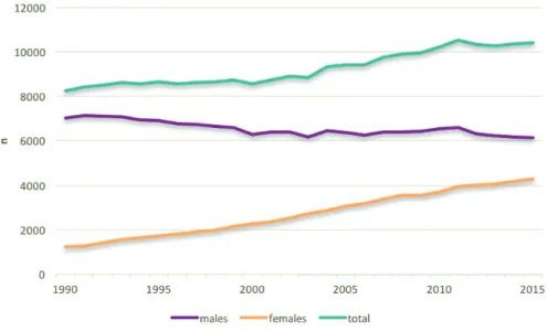
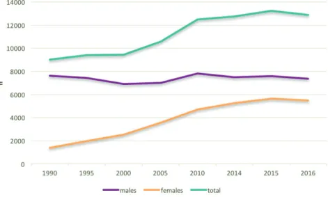
In the Netherlands, lung cancer is the leading cause of death among both genders: approximately 25% of cancer deaths among males and 20% of cancer deaths among females are caused by lung cancer (Figure 1) 4.
I
and 22.6% of females do smoke, which corresponds to 26.3% of the Dutch population over
18 years of age 11, 12. In 2015, the proportion of heavy smokers (people who smoke 20 or more
cigarettes per day) decreased since 2000 from 35.0% to 15.3%.
Figure 2: lung cancer incidence in the Netherlands, by gender 10
Aetiology
In approximately 85% of all lung cancer cases, cigarette smoking is the primary cause for developing lung cancer 6-8, 13. The amount and duration of cigarette smoking is strongly related
to the development of lung cancer 6-8, 13. In which, the duration of smoking is considered to be
the strongest link with lung cancer risk 14. Unadjusted lifetime risk for developing lung cancer
is approximately 6.4% 3. A current smoker who smoked one pack per day for 40 years has a 20
to 50 times higher risk to develop lung cancer than a never smoker 15. In general, one out of
nine smokers develops lung cancer 16. Most cases of lung cancers occur in moderate to heavy
smokers 16.
Other risk factors associated with lung cancer are: 1) family history of lung cancer, in which genetic factors can affect the risk for developing lung cancer regardless of exposure to cigarette
smoking 17, 18; 2) other lung diseases such as COPD, chronic bronchitis or emphysema which
may increase the risk for developing lung cancer independently from smoking 19-21; 3) passive
smoking 22, 23; 4) asbestosis and radon exposure 24-26; and 5) a medical history of treatment
with radiotherapy for a non-Hodgkin lymphoma or for breast cancer with increased risk for a second primary lung cancer
Histology
Lung cancer is defined as a malignant neoplasm of an unspecified part of an unspecified bronchus or lung according to the International Statistical Classification of Diseases and
Related Health Problems ( ICD-10; C34) 29. Lung cancer usually arises from uncontrolled cell
growth of the epithelium that lines in the bronchial tree and may spread to a site distant from the lungs and produce metastatic tumours in other parts of the body (e.g. brain, bones, liver or adrenal glands) 29.
There are two main types of lung cancer: small cell lung carcinoma (SCLC) and non-small cell lung carcinoma (NSCLC), accounting for approximately 15% and 85% of lung cancer
cases, respectively 1. These types are diagnosed based on the microscopic appearance of the
malignant cells. The distinction between SCLC and NSCLC is essential for staging, treatment and prognosis of the lung cancer 30.
Small cell lung carcinoma
Approximately 15% of all lung cancers are SCLCs 29, 31. They are characterised by small “blue”
malignant cells about twice the size of lymphocytes (Figure 3). The cytoplasm is sparse and nuclear features include finely dispersed chromatin without distinct nucleoli. SCLC is histologically divided into two subtypes: oat cell carcinoma and a combined small cell carcinoma (usually a combination of SCLC with adenocarcinoma, squamous cell carcinoma
or large cell carcinoma) 29, 30, 32. Large cell neuroendocrine carcinoma (LCNEC) is officially
classified under NSCLC, but its biological behaviour is similar to that of SCLC 32.
For SCLC and LCNEC there are standard immunohistochemical markers for lung origin
and/or neuroendocrine features which are useful for establishing the diagnosis 33. A
majority of the SCLC express the thyroid transcription factor (TTF-1), which can help in
distinguishing LCNEC from other neuroendocrine carcinomas 34. Other markers that can
be used to differentiate include CD56, chromogranin and synaptophysin 35. Up to two-thirds
of SCLC will be negative for chromogranin and synaptophysin and CD56 will be positive in
I
Figure 3: small cell carcinoma 29.
Non-small cell carcinoma
NSCLC are usually adenocarcinomas, squamous cell carcinomas (SQM) or large cell
carcinomas (LCC) 29, 30, 32. The distinguishing between the subtypes of NSCLC is necessary for
the guidance of treatment and prediction of the clinical course.
Adenocarcinoma
Adenocarcinomas are the most common type of lung cancer, accounting for approximately
half of all lung cancer cases 31, 38. The incidence of adenocarcinoma is increasing, which is
thought to be related to the introduction of low-tar filter cigarettes in the 1960s 38, 39. The use
of filters may have led to different inhalation behaviours, e.g., taking larger puffs and retaining smoke longer to compensate for the lower nicotine dose in the filter cigarettes. This might have led to increased carcinogenic damage in the peripheral lung zones, where the majority of the lung adenocarcinomas arise. Another explanation could be higher nitrate contents of the low-tar filter cigarettes. Also, lung cancer is increasing among females in whom adenocarcinoma
seems to be more common 26.
In the case of adenocarcinoma, the tumour tissue is commonly tested for the presence of a driver mutation (e.g. mutated epidermal growth factor, ALK translocation) and increasingly for other mutations (Figure 4) 40. This is necessary as it is possible to treat types of lung cancer
in an advanced stage based on the genotype (so-called, personalised, genotype-directed therapy) 41, 42.
used. Instead, the term “lepidic” is used, which describes non-invasive growth along intact
septae 32. Previous lesions classified as BAC are now classified under adenocarcinomas as:
I. Atypical adenomatous hyperplasia (AAH): ≤5mm. Previously recognised as
pre-invasive lesion for lung adenocarcinoma.
II. Adenocarcinoma in situ (AIS): a localised adenocarcinoma, smaller than 3 cm, in
which growth is restricted to tumour cells growing along alveolar structures (lepidic growth patter) and lacks any component of invasion. Most AIS are non-mucinous and just a small subset of such tumours are mucinous.
III. Minimally invasive adenocarcinoma: a new category which describes a small, solitary
adenocarcinoma (≤3cm) with predominantly lepidic growth patterns and 5mm invasion.
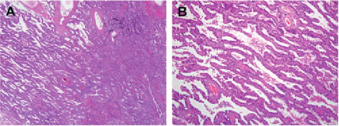
Figure 4: adenocarcinoma ( A: Lepidic predominant pattern with mostly lepidic growth and B: invasive
acinar adenocarcinoma) 29.
Squamous cell carcinoma
SQM used to be the most frequent type of lung cancer until the midst 1980s 29, 38. Currently, it
I
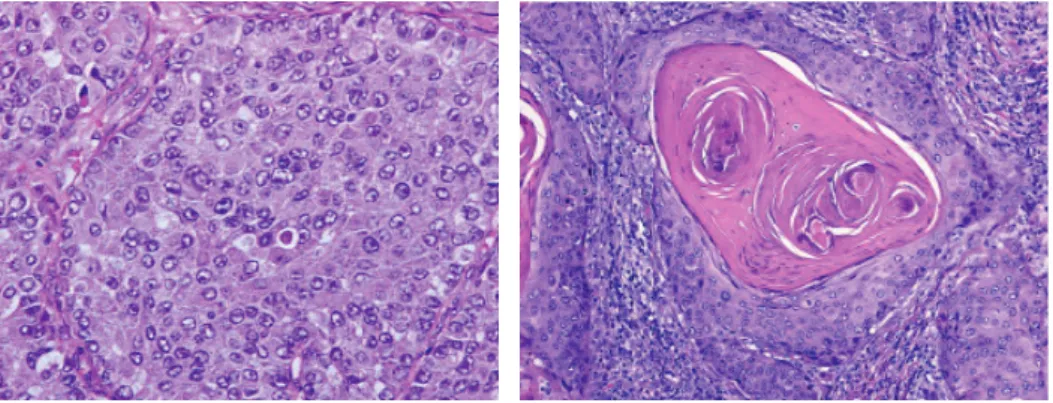
The diagnosis of SQM is mainly based on the presence of keratin production by the tumour cells and/or intercellular desmosomes, or by immunohistochemistry (expression of p40, p63, CK5, or CK5/6, desmoglein) 40, 46.
Figure 5: squamous cell carcinoma 29 and large cell carcinoma 29.
Large cell carcinoma
LCC comprises 3% of all lung cancers 1, 31. The incidence of LCC is decreasing, due to
reclassification of these tumours to mainly adenocarcinoma and squamous cell carcinoma
30. These tumours are mostly found in the lung periphery, although they may have a central
location. They frequently appear as large necrotic tumours.
LCC are malignant epithelial neoplasms lacking both glandular and squamous differentiation by light microscopy and immunohistochemistry, and lacking cytologic features of small cell
carcinoma (Figure 5) 40, 47.In other words, they are diagnosed by exclusion.
Other types of NSCLCs
Adenosquamous carcinomas are tumours which consist of at least 10% squamous cell
carcinoma cells and at least 10% adenocarcinoma cells 48.
Carcinoids are neuroendocrine lung cancers with neuroendocrine differentiation lower than
LCNEC and SCLC, and can further be divided into typical and atypical types 32. This type of
lung cancer can usually be diagnosed on the basis of light microscopy alone 29.
Non-small cell carcinoma, not otherwise specified (NSCLC NOS) is for cases where there is no evidence of squamous or adenocarcinomatous differentiation on immunohistochemistry
Clinical manifestation
Unfortunately, the majority of patients is in advanced stage of disease at the time of lung cancer diagnosis. Symptoms of lung cancer do result from local effects of the tumour, from regional or distant spread, and/or from distant effects not related to metastases (e.g. paraneoplastic syndromes) 49-51.
Local effects of lung cancer:
I. Coughing: reported in more than 50% of the lung cancer patients at presentation and
most frequently in those with a squamous cell and small cell carcinoma because of the central localization of the tumours 52-54.
II. Haemoptysis: reported in up to 30% of the patients with lung cancer 52-54.
III. Chest pain: reported in up to 40% of the patients with lung cancer 52-54.
IV. Dyspnoea: reported in approximately 50% of patients with lung cancer at presentation
52-54.
V. Other more general reported symptoms are: chest pain (20-49%), weight loss
(27-68%), weakness (0-10%) and obstruction of the superior vena cava (0-4%) 52-55.
The most frequent sites of distant metastasis at the time of diagnosis or during the course of the disease are the liver, adrenal glands, bones and/or brain 56.
Lung cancer can also lead to symptoms which are mediated by hormones, cytokines or
by an immune response against the tumour 36, 37. Some commonly observed effects are:
hypercalcemia (leading to anorexia, nausea, vomiting), syndrome of inappropriate antidiuretic hormone secretion (SIADH) in which the degree of hyponatremia leads to various symptoms, neurological paraneoplastic syndromes (e.g. difficulty to rise from a chair, dry mouth or stiff muscles), haematological symptoms such as thrombocytosis and Cushing’s syndrome, in which an ectopic production of adrenal corticotropin (ACTH) is observed.
I
Table 1: 7th edition TNM staging for lung cancer. Adapted from: Goldstraw et al (2007) 59.
Primary tumour (T)
T1 Tumour ≤3 cm diameter in greatest dimension, surrounded by lung or visceral pleura,
without invasion more proximal than lobar bronchus.
1a Tumour ≤2 cm in diameter in greatest dimension.
1b Tumour >2 cm but ≤3 cm in diameter in greatest dimension.
T2
Tumour >3 cm but ≤7 cm, or tumour with any of the following features:
- Involves main bronchus, ≥2 cm distal to carina;
- Invades visceral pleura;
- Associated with atelectasis or obstructive pneumonitis that extends to the
hilar region but does not involve the entire lung.
2a Tumour >3 cm but ≤5 cm in greatest dimension.
2b Tumour >5 cm but ≤7 cm in greatest dimension.
T3
Tumour >7 cm or any of the following:
- Directly invades any of the following: chest wall, diaphragm, phrenic nerve,
mediastinal pleura, parietal pericardium, main bronchus <2 cm from carina (without involvement of carina);
- Atelectasis or obstructive pneumonitis of the entire lung;
- Separate tumour nodules in the same lobe.
T4
Tumour of any size that invades the mediastinum, heart, great vessels, trachea, recurrent laryngeal nerve, esophagus, vertebral body, carina, or with separate tumour nodules in a different ipsilateral lobe.
Regional lymph nodes (N)
N0 No regional lymph node metastases.
N1 Metastasis in ipsilateral peribronchial and/or ipsilateral hilar lymph nodes and
intrapulmonary nodes, including involvement by direct extension.
N2 Metastasis in ipsilateral mediastinal and/or subcarinal lymph node(s).
N3 Metastasis in contralateral mediastinal, contralateral hilar, ipsilateral or contralateral
scalene, or supraclavicular lymph node(s).
Distant metastasis (M)
M0 No distant metastasis.
M1 Distant metastasis.
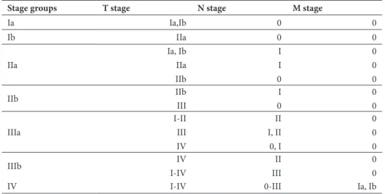
Table 2: stage grouping according to the seventh edition TNM staging. Adapted from: Goldstraw et al
(2007) 59.
Stage groups T stage N stage M stage
Ia Ia,Ib 0 0 Ib IIa 0 0 IIa Ia, Ib IIa IIb I I 0 0 0 0 IIb IIb III I 0 0 0 IIIa I-II III IV II I, II 0, I 0 0 0 IIIb IV I-IV II III 0 0
IV I-IV 0-III Ia, Ib
Table 3: stage distribution of lung cancers at time of diagnosis 3.
Stages Spread %
Ia- IIb Localised 18.2
IIIa-IIIb Regional 21.9
IV Distant 53.2
Unstaged Unknown 6.7
I
radiotherapy is recommended 11. For stage II resected tumours adjuvant chemotherapy is
recommended 65. For patients with compromised lung function or other co-morbidities (such
as heart failure) who cannot undergo curative surgery for lung cancer, stereotactic radiation may be applied 66, 67.
Patients with a stage III NSCLC and a good clinical performance are treated with concurrent chemoradiation 62, 68. Curative surgery plays a role in those patients with a down staging of the
tumour after concurrent chemoradiation.
Patients with stage IV NSCLC, are treated with systemic therapy (e.g. chemotherapy) or
symptom-based palliative approach (e.g. radiation for metastases) 62. Therapy is guided by the
mutation status of the tumour and by the clinical performance of the patient. Patients with metastasis may also benefit from resection of the metastasis (in case of isolated metastasis) as well as radiation (e.g. pain control). Local palliative measures (e.g. stenting of the vena cava, stenting of the oesophagus, coagulation of the bronchial vessels) may also be beneficial to control the pulmonary disease and increase the quality of life.
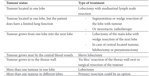
Table 4: overview of the NSCLC stage I or stage II treatment in the Netherlands 62.
Tumour status Type of treatment
Tumour located in one lobe Lobectomy with mediastinal lymph node
resection Tumour located in one lobe, but the patient
does have a limited lung function
- Segmentation or wedge resection of
the lobe with tumour
- Or stereotactic radiotherapy
Tumour grows from one lobe into the next lobe - Lobectomy of the main lobe with
wedge resection of the next lobe
- In case of central located tumour,
bilobectomy or pneumonectomy Tumour grows near by the central blood vessels Sleeve lobectomy
Tumour grows in to the thorax wall ‘En bloc’ resection of the thorax wall next to
surgical resection of the tumour
More than one tumour in one lobe Lobectomy
More than one tumour in different lobes Primary resection could be an option
Treatment of SCLC
At the time of diagnosis, SCLC is usually disseminated (extensive). As SCLC is very responsive
of chemotherapy and radiotherapy 62, 70. Surgery is only used in patients with a solitary
pulmonary nodule without metastasis or lymph node involvement 71. Patients without disease
progression and with a good clinical performance are recommended to receive a prophylactic
cranial radiation (PCI) within 60 days after chemotherapy 72.
For patients with extensive SCLC, chemotherapy is used as the only initial therapy 62. PCI
is considered in patients with a complete or partial response, or stable disease after the chemotherapy. Radiation therapy can also be used for symptom-based control.
Survival
In general, the 5-year survival rate for lung cancer is about 11-18% 3, 5, 31. Survival decreases
progressively with later stages of the disease. In Table 5 an overview is presented of the 5-year survival rate based upon the Surveillance, Epidemiology, and End Results (SEER) data of lung
cancer survival in the United States between 2007 and 2013 3.
In an analysis of surgical NSCLC cases from the IASLC lung cancer staging project, next to
stage, age and gender were prognostic factors for survival 73. Patients less than 70 years of
age had significantly a better overall survival compared to patients older than 70 years of age. BACs carried a better prognosis than all other subtypes of lung cancer. Squamous cell carcinomas were slightly favoured over adenocarcinomas and LCC, but only after adjusting for gender (male only) and stage. Adjusting for smoking status did not modify the effects of histology.
Furthermore, histologic grade has significant a prognostic value for the survival of NSCLC lung cancer patients, in which undifferentiated carcinoma have an elevated risk of death
compared to well-differentiated and moderate-differentiated carcinoma 74. The majority of
lung cancer patients who died perioperative are current smokers 75, 76. Non-smokers, former
smokers and recent quitters do have a significantly better prognosis than current smokers with lung cancer 75, 77.
I
Lung cancer screening
The majority of lung cancers are clinically diagnosed at an advanced disease stage. As a result, treatment options are limited which leads to a low 5-year lung cancer survival rate 3, 5, 31.
Preventing lung cancer by controlling the risk factors for lung cancer is called ‘primary prevention for lung cancer’. As approximately 85% of lung cancer cases are related to tobacco smoking, the abstinence of smoking and prevention of initiating the use of tobacco leads to
a decrease in lung cancer incidence 1. Currently, in the United States 15.1% of the population
is a prevalent smoker and in the Netherlands 26.3% of the population older than 18 years currently smokes 11, 12, 78. In low-income countries the prevalence of smoking is increasing 2, 31, 49, 79. Smoking cessation is an important prevention method for decreasing the incidence
of lung cancer in the long term 80, 81. However, the efficacy of the current smoking cessation
programmes aimed at the general population is insufficient 82, 83. Moreover, increasingly lung
cancers are being diagnosed in former smokers 84, 85. This underscores the need for early
detection and treatment (secondary prevention) of lung cancer.
Secondary prevention (screening) is detection of pre-clinical lung cancer lesions in asymptomatic persons, aiming to increase the opportunities for treatment and prevent progression of the cancer. The target population are people who are at high risk for developing lung cancer, but who are not already diagnosed with the disease.
Before the 1990s, chest X-ray and sputum cytology were studied in clinical trials as a potential
screening test for lung cancer and showed no significant lung cancer mortality reduction 86-89.
By the introduction of low-dose Computed Tomography (CT) scanning in the 1990s a new period started of investigating CT scanning as a screening test for lung cancer. Different single-arm studies of lung cancer screening with CT scanning showed that more lung cancers can be
detected in an early stage using this method 90-94. However, to overcome various biases, such
as, lead-time, length-time and overdiagnosis (discussed in more depth below), a randomised-controlled setting with a control group without intervention was necessary.
The largest randomised-controlled lung cancer CT screening trial is the National Lung
Screening Trial (NLST), which took place in the United States from 2002 until 2011 95, 96. The
primary aim was to investigate whether low-dose Computed Tomography (LDCT) leads to a lung cancer mortality reduction of at least 20% in subjects at a high risk for developing lung cancer. In total, 53,434 current or former smokers (who had smoked at least 30 pack years and did not quit smoking more than 15 years ago) aged between 55 and 74 years without symptoms or signs of lung cancer were enrolled. Participants received three annual screenings either with LDCT (screen group) or with standard chest radiography (control group). In 2011, the study demonstrated that LDCT screening led to a lung cancer mortality reduction in high-risk subjects by 18 to 20% compared to chest radiography screening. Fewer deaths
respectively) 96, 97. Furthermore, the all-cause mortality was 6.7% lower in the LDCT screening
group compared to the chest radiography group. The number of scanned individuals required to prevent 1 lung cancer death was 320 and the number to prevent 1 death overall was 219 over 6.5 years.
Based on the positive outcomes of the NLST, the United States Preventive Services Task Force (USPSTF) requested an independent review and a comparative modelling study to investigate
the effectiveness of lung cancer screening using CT scans 98, 99. In 2013, this led to a grade B
recommendation of annual CT lung cancer screening in subjects aged between 55 and 80 and who have a 30 pack year smoking history and who are current smokers or quit in the past 15
years 96, 98. A grade B recommendation is suggested for practice, which means that there is a
high certainty that the net benefit is moderate or there is a moderate certainty that the net benefit is moderate to substantial.
I
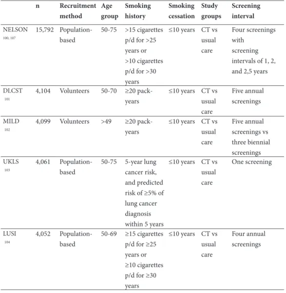
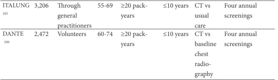
In Europe, seven randomised-controlled trials of LDCT have been conducted or are still
in progress at this moment (Table 6) 100-106. The main difference with the NLST is that the
European trials all used a control group in which no screening was offered. So far, four European trials have shown no significant lung cancer mortality reduction (Table 7), but it should be noted that these trials were strongly underpowered (e.g. small sample size) to be able to show a possible benefit. The mortality analyses of the NELSON, LUSI and UKLS studies are expected in the coming years.
Table 6: an overview of the seven European CT lung cancer screening trials and their study protocol. n Recruitment method Age group Smoking history Smoking cessation Study groups Screening interval NELSON 100, 107 15,792 Population-based 50-75 >15 cigarettes p/d for >25 years or >10 cigarettes p/d for >30 years ≤10 years CT vs usual care Four screenings with screening intervals of 1, 2, and 2,5 years DLCST 101 4,104 Volunteers 50-70 ≥20 pack-years ≤10 years CT vs usual care Five annual screenings MILD 102 4,099 Volunteers >49 ≥20 pack-years ≤10 years CT vs usual care Five annual screenings vs three biennial screenings UKLS 103 4,061 Population-based 50-75 5-year lung cancer risk, and predicted risk of ≥5% of lung cancer diagnosis within 5 years ≤10 years CT vs usual care One screening LUSI 104 4,052 Population-based 50-69 ≥15 cigarettes p/d for ≥25 years or ≥10 cigarettes p/d for ≥30 ≤10 years CT vs usual care Four annual screenings
ITALUNG 105 3,206 Through general practitioners 55-69 ≥20 pack-years ≤10 years CT vs usual care Four annual screenings DANTE 106 2,472 Volunteers 60-74 ≥20 pack-years ≤10 years CT vs baseline chest radio-graphy Four annual screenings
NELSON: The Dutch-Belgian Lung cancer Screening Trial; DLCST: Danish Lung Cancer Screening Trial; MILD: Multicentric Italian Lung Detection trial; UKLS: UK Lung cancer Screening pilot trial; LUSI: Lung tumour screening and intervention trial; ITALUNG; The Italian Lung Study; DANTE: Detection And screening of early lung cancer with Novel imaging Technology.
Table 7: an overview of the seven European CT lung cancer screening trials and their end-point analysis. NELSON DLCST 108, 109 MILD 99 UKLS LUSI ITALUNG 110 DANTE 111
LC mortality, RR (95%CI) - (0.66-1.60)1.03 1.64 1 (0.73-4.01) - - (0.47-1.03)0.70 (0.69-1.43)0.99 All-cause mortality, RR (95%CI) - 1.02 (0.82-1.27) 1.40 2 (0.82-2.38) - - (0.67-1.03)0.83 (0.77-1.17)0.95
1There was no significant difference between the annual and biennial screening arm (p=0.21). This
presents the HR when comparing the two LDCT arms together with the control group; 2There was no
significant difference between the annual and biennial screening arm (p=0.13). This presents the HR
Table 6: continued. an overview of the seven European CT lung cancer screening trials and their study
I
The most important benefit of lung cancer screening is the reduction in lung cancer mortality (Table 8). This has been demonstrated by the NLST and has been an important factor in the
implementation of LDCT screening for lung cancer in the United States 98. The lung cancer
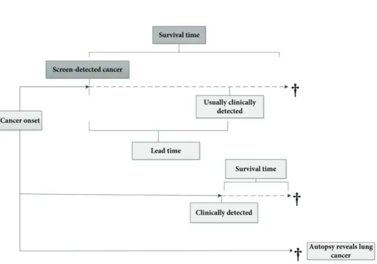
mortality rate is widely accepted as a measurement to determine the effectiveness of a lung cancer screening programme. Case-specific survival of lung cancer (or survival rate) is not recommended since it refers to the number of people with lung cancer remaining alive at a certain point in time after diagnosis. Moreover, survival rate does not adjust for the effects of lead-time, length-time and overdiagnosis biases (Figure 7 and Figure 8):
- Lead-time bias: length of time between early diagnosis of cancer by screening and the
time in which the diagnosis would have been made without screening. In this case, the survival time is increased without affecting the actual course of the disease. In other words, there is no benefit of screening.
- Overdiagnosis bias: detection of tumours by screening, which may have remained
subclinical before death from another cause.
- Length-time bias: screening is most likely to detect relatively slow-growing tumours,
because they have a longer interval of being visible to be detectable by CT scan and have a longer asymptomatic phase. It gives the appearance that screening prolongs life. Therefore, in mortality analyses mortality of both the screen group and control group should be investigated.
Other benefits of lung cancer screening are the reduction in all-cause mortality and increase
of early staged lung cancers with more favourable treatment options 96, 112. Furthermore,
I
However, screening might unintentionally expose the screened population to a variety of harms. One of the primary potential harm is a false positive test result: a benign nodule identified by CT-scanning which may leads to unnecessary invasive investigation. The
false-positive rate across the three screening rounds in the NLST was quite high (23.3%) 96. Another
example is the delay of lung cancer diagnosis by a false negative test result 115. Moreover,
screening will also lead to detection of lung cancers which may have not affected the patient’s lifetime if left untreated (overdiagnosis). True extent of overdiagnosis in lung cancer is difficult to determine because most of the current information what we know about lung cancer is derived from symptomatic patients. One study using excess-incidence reported that
more than 18% of all lung cancers detected by LCDT in the NLST are indolent 116. While,
another study using Microsimulation SCreening Analysis (MISCAN) Lung model with the
NLST data estimated that at year eight overdiagnosis rate to be 12.5% compared to 18.5%117.
Hence, it is important to maximise the benefits while simultaneously minimizing the harms.
Table 8: benefits and harms of lung cancer screening.
Benefits Harms
Overall mortality and lung cancer mortality reduction
Complications of screen result and diagnostic work-up (false positives, invasive diagnostic work-up associated with morbidity and mortality)
Reduction in lung cancer incidence and advanced stage lung cancers
Delay of lung cancer diagnosis by a false negative test result
DALY/QALY/life-years gained Overdiagnosis
Increase of curative treatment options Radiation exposure (induces the risk for
developing cancer)
A teachable moment for smoking cessation Psychological consequences (patient distress,
anxiety) Improvement in diagnostic procedures and
cancer treatment
Possible negative effect on smoking cessation and false reassurance
The NELSON trial Study design
The largest European lung cancer screening trial, the NELSON trial was initiated in the
Netherlands and in Belgium in 2003 107.
The primary aims of this trial are:
I. To establish whether LDCT screening in high-risk subjects would lead to a reduction
of ≥25% in lung cancer mortality;
II. To estimate the impact of lung cancer screening on health-related quality of life and
smoking cessation;
III. To estimate cost-effectiveness of LDCT screening for lung cancer.
Study participants were randomised (1:1) into no screening (control group) or screening using LDCT. Screening took place at baseline (round 1), after one year (round 2), after three years (round 3) and after five and half years (round 4). Participants of both groups are followed-up and the difference in lung cancer mortality between the study groups will be determined ten years after randomisation (Figure 9).
I
The NELSON trial Study design
The largest European lung cancer screening trial, the NELSON trial was initiated in the
Netherlands and in Belgium in 2003 107.
The primary aims of this trial are:
I. To establish whether LDCT screening in high-risk subjects would lead to a reduction
of ≥25% in lung cancer mortality;
II. To estimate the impact of lung cancer screening on health-related quality of life and
smoking cessation;
III. To estimate cost-effectiveness of LDCT screening for lung cancer.
Study participants were randomised (1:1) into no screening (control group) or screening using LDCT. Screening took place at baseline (round 1), after one year (round 2), after three years (round 3) and after five and half years (round 4). Participants of both groups are followed-up and the difference in lung cancer mortality between the study groups will be determined ten years after randomisation (Figure 9).
Figure 9: study design of the NELSON trial.
Population-based recruitment method
Between the second half of 2003 and the second half of 2005, addresses of approximately 600,000 subjects aged 50-74 years were obtained from seven districts in the Netherlands and
14 municipalities around Leuven (Belgium) 100. These subjects received a questionnaire about
their general health status, medical check-ups and history, physical activity, body weight and length, smoking history, alcohol consumption, their own medical history and family
history of cancer, level of education and their opinions on screening programmes 100, 118. The
questionnaire did not contain any information about the upcoming lung cancer screening trial.
The information obtained from this first questionnaire was used to decide who met the
inclusion- and/or exclusion criteria of the trial (Table 9) 100. Respondents who were eligible
received an information leaflet about the NELSON trial, an invitation to participate, an informed consent form for participating in the NELSON trial and a second questionnaire (about smoking habits and exposure to asbestos in more detail).
Finally, those subjects who provided the informed consent and completed the second questionnaire (initially n=15,822, later adjusted to 15,792) were randomised (1:1) to either the screening group or the control group.
The inclusion criteria were:
I. Aged between 50 and 74 at randomization;
II. Smoking history >15 cigarettes per day during >25 years or >10 cigarettes per day
during >30 years;
III. Smoking cessation ≤ 10 years ago.
The exclusion criteria were:
I. A moderate or poor self-reported health who were unable to climb two flights of stairs;
II. A body weight ≥140 kilogram;
III. A lung cancer diagnosis <5 years ago or ≥5 years ago but still under treatment;
IV. A current or past renal cancer, breast cancer or melanoma;
V. A CT chest examination <1 year ago;
VI. A uncompleted informed consent. Screening procedure and protocol
Screen group participants were invited by mail for an LDCT scan at one of the four screening sites: University Medical Centre Groningen, University Medical Centre Utrecht and Kennemer Gasthuis Haarlem in the Netherlands or at University Hospital Gasthuisberg Leuven in
Belgium 107, 119. For the LDCT examination, 16-detector, or in later rounds 64-detector CT
scanners in low-dose settings were used, without the administration of intravenous contrast
Images were analysed using semi-automatic software (LungCARE, version Somaris/5 VA70C-W, Siemens Medical Solutions), in which the semi-automatic segmentation of nodules and determination of the nodule volume took place. In case the software could not segment
the nodule accurately, the radiologist measured the diameter manually 107.
A nodule was defined as a small, spherical, non-linear circumscribed focus of abnormal tissue. The nodule characteristics (e.g. diameter, volume, density, location, lung segment and surface characteristics as smooth, speculated or other) were recorded and uploaded immediately in
the NELSON Nodule Management System (NMS) 107.
In the first two rounds, two radiologists independently reviewed the images in NMS. In case of a discrepancy, a third expert reader made the final decision 107, 120. However, for the last two
rounds a single reading was performed, as Wang et al. showed that there was no benefit for
double reading consensus with the use of semi-automated software 121.
The screening test had three possible results, depending on the presence of one or more
nodule(s), nodule volume and volume doubling time (VDT) 119:
I. Negative: no nodule detected, screen result not suspicious for lung cancer. No further
diagnostic tests warranted;
II. Indeterminate: a small abnormality identified for which at this moment no further
investigation is needed, however, in order to see whether there has been a change in this abnormality over time a follow-up scan will be made;
III. Positive: abnormalities suspicious for lung cancer. Participant referred to a
pulmonologist for a diagnostic work-up.
In the case of newly detected solid nodules and the solid component of the part-solid nodules, the volume determined the screening outcome. In the case of previously detected nodules, the volume growth (change in volume of the nodule) and the VDT of the nodule determined the outcome.
I. Negative screening outcome: a newly detected nodule volume of <50mm3 or, in the
I
for the next screening round. For those in case of the fourth round, this was the end of the screening programme.
All lung cancers were staged using the 7th TNM staging system 57, 59.
Data collection
Prospectively, all relevant medical data about the participants in the two groups is being collected. Relevant medical data contains information about the diagnosis, treatment and follow-up of the lung cancer, and about the cause of death. Furthermore, it contains information about the diagnostic work-up performed in the participants with positives screen test results.
Information about the participants from whom all relevant medical data needs to be collected, is obtained through data linkages with the Dutch Cancer Registry (NKR) and the Belgian Cancer Registry.
After collecting medical data it was first verified whether the participant was indeed diagnosed with lung cancer during the course of the study or at the time of autopsy.
End point verification
Linkages with Statistics Netherlands provided the cause of death of deceased participants. However, a clinical expert death review committee has been formed in the NELSON trial in order to accurately verify the cause of death of the study participants. The committee consists of two main reviewers: a pulmonologist-oncologist and a pathologist specialised in lung oncology. Only in case when no consensus is reached between the two reviewers a third reviewer (an epidemiologist specialised in screening) is consulted. This committee reviews the blinded medical files of the deceased participants diagnosed with lung cancer,
Research questions
In this thesis three research topics are addressed: 1) the optimization of the NELSON screening rounds; 2) the implications and generalizability of the NELSON trial results; and 3) the cause of death verification process of the deceased NELSON study participants
Part I: the optimization of the NELSON screening rounds
I. What is the added value of a fourth screening round with an interval of 2.5 years after
the previous three screening rounds?
II. Which NELSON subgroups with different risks for detecting lung cancer can be
identified based on their previous screening history? Part II: interim stage shift results in the NELSON trial
III. What is the level of cancer and treatment shift between the two study groups in a
selection of the study cohort?
Part III: the cause of death of the NELSON study participants
IV. What are the differences in characteristics and mortality profile of NELSON
participants and eligible non-responders?
V. What is the outcome of the Cause of Death verification process in the NELSON trial,
I
Outline of this thesis
The research questions are divided into three parts. Part 1 consists of two chapters that address the “Optimization of the NELSON screening rounds” by determining the added value of a fourth screening round (Chapter II), and in which NELSON subgroups with various risks for detecting lung cancer can be identified based on their previous screening results (Chapter III). Part 2 consists of one chapter, which presents the interim stage shift results of the NELSON trial (Chapter IV). Part 3 addresses the difference in characteristics and mortality profile of the NELSON participants and eligible non-responders (Chapter V) and makes a comparison between the cause of death verification process in the NELSON trial and the official death certificates (Chapter VI). This is essential for the primary end results of this trial (lung cancer specific mortality).
A general discussion is presented in Chapter VII, in which the published articles referred to in this thesis are reviewed in order to interpret important results, answer the research questions and to formulate general conclusions and recommendations.
References
1. Torre LA, Siegel RL, Jemal A. Lung Cancer Statistics. Adv Exp Med Biol. 2016;893:1-19.
2. Islami F, Torre LA, Jemal A. Global trends of lung cancer mortality and smoking prevalence.
Transl Lung Cancer Res. 2015;4(4):327-38.
3. Howlader N, Noone AM, Krapcho M, Miller D, Bishop K, Kosary CL, Yu M, Ruhl J, Tatalovich
Z, Mariotto A, Lewis DR, Chen HS, Feuer EJ, Cronin KA (eds). SEER Cancer Statistics Review, 1975-2014, National Cancer Institute. Bethesda, MD, https://seer.cancer.gov/csr/1975_2014/, based on November 2016 SEER data submission, posted to the SEER web site, April 2017. Access date 19-07-2017.
4. Dataset regarding lung cancer deaths provided by Statistics Netherlands (CBS), publically
accesable from statline.cbs.nl, access date 04-04-2017.
5. Siegel RL, Miller KD, Jemal A. Cancer Statistics, 2017. CA Cancer J Clin. 2017;67(1):7-30.
6. Wynder EL, Graham EA. Landmark article May 27, 1950: Tobacco Smoking as a possible
etiologic factor in bronchiogenic carcinoma. A study of six hundred and eighty-four proved cases. By Ernest L. Wynder and Evarts A. Graham. Jama. 1985;253(20):2986-94.
7. Wynder EL, Graham EA. Tobacco smoking as a possible etiologic factor in bronchiogenic
carcinoma; a study of 684 proved cases. J Am Med Assoc. 1950;143(4):329-36.
8. Bach PB. Smoking as a factor in causing lung cancer. Jama. 2009;301(5):539-41.
9. Thun M, Peto R, Boreham J, Lopez AD. Stages of the cigarette epidemic on entering its second
century. Tob Control. 2012;21(2):96-101.
10. Netherlands Comprehensive Cancer Organisation, IKNL. Publicaly accessible data available on
www cijfersoverkanker nl, 2016. Access date 19-07-2017 by www.cijfersoverkanker.nl
11. Billiet C, Decaluwe H, Peeters S, Vansteenkiste J, Dooms C, Haustermans K, et al. Modern
post-operative radiotherapy for stage III non-small cell lung cancer may improve local control and survival: a meta-analysis. Radiother Oncol. 2014;110(1):3-8.
12. van Laar MW, Nationale Drug Monitor. Published in 2016 by Trimbos Institute. Accesable from
www.trimbos.nl/kerncijfers/nationale-drug-monitor. Access date 23-08-2017.
13. Alberg AJ, Samet JM. Epidemiology of lung cancer. Chest. 2003;123(1 Suppl):21S-49S.
14. Pesch B, Kendzia B, Gustavsson P, Jockel KH, Johnen G, Pohlabeln H, et al. Cigarette smoking
I
20. Yu YH, Liao CC, Hsu WH, Chen HJ, Liao WC, Muo CH, et al. Increased lung cancer risk among
patients with pulmonary tuberculosis: a population cohort study. J Thorac Oncol. 2011;6(1):32-7.
21. Young RP, Hopkins RJ, Christmas T, Black PN, Metcalf P, Gamble GD. COPD prevalence
is increased in lung cancer, independent of age, sex and smoking history. Eur Respir J. 2009;34(2):380-6.
22. Besaratinia A, Pfeifer GP. Second-hand smoke and human lung cancer. Lancet Oncol.
2008;9(7):657-66.
23. Brennan P, Buffler PA, Reynolds P, Wu AH, Wichmann HE, Agudo A, et al. Secondhand smoke
exposure in adulthood and risk of lung cancer among never smokers: a pooled analysis of two large studies. Int J Cancer. 2004;109(1):125-31.
24. Villeneuve PJ, Parent ME, Harris SA, Johnson KC, Canadian Cancer Registries Epidemiology
Research G. Occupational exposure to asbestos and lung cancer in men: evidence from a population-based case-control study in eight Canadian provinces. BMC Cancer. 2012;12:595.
25. Darby S, Hill D, Auvinen A, Barros-Dios JM, Baysson H, Bochicchio F, et al. Radon in homes
and risk of lung cancer: collaborative analysis of individual data from 13 European case-control studies. Bmj. 2005;330(7485):223.
26. Kligerman S, White C. Epidemiology of lung cancer in women: risk factors, survival, and
screening. AJR Am J Roentgenol. 2011;196(2):287-95.
27. Lorigan P, Radford J, Howell A, Thatcher N. Lung cancer after treatment for Hodgkin’s
lymphoma: a systematic review. Lancet Oncol. 2005;6(10):773-9.
28. Kirova YM, Gambotti L, De Rycke Y, Vilcoq JR, Asselain B, Fourquet A. Risk of second
malignancies after adjuvant radiotherapy for breast cancer: a large-scale, single-institution review. Int J Radiat Oncol Biol Phys. 2007;68(2):359-63.
29. Travis WD. Pathology of lung cancer. Clin Chest Med. 2011;32(4):669-92.
30. Travis WD, Brambilla E, Noguchi M, Nicholson AG, Geisinger KR, Yatabe Y, et al. International
association for the study of lung cancer/american thoracic society/european respiratory society international multidisciplinary classification of lung adenocarcinoma. J Thorac Oncol. 2011;6(2):244-85.
31. Torre LA, Siegel RL, Jemal A. Lung Cancer Statistics. Adv Exp Med Biol. 2016;893(0065-2598
(Print)):1-19.
32. Travis WD, Brambilla E, Nicholson AG, Yatabe Y, Austin JH, Beasley MB, et al. The 2015 World
Health Organization Classification of Lung Tumors: Impact of Genetic, Clinical and Radiologic Advances Since the 2004 Classification. J Thorac Oncol. 2015;10(9):1243-60.
33. Karachaliou N, Pilotto S, Lazzari C, Bria E, de Marinis F, Rosell R. Cellular and molecular
biology of small cell lung cancer: an overview. Transl Lung Cancer Res. 2016;5(1):2-15.
34. Kitamura H, Yazawa T, Sato H, Okudela K, Shimoyamada H. Small cell lung cancer: significance
of RB alterations and TTF-1 expression in its carcinogenesis, phenotype, and biology. Endocr Pathol. 2009;20(2):101-7.
36. Bentea G, Sculier C, Grigoriu B, Meert AP, Durieux V, Berghmans T, et al. Autoimmune paraneoplastic syndromes associated to lung cancer: A systematic review of the literature: Part 3: Neurological paraneoplastic syndromes, involving the central nervous system. Lung Cancer. 2017;106:83-92.
37. Richardson GE, Johnson BE. Paraneoplastic syndromes in lung cancer. Curr Opin Oncol.
1992;4(2):323-33.
38. Janssen-Heijnen ML, Coebergh JW. The changing epidemiology of lung cancer in Europe. Lung
Cancer. 2003;41(3):245-58.
39. Janssen-Heijnen ML, Coebergh JW, Klinkhamer PJ, Schipper RM, Splinter TA, Mooi WJ. Is there
a common etiology for the rising incidence of and decreasing survival with adenocarcinoma of the lung? Epidemiology. 2001;12(2):256-8.
40. Rekhtman N, Ang DC, Sima CS, Travis WD, Moreira AL. Immunohistochemical algorithm for
differentiation of lung adenocarcinoma and squamous cell carcinoma based on large series of whole-tissue sections with validation in small specimens. Mod Pathol. 2011;24(10):1348-59.
41. Kumarakulasinghe NB, van Zanwijk N, Soo RA. Molecular targeted therapy in the treatment of
advanced stage non-small cell lung cancer (NSCLC). Respirology. 2015;20(3):370-8.
42. Hensing T, Chawla A, Batra R, Salgia R. A personalized treatment for lung cancer: molecular
pathways, targeted therapies, and genomic characterization. Adv Exp Med Biol. 2014;799:85-117.
43. Funai K, Yokose T, Ishii G, Araki K, Yoshida J, Nishimura M, et al. Clinicopathologic characteristics
of peripheral squamous cell carcinoma of the lung. Am J Surg Pathol. 2003;27(7):978-84.
44. Khuder SA. Effect of cigarette smoking on major histological types of lung cancer: a
meta-analysis. Lung Cancer. 2001;31(2-3):139-48.
45. Yun YH, Lim MK, Jung KW, Bae JM, Park SM, Shin SA, et al. Relative and absolute risks of
cigarette smoking on major histologic types of lung cancer in Korean men. Cancer Epidemiol Biomarkers Prev. 2005;14(9):2125-30.
46. Kim MJ, Shin HC, Shin KC, Ro JY. Best immunohistochemical panel in distinguishing
adenocarcinoma from squamous cell carcinoma of lung: tissue microarray assay in resected lung cancer specimens. Ann Diagn Pathol. 2013;17(1):85-90.
47. Rossi G, Mengoli MC, Cavazza A, Nicoli D, Barbareschi M, Cantaloni C, et al. Large cell
I
52. Hyde L, Hyde CI. Clinical manifestations of lung cancer. Chest. 1974;65(3):299-306.
53. Chute CG, Greenberg ER, Baron J, Korson R, Baker J, Yates J. Presenting conditions of 1539
population-based lung cancer patients by cell type and stage in New Hampshire and Vermont. Cancer. 1985;56(8):2107-11.
54. Spiro SG, Gould MK, Colice GL, American College of Chest P. Initial evaluation of the patient
with lung cancer: symptoms, signs, laboratory tests, and paraneoplastic syndromes: ACCP evidenced-based clinical practice guidelines (2nd edition). Chest. 2007;132(3 Suppl):149S-60S.
55. Eren S, Karaman A, Okur A. The superior vena cava syndrome caused by malignant disease.
Imaging with multi-detector row CT. Eur J Radiol. 2006;59(1):93-103.
56. Riihimaki M, Hemminki A, Fallah M, Thomsen H, Sundquist K, Sundquist J, et al. Metastatic
sites and survival in lung cancer. Lung Cancer. 2014;86(1):78-84.
57. Marshall HM, Leong SC, Bowman RV, Yang IA, Fong KM. The science behind the 7th edition
Tumour, Node, Metastasis staging system for lung cancer. Respirology. 2012;17(2):247-60.
58. Little AG, Gay EG, Gaspar LE, Stewart AK. National survey of non-small cell lung cancer in the
United States: epidemiology, pathology and patterns of care. Lung Cancer. 2007;57(3):253-60.
59. Goldstraw P, Crowley J, Chansky K, Giroux DJ, Groome PA, Rami-Porta R, et al. The IASLC
Lung Cancer Staging Project: proposals for the revision of the TNM stage groupings in the forthcoming (seventh) edition of the TNM Classification of malignant tumours. J Thorac Oncol. 2007;2(8):706-14.
60. Villalobos P, Wistuba, II. Lung Cancer Biomarkers. Hematol Oncol Clin North Am. 2017;31(1):13-29.
61. Minuti G, D’Incecco A, Cappuzzo F. Targeted therapy for NSCLC with driver mutations. Expert
Opin Biol Ther. 2013;13(10):1401-12.
62. Dutch Guidelines regarding lung cancer. Adapted from www.oncoline.nl Access date
04-05-2017.
63. Vansteenkiste J, Crino L, Dooms C, Douillard JY, Faivre-Finn C, Lim E, et al. 2nd ESMO
Consensus Conference on Lung Cancer: early-stage non-small-cell lung cancer consensus on diagnosis, treatment and follow-up. Ann Oncol. 2014;25(8):1462-74.
64. Scott WJ, Allen MS, Darling G, Meyers B, Decker PA, Putnam JB, et al. Video-assisted thoracic
surgery versus open lobectomy for lung cancer: a secondary analysis of data from the American College of Surgeons Oncology Group Z0030 randomized clinical trial. J Thorac Cardiovasc Surg. 2010;139(4):976-81; discussion 81-3.
65. Group NM-aC, Arriagada R, Auperin A, Burdett S, Higgins JP, Johnson DH, et al. Adjuvant
chemotherapy, with or without postoperative radiotherapy, in operable non-small-cell lung cancer: two meta-analyses of individual patient data. Lancet. 2010;375(9722):1267-77.
66. Solda F, Lodge M, Ashley S, Whitington A, Goldstraw P, Brada M. Stereotactic radiotherapy
(SABR) for the treatment of primary non-small cell lung cancer; systematic review and comparison with a surgical cohort. Radiother Oncol. 2013;109(1):1-7.
67. van den Berg LL, Klinkenberg TJ, Groen HJ, Widder J. Patterns of Recurrence and Survival after
2015;10(5):826-68. Auperin A, Le Pechoux C, Rolland E, Curran WJ, Furuse K, Fournel P, et al. Meta-analysis of concomitant versus sequential radiochemotherapy in locally advanced non-small-cell lung cancer. J Clin Oncol. 2010;28(13):2181-90.
69. Califano R, Abidin AZ, Peck R, Faivre-Finn C, Lorigan P. Management of small cell lung cancer:
recent developments for optimal care. Drugs. 2012;72(4):471-90.
70. Pijls-Johannesma M, De Ruysscher D, Vansteenkiste J, Kester A, Rutten I, Lambin P. Timing of
chest radiotherapy in patients with limited stage small cell lung cancer: a systematic review and meta-analysis of randomised controlled trials. Cancer Treat Rev. 2007;33(5):461-73.
71. Schneider BJ, Saxena A, Downey RJ. Surgery for early-stage small cell lung cancer. J Natl Compr
Canc Netw. 2011;9(10):1132-9.
72. Auperin A, Arriagada R, Pignon JP, Le Pechoux C, Gregor A, Stephens RJ, et al. Prophylactic
cranial irradiation for patients with small-cell lung cancer in complete remission. Prophylactic Cranial Irradiation Overview Collaborative Group. N Engl J Med. 1999;341(7):476-84.
73. Chansky K, Sculier JP, Crowley JJ, Giroux D, Van Meerbeeck J, Goldstraw P, et al. The
International Association for the Study of Lung Cancer Staging Project: prognostic factors and pathologic TNM stage in surgically managed non-small cell lung cancer. J Thorac Oncol. 2009;4(7):792-801.
74. Sun Z, Aubry MC, Deschamps C, Marks RS, Okuno SH, Williams BA, et al. Histologic grade is
an independent prognostic factor for survival in non-small cell lung cancer: an analysis of 5018 hospital- and 712 population-based cases. J Thorac Cardiovasc Surg. 2006;131(5):1014-20.
75. Sardari Nia P, Weyler J, Colpaert C, Vermeulen P, Van Marck E, Van Schil P. Prognostic value of
smoking status in operated non-small cell lung cancer. Lung Cancer. 2005;47(3):351-9.
76. Kobayashi N, Toyooka S, Soh J, Ichimura K, Yanai H, Suehisa H, et al. Risk factors for recurrence
and unfavorable prognosis in patients with stage I non-small cell lung cancer and a tumor diameter of 20 mm or less. J Thorac Oncol. 2007;2(9):808-12.
77. Zhou W, Heist RS, Liu G, Park S, Neuberg DS, Asomaning K, et al. Smoking cessation before
diagnosis and survival in early stage non-small cell lung cancer patients. Lung Cancer. 2006;53(3):375-80.
78. Jamal A, King BA, Neff LJ, Whitmill J, Babb SD, Graffunder CM. Current Cigarette Smoking
Among Adults - United States, 2005-2015. MMWR Morb Mortal Wkly Rep. 2016;65(44):1205-11.
I
83. Cahill K, Lancaster T. Workplace interventions for smoking cessation. Cochrane Database Syst
Rev. 2014(2):CD003440.
84. Yang P, Allen MS, Aubry MC, Wampfler JA, Marks RS, Edell ES, et al. Clinical features of
5,628 primary lung cancer patients: experience at Mayo Clinic from 1997 to 2003. Chest. 2005;128(1):452-62.
85. Mong C, Garon EB, Fuller C, Mahtabifard A, Mirocha J, Mosenifar Z, et al. High prevalence
of lung cancer in a surgical cohort of lung cancer patients a decade after smoking cessation. J Cardiothorac Surg. 2011;6:19.
86. Manser R. Screening for lung cancer: a review. Curr Opin Pulm Med. 2004;10(4):266-71.
87. Manser RL, Irving LB, Byrnes G, Abramson MJ, Stone CA, Campbell DA. Screening for lung
cancer: a systematic review and meta-analysis of controlled trials. Thorax. 2003;58(9):784-9.
88. Humphrey LL, Teutsch S, Johnson M, Force USPST. Lung cancer screening with sputum
cytologic examination, chest radiography, and computed tomography: an update for the U.S. Preventive Services Task Force. Ann Intern Med. 2004;140(9):740-53.
89. Eddy DM. Screening for lung cancer. Ann Intern Med. 1989;111(3):232-7.
90. International Early Lung Cancer Action Program I, Henschke CI, Yankelevitz DF, Libby DM,
Pasmantier MW, Smith JP, et al. Survival of patients with stage I lung cancer detected on CT screening. N Engl J Med. 2006;355(17):1763-71.
91. Sone S, Li F, Yang ZG, Honda T, Maruyama Y, Takashima S, et al. Results of three-year mass
screening programme for lung cancer using mobile low-dose spiral computed tomography scanner. Br J Cancer. 2001;84(1):25-32.
92. Swensen SJ, Jett JR, Hartman TE, Midthun DE, Sloan JA, Sykes AM, et al. Lung cancer screening
with CT: Mayo Clinic experience. Radiology. 2003;226(3):756-61.
93. Sobue T, Moriyama N, Kaneko M, Kusumoto M, Kobayashi T, Tsuchiya R, et al. Screening for
lung cancer with low-dose helical computed tomography: anti-lung cancer association project. J Clin Oncol. 2002;20(4):911-20.
94. Nawa T, Nakagawa T, Kusano S, Kawasaki Y, Sugawara Y, Nakata H. Lung cancer screening using
low-dose spiral CT: results of baseline and 1-year follow-up studies. Chest. 2002;122(1):15-20.
95. Aberle Dr Fau - Berg CD, Berg Cd Fau - Black WC, Black Wc Fau - Church TR, Church Tr Fau
- Fagerstrom RM, Fagerstrom Rm Fau - Galen B, Galen B Fau - Gareen IF, et al. The National Lung Screening Trial: overview and study design. (1527-1315 (Electronic)).
96. National Lung Screening Trial Research T, Aberle DR, Adams AM, Berg CD, Black WC, Clapp
JD, et al. Reduced lung-cancer mortality with low-dose computed tomographic screening. N Engl J Med. 2011;365(5):395-409.
97. Patz EF, Jr., Greco E, Gatsonis C, Pinsky P, Kramer BS, Aberle DR. Lung cancer incidence and
mortality in National Lung Screening Trial participants who underwent low-dose CT prevalence screening: a retrospective cohort analysis of a randomised, multicentre, diagnostic screening trial. Lancet Oncol.May;17(5)(1474-5488 (Electronic)):590-9.
98. de Koning HJ, Meza R, Plevritis SK, ten Haaf K, Munshi VN, Jeon J, et al. Benefits and harms of
99. Humphrey LL, Deffebach M, Pappas M, Baumann C, Artis K, Mitchell JP, et al. Screening for lung cancer with low-dose computed tomography: a systematic review to update the US Preventive services task force recommendation. Ann Intern Med. 2013;159(6):411-20.
100. van Iersel CA, de Koning HJ, Draisma G, Mali WP, Scholten ET, Nackaerts K, et al. Risk-based selection from the general population in a screening trial: selection criteria, recruitment and power for the Dutch-Belgian randomised lung cancer multi-slice CT screening trial (NELSON). Int J Cancer. 2007;120(4):868-74.
101. Pedersen JH, Ashraf H, Dirksen A, Bach K, Hansen H, Toennesen P, et al. The Danish randomized lung cancer CT screening trial--overall design and results of the prevalence round. J Thorac Oncol. 2009;4(5):608-14.
102. Pastorino U, Rossi M, Rosato V, Marchiano A, Sverzellati N, Morosi C, et al. Annual or biennial CT screening versus observation in heavy smokers: 5-year results of the MILD trial. Eur J Cancer Prev. 2012;21(3):308-15.
103. Baldwin DR, Duffy SW, Wald NJ, Page R, Hansell DM, Field JK. UK Lung Screen (UKLS) nodule management protocol: modelling of a single screen randomised controlled trial of low-dose CT screening for lung cancer. Thorax. 2011;66(4):308-13.
104. Becker N, Motsch E, Gross ML, Eigentopf A, Heussel CP, Dienemann H, et al. Randomized study on early detection of lung cancer with MSCT in Germany: study design and results of the first screening round. J Cancer Res Clin Oncol. 2012;138(9):1475-86.
105. Lopes Pegna A, Picozzi G, Mascalchi M, Maria Carozzi F, Carrozzi L, Comin C, et al. Design, recruitment and baseline results of the ITALUNG trial for lung cancer screening with low-dose CT. Lung Cancer. 2009;64(1):34-40.
106. Infante M, Cavuto S, Lutman FR, Brambilla G, Chiesa G, Ceresoli G, et al. A randomized study of lung cancer screening with spiral computed tomography: three-year results from the DANTE trial. Am J Respir Crit Care Med. 2009;180(5):445-53.
107. van Klaveren RJ, Oudkerk M, Prokop M, Scholten ET, Nackaerts K, Vernhout R, et al. Management
of lung nodules detected by volume CT scanning. N Engl J Med. 2009;361(23):2221-9.
108. Saghir Z, Dirksen A, Ashraf H, Bach KS, Brodersen J, Clementsen PF, et al. CT screening for lung cancer brings forward early disease. The randomised Danish Lung Cancer Screening Trial: status after five annual screening rounds with low-dose CT. Thorax. 2012;67(4):296-301. 109. Wille MM, Dirksen A, Ashraf H, Saghir Z, Bach KS, Brodersen J, et al. Results of the
I
113. Ashraf H, Tonnesen P, Holst Pedersen J, Dirksen A, Thorsen H, Dossing M. Effect of CT screening on smoking habits at 1-year follow-up in the Danish Lung Cancer Screening Trial (DLCST). Thorax. 2009;64(5):388-92.
114. van der Aalst CM, van den Bergh KA, Willemsen MC, de Koning HJ, van Klaveren RJ. Lung cancer screening and smoking abstinence: 2 year follow-up data from the Dutch-Belgian randomised controlled lung cancer screening trial. Thorax. 2010;65(7):600-5.
115. Horeweg N, Scholten ET, de Jong PA, van der Aalst CM, Weenink C, Lammers JW, et al. Detection of lung cancer through low-dose CT screening (NELSON): a prespecified analysis of screening test performance and interval cancers. Lancet Oncol. 2014;15(12):1342-50.
116. Patz EF, Jr., Pinsky P, Gatsonis C, Sicks JD, Kramer BS, Tammemagi MC, et al. Overdiagnosis in low-dose computed tomography screening for lung cancer. JAMA Intern Med. 2014;174(2):269-74.
117. Ten Haaf K, de Koning HJ. Overdiagnosis in lung cancer screening: why modelling is essential. J Epidemiol Community Health. 2015;69(11):1035-9.
118. van der Aalst CM, van Iersel CA, van Klaveren RJ, Frenken FJ, Fracheboud J, Otto SJ, et al. Generalisability of the results of the Dutch-Belgian randomised controlled lung cancer CT screening trial (NELSON): does self-selection play a role? Lung Cancer. 2012;77(1):51-7. 119. Xu DM, Gietema H, de Koning H, Vernhout R, Nackaerts K, Prokop M, et al. Nodule
management protocol of the NELSON randomised lung cancer screening trial. Lung Cancer. 2006;54(2):177-84.
120. Xie X, Zhao Y Fau - Snijder RA, Snijder Ra Fau - van Ooijen PMA, van Ooijen Pm Fau - de Jong PA, de Jong Pa Fau - Oudkerk M, Oudkerk M Fau - de Bock GH, et al. Sensitivity and accuracy of volumetry of pulmonary nodules on low-dose 16- and 64-row multi-detector CT: an anthropomorphic phantom study. (1432-1084 (Electronic)).
121. Wang Y, van Klaveren RJ, van der Zaag-Loonen HJ, de Bock GH, Gietema HA, Xu DM, et al. Effect of nodule characteristics on variability of semiautomated volume measurements in pulmonary nodules detected in a lung cancer screening program. Radiology. 2008;248(2):625-31.
122. Horeweg N, van Klaveren RJ, Groen HJ, Lammers JW, Weenink C, Nackaerts K, et al. Blinded and uniform cause of death verification in a lung cancer CT screening trial. Lung Cancer. 2012;77(3):522-5.
II
ABSTRACT Background
In the USA annual lung cancer screening is recommended. However, the optimal screening strategy (eg, screening interval, screening rounds) is unknown. This study provides results of the fourth screening round after a 2.5-year interval in the Dutch-Belgian Lung Cancer Screening trial (NELSON).
Methods
Europe’s largest, sufficiently powered randomised lung cancer screening trial was designed to determine whether low-dose CT screening reduces lung cancer mortality by ≥25% compared with no screening after 10 years of follow-up. The screening arm (n=7,915) received screening at baseline, after 1 year, 2 years and 2.5 years. Performance of the NELSON screening strategy in the final fourth round was evaluated. Comparisons were made between lung cancers detected in the first three rounds, in the final round and during the 2.5-year interval.
Results
In round 4, 46 cancers were screen-detected and there were 28 interval cancers between the third and fourth screenings. Compared with the second round screening (1-year interval), in round 4 a higher proportion of stage IIIb/IV cancers (17.3% vs 6.8%, p=0.02) and higher proportions of squamous-cell, bronchoalveolar and small-cell carcinomas (p=0.001) were detected. Compared with a 2-year interval, the 2.5-year interval showed a higher non-significant stage distribution (stage IIIb/IV 17.3% vs 5.2%, p=0.10). Additionally, more interval cancers manifested in the 2.5-year interval than in the intervals of previous rounds (28 vs 5 and 28 vs 19).
Conclusions
A 2.5-year interval reduced the effect of screening: the interval cancer rate was higher compared with the 1-year and 2-year intervals, and proportion of advanced disease stage in the final round was higher compared with the previous rounds.
2.1. INTRODUCTION
Lung cancer remains the leading cause of cancer death worldwide, mainly due to its advanced
stage at the time of diagnosis 1. Based on the results of the National Lung Screening Trial
(NLST), the US Preventive Services Task Force recommends annual lung cancer screening
with CT 2, 3. People eligible for screening are aged 55 years through 80 years, have smoked at
least 30 pack-years, and currently smoke or have quit within the past 15 years 4, 5. However,
little is known about the effect of longer screening intervals in lung cancer screening trials: thus far, only the Multicentric Italian Lung Detection trial which consisted of two low-dose CT (LDCT) arms (annual vs biennial screening), reported no differences in mortality or in
screening test performances between the two arms (n=1,190 and n=1,186) 6, 7.
The Dutch-Belgian Lung Cancer Screening trial (NELSON) is the largest European randomized lung cancer screening trial, which was designed to investigate whether LDCT screening reduces lung cancer mortality by ≥25% compared with no screening after 10
years of follow-up 8, 9. The trial randomised (1:1) 15,822 current or former smokers into a
screening group and a control group. Compared with the NLST control group who received screening by chest radiography, NELSON control group participants received no screening. Furthermore, the NELSON screening group received LDCT screening at baseline (round 1), after 1 year (round 2), after 3 years (round 3) and after 5.5 years after baseline (round
4), whereas the NLST provided three annual screenings 10. The use of variable screening
intervals in one LCDT arm in the sufficiently powered NELSON trial is unique and presents an opportunity to investigate the influence of the intervals on the screening test performances (eg, lung cancer detection rate, false-positive (FP) rate) and the characteristics of screening-detected lung cancers.
Analyses of the first three rounds of the NELSON trial indicated that a 2-year interval between the second and the third screening rounds did not lead to a significantly higher proportion of advanced stage lung cancers compared with a 1-year screening interval between the first
and second rounds 11. Furthermore, the lung cancer detection rate was relatively stable across
the first three rounds 11-13. Analyses also indicated that, despite the 2-year interval between
II
2.2. METHODS NELSON trial
Details of the design and conduct of the NELSON trial have been reported previously 8, 9. In
brief, eligible participants were selected after completing questionnaires about general health, lifestyle and smoking habits. Based on this information, persons aged 50–75 years, who had smoked ≥15 cigarettes per day for ≥25 years or ≥10 cigarettes per day for ≥30 years, and who were current smokers or former smokers with cessation ≤10 years ago, were invited to participate in the NELSON trial. Eventually, 15 822 eligible high-risk subjects for developing lung cancer participated in this population-based randomised trial. The primary aim of NELSON is to determine whether LDCT screening reduces lung cancer mortality by ≥25%
compared with no screening after 10 years of follow-up 8 .
To perform a fourth screening round an additional informed consent was obtained, as the original protocol consisted of only three screening rounds. The final screening round was conducted from November 2009 through March 2012.
Study population
For this study, all 7,915 participants randomised to the screening arm were included. Screening procedures
Screening group participants were invited to one of the four screening sites (University Medical Centre Groningen, University Medical Centre Utrecht and Kennemer Gasthuis Haarlem in the Netherlands, and University Hospital Gasthuisberg Leuven in Belgium). For the screening, 16-detector or, in later rounds 64-detector CT scanners in low-dose setting were used, without the administration of intravenous contrast media. Images were analysed using semiautomated software (LungCARE, version Somaris/5 VA70C-W, Siemens Medical
Solutions) 9, 15. The analysis included the semiautomated segmentation of nodules and
determination of the nodule volume. In case the software was not able to segment a nodule
accurately, the diameter was measured manually by the radiologist 16. In the first two rounds,
two radiologists independently reviewed the images. In case of a discrepancy, a third expert reader made the final decision. In the last two rounds, a single reading was performed by a
radiologist with at least 6 years of experience in thoracic imaging. Wang et al 17 showed that
there was no benefit for double reading consensus with the use of semiautomated software. More detailed descriptions of the equipment and execution of the screening examination have been provided in previous reports 9, 15, 18
Screening outcomes and the nodule management protocol
The screening test had three possible results, depending on the presence of nodules, nodule
volume and volume doubling time (VDT): negative, indeterminate or positive 10. Negative
results led to invitation to the next screening round, or in case of the final round to the end of the screening programme. Indeterminate results led to invitation for a repeat scan (after 6–8 weeks or after 12 months, depending on nodule size and screening round) in ord