PLEASE SCROLL DOWN FOR ARTICLE
Access details: Access Details: [subscription number 918588849]
Publisher Taylor & Francis
Informa Ltd Registered in England and Wales Registered Number: 1072954 Registered office: Mortimer House,
37-41 Mortimer Street, London W1T 3JH, UK
Traffic Injury Prevention
Publication details, including instructions for authors and subscription information:
http://www.informaworld.com/smpp/title~content=t713456148
The Anatomy and Biomechanics of Acute and Chronic Whiplash Injury
Gunter P. Siegmundab; Beth A. Winkelsteinc; Paul C. Ivancicd; Mats Y. Svenssone; Anita Vasavadaf a MEA Forensic Engineers & Scientists, Richmond, British Columbia, Canada b School of Human Kinetics, University of British Columbia, Vancouver, British Columbia, Canada c Departments of Bioengineering and Neurosurgery, University of Pennsylvania, Philadelphia, Pennsylvania, USA d Department of Orthopaedics and Rehabilitation, Yale University School of Medicine, New Haven, Connecticut, USA e Vehicle Safety Division, Department of Applied Mechanics, Chalmers University of Technology, Göteborg, Sweden f School of Chemical Engineering and Bioengineering, Washington State University, Pullman, WashingtonTo cite this Article Siegmund, Gunter P. , Winkelstein, Beth A. , Ivancic, Paul C. , Svensson, Mats Y. and Vasavada, Anita(2009) 'The Anatomy and Biomechanics of Acute and Chronic Whiplash Injury', Traffic Injury Prevention, 10: 2, 101 — 112
To link to this Article: DOI: 10.1080/15389580802593269 URL: http://dx.doi.org/10.1080/15389580802593269
Full terms and conditions of use: http://www.informaworld.com/terms-and-conditions-of-access.pdf This article may be used for research, teaching and private study purposes. Any substantial or systematic reproduction, re-distribution, re-selling, loan or sub-licensing, systematic supply or distribution in any form to anyone is expressly forbidden.
The publisher does not give any warranty express or implied or make any representation that the contents will be complete or accurate or up to date. The accuracy of any instructions, formulae and drug doses should be independently verified with primary sources. The publisher shall not be liable for any loss, actions, claims, proceedings, demand or costs or damages whatsoever or howsoever caused arising directly or indirectly in connection with or arising out of the use of this material.
ISSN: 1538-9588 print / 1538-957X online DOI: 10.1080/15389580802593269
T h e A n a t o m y a n d B i o m e c h a n i c s o f A c u t e
a n d C h r o n i c W h i p l a s h I n j u r y
GUNTER P. SIEGMUND,
1,2BETH A. WINKELSTEIN,
3PAUL C. IVANCIC,
4MATS Y. SVENSSON,
5and ANITA VASAVADA
61MEA Forensic Engineers & Scientists, Richmond, British Columbia, Canada
2School of Human Kinetics, University of British Columbia, Vancouver, British Columbia, Canada
3Departments of Bioengineering and Neurosurgery, University of Pennsylvania, Philadelphia, Pennsylvania, USA 4Department of Orthopaedics and Rehabilitation, Yale University School of Medicine, New Haven, Connecticut, USA 5Vehicle Safety Division, Department of Applied Mechanics, Chalmers University of Technology, G¨oteborg, Sweden 6School of Chemical Engineering and Bioengineering, Washington State University, Pullman, Washington
Whiplash injury is the most common motor vehicle injury, yet it is also one of the most poorly understood. Here we examine the evidence supporting an organic basis for acute and chronic whiplash injuries and review the anatomical sites within the neck that are potentially injured during these collisions. For each proposed anatomical site—facet joints, spinal ligaments, intervertebral discs, vertebral arteries, dorsal root ganglia, and neck muscles—we present the clinical evidence supporting that injury site, its relevant anatomy, the mechanism of and tolerance to injury, and the future research needed to determine whether that site is responsible for some whiplash injuries. This article serves as a snapshot of the current state of whiplash biomechanics research and provides a roadmap for future research to better understand and ultimately prevent whiplash injuries.
Keywords Whiplash injury; Biomechanics; Neck; Injury mechanisms; Tolerance; Acute and chronic injury
INTRODUCTION
Neck sprains and strains—commonly known as whiplash injuries—are the most common motor vehicle injuries treated in U.S. hospital emergency departments (Quinlan et al. 2004). Incidence rates for whiplash injury range from 28 to 834 per 100,000 each year (Cassidy et al. 2000; Ostremski et al. 1989), and data stratified on gender and age show that females aged 20 to 24 have the highest incidence (∼965 per 100,000 annu-ally; Quinlan et al. 2004). Chronicity rates for whiplash patients also vary widely. In one study, 66 percent of subjects had resid-ual neck pain after two years (Norris and Watt 1983), whereas in another study only 6 percent of subjects had residual neck pain after one month (Schrader et al. 1996). Such wide-ranging incidence and chronicity rates may stem from differing sam-ple sizes, sampling methods, and injury definitions, but despite these differences, acute and chronic whiplash injuries are by a wide margin the most frequent automobile-related injury (Viano 2003). It also remains one of the most poorly understood auto-motive injuries.
Received 12 October 2008; accepted 2 November 2008.
Address correspondence to: Gunter P. Siegmund, Ph.D., P.Eng., 11-11151 Horseshoe Way, Richmond, BC, Canada V7A 4S5. E-mail: gunter.siegmund@ meaforensic.com
Clinically, whiplash patients present with neck, shoulder, or back pain; headaches; dizziness; paresthesias; vertigo; or cogni-tive/psychological symptoms (Evans 1992; Radanov et al. 1995; Sterner and Gerdle 2004). The source of the initial symptoms is often uncertain (Binder 2007), but it is generally presumed these initial symptoms have an organic basis. Multiple anatom-ical sites in the neck have been postulated for this initial in-jury, including the facet joints, spinal ligaments, intervertebral discs, vertebral arteries, dorsal root ganglia, and neck muscles (Figure 1, Table I). Some chronic pain also appears to be or-ganic in nature (Lord et al. 1996a; Sterling 2006), although late whiplash syndrome is viewed by some not as a chronic injury but rather as a self-perpetuating cycle of maladaptive behav-iors possibly initiated by an acute organic lesion (Ferrari and Schrader 2001).
Despite these disparate views regarding their origin, some symptoms of whiplash injury likely have organic bases that are related in some way to the forces transmitted through the neck and the strains experienced by tissues in the neck during a col-lision exposure. Indirect evidence supporting this premise is the 31 to 75 percent reduction in whiplash injuries reported for collisions in vehicles with new anti-whiplash seats designed to reduce these forces (Farmer et al. 2003; Jakobsson and Norin 2004; Viano and Olsen 2001). If there was no underlying in-jury caused by the collision exposure, then these new seats 101
Figure 1 Cross section of the neck showing the anatomical arrangement of the proposed sites of whiplash injury. The shaded areas show muscle (pink), spinal ligament (aqua), facet joints (blue), dorsal root ganglia (yellow), vertebral arteries (red), and intervertebral disc (grey). (Adapted from Rohen and Yokochi 1993.)
would presumably have little or no effect on the rate of injury. Moreover, some whiplash injuries likely do not resolve for or-ganic reasons rather than psychosocial ones. This latter proposi-tion is supported by the delayed recovery and higher chronicity rates for patients with more severe initial symptoms (Scholten-Peeters et al. 2003; Suissa et al. 2001; Williams et al. 2007). What remains unclear, however, is whether chronic pain origi-nates from the acutely injured tissue or whether other physio-logic processes account for the persistence of pain.
Here we review the evidence supporting an organic basis for acute and chronic whiplash injuries. For each proposed anatom-ical site of whiplash injury—facet joints, spinal ligaments, inter-vertebral discs, inter-vertebral arteries, dorsal root ganglia, and neck muscles—we present the clinical evidence supporting that in-jury, the relevant anatomy, the mechanism of and tolerance to injury, and the future research needed to definitively determine whether that site is responsible for some whiplash injuries.
FACET JOINT AND CAPSULAR LIGAMENT Clinical Evidence of Injury
The cervical facet joints are the most common source of neck pain (Aprill and Bogduk 1992; Barnsley et al. 1994). Medial branch blocks and provocative testing have also implicated the facet joint in neck pain, particularly in chronic whiplash patients (Barnsley et al. 1993; Bogduk and Marsland 1988). This strong clinical evidence of facet-mediated neck pain has led to the
development of diagnostic tests (e.g., facet blocks) and treatment procedures (e.g., radiofrequency neurotomies) that can reduce or eliminate pain for a period of time (Lord et al. 1996b).
Relevant Anatomy
There are two facet joints between each pair of cervical vertebra from C2 to C7. The facet joint is a synovial joint enclosed by a thin, loose ligament known as the facet capsule. A synovial fold on the inner capsule extends between the margins of the articu-lating bony surfaces. The facet capsule itself lacks the stiffness to alter the intervertebral kinematics and instead follows the motions of its surrounding bony vertebrae (Winkelstein et al. 2000).
Cervical facet joints are innervated by the medial branches of the dorsal primary ramus from the two levels surrounding each joint (Lang 1993). Several histologic and anatomic studies have identified mechanoreceptors and unmyelinated nocicep-tors in the cervical facet joint (Giles and Harvey 1987; Inami et al. 2001; Kallakuri et al. 2004; McLain 1994; Ohtori et al. 2003). Though the size of the receptive fields of these pain fibers remains unknown, it has been proposed that each fiber innervates an area large enough to collectively cover the entire joint (Cavanaugh 2000). The facet capsule also contains Aδ- and C-fibers, both of which transmit nociceptive singals; i.e., pain (Cavanaugh 2000; Cavanaugh et al. 1989; Giles and Harvey 1987; Inami et al. 2001; Kallakuri et al. 2004; McLain 1994; Ohtori et al. 2003). Nociceptors reactive for substance P and calcitonin gene-related peptide have also been identified in the cervical facet capsules (Inami et al. 2001; Kallakuri et al. 2004; McLain 1994; Ohtori et al. 2003). Both of these neuropeptides are neurotransmitters and nociceptive neuromodulators (Ma and Eisenach 2003; Munglani et al. 1996). Thus, the cervical facet joints have the necessary anatomical features to initiate and potentially modulate more widespread neck pain.
Injury Mechanism and Tolerance
The motion of the facet joint articular processes and the capsule during whiplash-like impacts have been characterized in both human volunteers and cadaveric specimens (Cusick et al. 2001; Kaneoka et al. 1999; Pearson et al. 2004). Based on documented joint motion, two mechanisms of facet joint injury have been proposed: pinching of the synovial fold and excessive strain of the capsule. Ono et al. (1997) and Kaneoka et al. (1999) observed that the cervical vertebrae rotate about a higher instantaneous Table I Summary of the potential location, type, and duration of the injuries sustained at each proposed anatomical site of whiplash injury
Possible duration of injury or related pain Anatomical
site
Specific site
or level Type of injury <1 Month 1–6 Months >6 Months Facet joint C2/3 to C7/T1 Synovial fold pinching;
excess capsule strain
Yes Yes Yes
Ligaments Occiput to T1 Excess strain Yes Yes Yes Vertebral artery Occiput to C6 Excess strain/pinching Yes Yes Yes Nerve root C3 to T1 Cell membrane dysfunction Yes Yes Yes Muscles Multiple muscles, each with
associated tendons and fascia
Excess strain while active Yes No, but may mediate other pain sources
No, but may mediate other pain sources
center during a whiplash exposure than during normal voluntary motion and proposed that this abnormal motion compresses the posterior facet surfaces together, pinching the synovial fold. Al-though the synovial folds are innervated by nociceptors (Inami et al. 2001), no further work attempting to isolate this potential mechanism of whiplash pain has been performed.
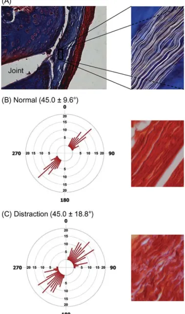
Excessive facet capsule strain during whiplash has been demonstrated by numerous groups (Luan et al. 2000; Pearson et al. 2004; Yang and King 2003; Yoganandan et al. 2002). Peak strains of 29 to 40 percent have been measured in the C6/C7 capsule of cadaveric specimens exposed to whiplash dynamics, whereas peak strains experienced during normal bending are only 6±5 percent (Panjabi et al. 1998a; Pearson et al. 2004). Head-turned postures can double peak capsule strain during sim-ulated whiplash loading (Siegmund et al. 2008b). Prior to the occurence of tissue failure, partial ruptures of the facet capsule have been observed in both tension and shear loading of this joint (Siegmund et al. 2001; Winkelstein et al. 2000). Further, the maximum capsule strains at partial rupture (35–65 percent) do not exceed those strains observed in some capsules during the simulated whiplash loading (Siegmund et al. 2001; Winkelstein et al. 2000). These data suggest that capsule elongation during whiplash is a potential mechanism of injury in some individuals. More recently, in vivo animal models have related facet joint biomechanics to afferent activity and pain symptoms. In a goat model, afferents in the facet capsule are activated by tensile loading of the C5/C6 facet joint (Lu et al. 2005a, 2005b). Cap-sule strains of 10±3 percent activated nociceptive afferents, whereas strains of 44 to 47 percent were sufficient to saturate the mechanoreceptors and nociceptors. Similar strains in the C6/C7 capsule of the rat during joint distraction also produce persistent pain symptoms (Dong et al. 2008; Lee et al. 2004; Lee et al. 2004; Quinn et al. 2007). More importantly, however, the intensity and duration of persistent pain in the rat depend upon the magnitude of strain in the capsule. A maximum principal strain of about 21 percent is associated with persistent sensitiv-ity (Dong et al. 2008; Lee et al. 2004; Lee et al. 2004b). These strains are consistent with those detected in the human capsule during whiplash simulations (Pearson et al. 2004; Siegmund et al. 2001; Sundararajan et al. 2004; Winkelstein et al. 2000). For the same levels of joint distraction that produce pain, the fiber organization in the capsular ligament is also altered (Quinn et al. 2007), indicating that collagen in the capsule is being disor-ganized by the joint distraction, despite the absence of complete ligament failure (Figure 2).
Physiologic responses can contribute to pain in the absence of major mechanical failure. For instance, Lu et al. (2005a, 2005b) reported persistent after-discharges from afferents after joint loads were removed. At the cellular level, both neurons and other cells in the dorsal root ganglia demonstrate sensitive responses to painful and non-painful joint loading (Lee et al. 2008). Persistent increased expression of binding protein (BiP), a marker of cellular stress response (Dong et al. 2008), occurs predominantly in neurons of the dorsal root ganglia follow-ing painful facet joint loadfollow-ing similar to that which develops in
Figure 2 Facet capsule histology demonstrating collagen fiber organization in the rat facet capsular ligament. (A) Masson trichrome staining demonstrating a facet joint with its enclosing capsule (blue stain) and surrounding muscle (red). The inset shows a closeup of the facet capsule ligament with fibers— outlined in white—showing their typical undulation. (B), (C) Following a joint distraction sufficient to produce persistent pain symptoms, the collagen fiber organization (measured by angular deviation) is significantly larger. Shown here are representative histograms of angular deviation and sample tissue from (B) normal and (C) distracted ligaments.
whiplash. Inflammatory responses in the spinal cord are induced and sustained following painful joint loading and depend on the strain imposed on the capsule (Lee et al. 2004, 2008). These local and more widespread neuro-inflammatory cascades con-tribute to a variety of other chronic pain syndromes (DeLeo and Yezierski 2001). Their induction, persistence, and relationship to joint/capsule mechanics in painful whiplash loading supports the facet joint’s involvement in whiplash pain.
Future Directions
Continued biomechanical research is needed to define how col-lagen injury during subfailure ligament loading initiates pain re-sponses, their temporal response, and how such scenarios may be produced during whiplash. Moreover, continued research is needed to identify and define the specific physiologic path-ways (electrophysiologic, immunologic, and otherwise) that are responsible for chronic pain following this joint’s injury. Us-ing this information, better diagnosis and treatment for facet-mediated, or at least facet-initiated, whiplash pain can be developed.
LIGAMENT AND DISC Clinical Evidence of Injury
Magentic resonance and autopsy studies of whiplash patients have documented injuries to the neck ligaments and interver-tebral discs in addition to the facet joints (Jonsson et al. 1991; Kaale et al. 2005a, 2005b; Krakenes and Kaale 2006; Pettersson et al. 1997). Whiplash-related symptoms may be due, in part, to injuries of cervical ligaments and discs and their embedded mechanoreceptive and nociceptive nerve endings. Ligament in-juries may cause acute neck pain and lead to chronic spinal instability, and injured mechanoreceptors may corrupt normal sensory signals and could lead to abnormal muscle response pat-terns and decreased neck mobility and proprioception (Panjabi 2006).
Relevant Anatomy
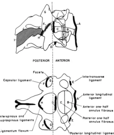
The cervical vertebrae are joined by multiple ligaments. The main ligaments below the axis include the anterior and poste-rior longitudinal, capsular, interspinous, and supraspinous liga-ments and the ligamentum flavum (Figure 3). The anterior and posterior longitudinal ligaments are thin sheets of tissue that span the anterior and posterior surfaces of the vertebral bodies, respectively, and blend with the underlying annular fibers. The capsular ligaments, as described earlier, encase the facet joints. The interspinous ligaments join adjacent spinous processes and are not present in all adults. When present, these ligaments are thin, weak tissues of high collagen content that blend posteriorly with the supraspinous ligament. The ligamentum flavum is the most elastic tissue in the human body—comprised of up to 80 percent elastin—and joins adjacent laminae bilaterally (Yahia et al. 1990). The intervertebral disc, located between adjacent vertebral bodies, consists of a central nucleus pulposus encased by annulus fibrosis fibers.
Ligaments of the upper cervical spine—occiput through the axis—have unique functional and structural anatomy. Alar and transverse ligaments play key roles in providing stability in this region due to the absence of intervertebral discs and the horizontal alignment of the facet joints (Dvorak et al. 1988). These ligaments have a high collagen and low elastin content, predisposing them to partial or complete rupture at low strains during high-speed elongation (Panjabi et al. 1998b).
Ligaments provide joint position sense during normal mo-tion and combined with discs provide passive stability and
ab-Figure 3 Ligaments of the middle and lower cervical spine (from White and Panjabi 1990).
sorb energy during high-speed trauma. The specific function of each cervical ligament and disc in resisting whiplash loading is dependent upon its specific anatomical location, orientation, geometry, and unique mechanical properties.
Injury Mechanism and Tolerance
Spinal ligaments and annular fibers encapsulating the discs can partially or completely rupture when stretched beyond their physiological limit. The whiplash-related response of the cer-vical ligaments and discs have been quantified for frontal, side, and rear impacts using a whole cadaveric cervical spine model with muscle force replication and a surrogate head (Figure 4A; Ivancic et al. 2005). During rear impacts with the head facing for-ward, dynamic strains in the anterior longitudinal ligament and annular fibers above physiological levels (Ivancic et al. 2004; Panjabi et al. 2004a) and increased joint laxity (Ito et al. 2004) were observed. The C5/C6 disc was found to be at highest risk of injury during both frontal and rear impacts (Ito et al. 2005). In addition to the C5/C6 disc, excessive strains were observed in superior discs, including C2/C3, during frontal impacts. The disc injuries occurred at lower impact accelerations during rear impacts compared to frontal impacts. During frontal impacts, the supraspinous ligament, interspinous ligament, and ligamen-tum flavum at C2/C3 through C7/T1 are at risk for injury due to excessive strain (Panjabi et al. 2004b). The T1 horizontal accel-eration at which ligament and/or disc injuries were detected in those studies using pre- and post-impact flexibility tests was 5g
Figure 4 (A) Schematic of the biofidelic whole cervical spine model with surrogate head and muscle force replication used to simulate whiplash, showing the position of vertebral artery transducer (VAT) and vertebral artery (VA) cable passing through the foramen and attaching to the occiput. (B) Lateral and (C) top schematic views of the vertebral artery transducer: frame (a), Hall effect sensor (b), movable carriage (c) carrying two rare earth magnets (d), vertebral artery cable (e), and tension spring (f). (Adapted from Ivancic et al., 2006.)
for rear impacts, 6.5gfor side impacts, and 8gfor frontal im-pacts. For all impact configurations, the spinal levels at greatest risk of ligament and/or disc injury were C3/C4 through C7/T1. Injuries to the alar and transverse ligaments of the upper cervical spine are reportedly more severe in individuals who have their head rotated at impact (Kaale et al. 2005a). These injuries have not been reproduced in cadaveric studies at impact accelerations (applied at T1) up to 8g (Hartwig et al. 2004; Maak et al. 2006). This suggests that the upper cervical spine symptomatology reported by some whiplash patients may be due to impacts causing T1 accelerations in excess of 8gor from some other anatomical structure.
Future Directions
Additional research is needed to further our understanding of ligament and disc injury mechanisms during whiplash and to in-vestigate preventative mechanisms. Biomechanical studies are needed to correlate increased ligament and disc laxity with spe-cific ligament injuries for each impact configuration. Future work is also needed to correlate the severity of ligament and disc injuries, in the form of biomechanical instability, with the onset of neck pain and, ultimately, to link specific ligament injuries to neck pain, or pain patterns, in whiplash patients. Biomechanical studies of simulated whiplash are needed to determine whether dynamic neck loads and high-speed ligament and disc strains are reduced by implementing specific injury prevention sys-tems; e.g., active head restraint or energy-absorbing seat. These results may be correlated with those of epidemiological studies that investigate the effectiveness of injury prevention systems in reducing neck injury in real-life automobile collisions.
VERTEBRAL ARTERY Clinical Evidence of Injury
Altered blood flow rates due to spasm and/or narrowing of ver-tebral arteries in whiplash patients have been associated with chronic symptoms of headache, blurred vision, tinnitus, dizzi-ness, and vertigo (Reddy et al. 2002; Seric et al. 2000). Intimal tears of the vertebral artery are most common at the primary site of cervical axial rotation, the atlanto-axial joint (Barton and Margolis 1975; Chung and Han 2002; Davis and Zimmerman 1983; Pollanen et al. 1996; Sherman et al. 1981; Stahmer et al. 1997; Taneichi et al. 2005). Vertebral artery injury causing inadequate perfusion of the brainstem and surrounding tissues could explain some of the whiplash-related symptoms (e.g., headache, dizziness, and vertigo).
Relevant Anatomy
The vertebral arteries supply blood to the head, brain, and neck tissues. The vertebral arteries enter the spine at the C6 transverse processes bilaterally and run superiorly in the transverse fora-men of each cervical vertebra. After exiting C1, the vertebral arteries travel along the C1 posterior arch and enter the fora-men magnum of the skull. The vertebral artery is a viscoelastic structure: the adventitia is composed primarily of collagen fibers and the media consists of collagen as well as more substantial portions of smooth muscle and elastic fibers. It is encased in a fi-brous tunnel and affixed to adjacent structures via a traneculated collagen network (Chopard et al. 1992).
Injury Mechanism and Tolerance
Coupled extension and axial rotation of the upper cervical spine has been hypothesized to cause vertebral artery injury (Barton and Margolis 1975; Chung and Han 2002; Davis and Zimmerman 1983; Sherman et al. 1981). Vertebral artery elon-gation causes a decrease in the vessel diameter due to Poisson’s effect and could cause transient vascular compromise (Dobrin 1978). Alternatively, stretching or pinching of the vessel along a turn in its circuitous course is also possible (Barton and Mar-golis 1975). These mechanisms can also precipitate tearing of the intimal layer of the vertebral artery (Chung and Han 2002). Cadaveric neck models have demonstrated coupled extension and axial rotation during side and rear impacts with the head turned but not during frontal or rear impacts with the head facing forward (Carlson et al. 2007; Ivancic et al. 2006). In those stud-ies, average vertebral artery elongation was measured between the occiput and C6 vertebra using a custom transducer mounted in a cadaveric neck (Figure 4B). Peak vertebral artery elonga-tion of 30.5 mm during head-turned rear impacts and 17.4 mm during side impacts significantly exceeded physiological elon-gation limits. Moreover, peak elonelon-gation occurred early—about 85 ms following the onset of T1 acceleration—with elongation rates reaching 1340 mm/s during head-turned rear impacts and 610 mm/s during side impacts. The magnitude, rate, and timing of vertebral artery elongation are thus sufficient to potentially cause vertebral artery injury.
Future Directions
Further biomechanical research is needed to determine the strain distribution throughout the vertebral artery during physiological movements and whiplash-related loading rates from different initial neck postures and in various impact directions.
DORSAL ROOT GANGLION AND DORSAL ROOT Clinical Evidence of Injury
The dorsal root ganglion contains the cell bodies of most pe-ripheral sensory nerves at each spinal level. Direct injury to cell bodies within the dorsal root ganglion could thus explain many of the typical whiplash symptoms (e.g., neck pain, cervico-genic headache, vertigo, vision disturbance, and neurological symptoms in the upper extremities). Generalized hypersensitiv-ity to pressure acutely and chronically and decreased thermal pain thresholds in the skin over the cervical spine can be ex-plained by impaired local sensory processing (Greening et al. 2005; Kasch et al. 2001b; Scott et al. 2005; Sterling et al. 2003, 2006; Sterner et al. 2001). In addition, increased electrical ac-tivity in the spinal cord and widespread reductions in electrical and pressure thresholds after whiplash suggest altered central pain processing (Banic et al. 2004; Curatolo et al. 2001; Kasch et al. 2001a; Scott et al. 2005). Increased sensitivity to pain (hy-peralgesia) and larger areas of referred pain are also reported for whiplash patients (Koelbaek Johansen et al. 1999). These studies documenting both local and referred pain after whiplash injury provide clinical evidence for altered sensory transmission and pain pathways in the central nervous system.
Relevant Anatomy
The anterior and posterior rootlets coming off the spinal cord combine to form dorsal and ventral nerve roots, which make up the spinal nerves at each spinal level. The location, direction, and number of nerve rootlets vary at each cervical level. The dorsal and ventral roots come together in the region of the neural foramen and continue more distally into the periphery as the spinal nerve to innervate structures outside the spinal column. Posterior rootlets making up the dorsal root are the sensory (afferent) fibers, whereas the anterior rootlets making up the ventral root are the effector (efferent) fibers. Cell bodies of peripheral afferents are housed in the dorsal root ganglion, which has been shown to be particularly sensitive to loading—even slight compression of normal dorsal root ganglia can produce sustained electrical activity and pain (Howe et al. 1977). Unlike peripheral nerves, the nerve roots themselves are not enclosed by a thick epineurial sheath, and thus they lack the mechanical strength of their peripheral counterparts, potentially exposing nerve roots to increased risk of injury when loaded.
Injury Mechanism and Tolerance
Movements of the cervical spine in flexion, extension, and lateral bending cause the volume of the spinal canal to change. During normal voluntary neck motions, blood volumes in the internal and external vertebral venous plexa can easily move to
compen-Figure 5 Pressure and displacement during a whiplash extension experiment using a pull-force on the porcine head-plate of 600 N. Graphs show (A) the pressure in the CNS at the skull, C4 and T1 vertebral levels, and (B) the angular and linear X-displacement of the head center of mass. (Adapted from Svensson et al., 2000.)
sate for these volume changes. During rapid whiplash-induced motions, however, resistance to blood flow and the inertia of the fluid mass itself can generate transient pressure gradients be-tween the inside and outside of the spinal canal (Aldman 1986). These pressure gradients can directly load the spinal ganglia and nerve roots, potentially leading to whiplash-related symptoms.
Whiplash experiments carried out on anesthetized pigs in extension, flexion, and lateral bending revealed a transient pres-sure drop inside the spinal canal during rapid motion in all directions (Figure 5; Svensson et al. 2000). Follow-up histology showed leakage of the plasma membrane of spinal ganglia nerve cells consistent with cellular injury ( ¨Ortengren et al. 1996). Eichberger et al. (2000) reported similar pressure recordings in cadavers exposed to whiplash and Schmitt et al. (2003) have since recreated the pressure pattern in a computational fluid dy-namics model of the human cervical spine. These experimental findings are supported by an autopsy study of individuals who had sustained severe inertial neck loading (Taylor et al. 1998). Interstitial hemorrhage in the cervical dorsal root ganglia was observed in those autopsies despite an absence of injury to other structures surrounding the ganglia.
The relationship between the head-neck motion and the pres-sure magnitude in the spinal canal is quantified by the neck in-jury criterion (NIC; Bostr¨om et al. 2000). NIC is related to the relative horizontal acceleration and velocity of the head with respect to the torso, and a low NIC equates to a low risk of long-term neck injury (Krafft et al. 2003). Because many other loads and strains within the neck tissues also vary with NIC, this relationship between NIC and long-term neck injuries is not proof that dorsal root ganglion injuries explain all long-term whiplash injuries.
Deformation of the nerve roots themselves is another poten-tial mechanism for producing persistent neck pain. The neural
foramina change shape and decrease their diameter during ex-treme neck motions (Carter et al. 2000; Krivickas and Wilbourn 2000; Yoo et al. 1992). This can compress the nerve root within the intervertebral foramen during whiplash motions. Nuckley et al. (2004) reported a 20 percent decrease in area for the C4-C7 intervertebral foramina of cadaveric cervical spines in extension. The intervertebral foramen at C5/C6 narrowed by as much as 1.8 mm during simulated rear impacts of a cadaveric head-neck model using horizontal T1 accelerations up to 8g (Panjabi et al. 2006; Tominaga et al. 2006). This dynamic nar-rowing of the foramen during whiplash may compress the nerve roots and ganglia in the lower cervical spine, particularly in individuals with congenitally narrow foramen or those with os-teophytes.
Transient loads on the cervical dorsal nerve roots have pro-duced significantly elevated pain symptoms in a rat model (Hubbard and Winkelstein 2005; Hubbard et al. 2008; Rothman et al. 2005). Wallerian degeneration, disrupted axonal trans-port, and altered neuronal responses in the dorsal root ganglion are also produced (Hubbard and Winkelstein 2008). These data further support direct and indirect relationships between tissue loading, neuronal function, and altered physiology locally, in the dorsal root ganglia and throughout the nervous system for painful loading conditions.
Future Directions
Refined finite element and fluid dynamics models of the human head and neck may lead to better understanding of the flow and pressure phenomena that appear to result in ganglion dysfunc-tion. This improved understanding would enable the develop-ment of more accurate injury criteria and tolerance limits for ganglion injury and would guide the development of improved crash dummies and performance requirements for injury pro-tection systems in vehicles. Additional work is also needed to establish the link between the observed pressure transients and the generation and time course of ganglion dysfunction. The in-fluence of nerve cell membrane dysfunction on nerve function and pain sensitization also needs to be investigated following experimentally induced ganglion injury.
MUSCLE
Clinical Evidence of Injury
Muscle or myofascial pain is a common symptom reported by whiplash patients (Evans 1992), although evidence of direct injury to muscle remains inconclusive. Injury-related muscle soreness is associated with a rise in serum creatine kinase de-tected at 3 to 24 h after high-intensity exercise and may persist for up to 9 days (Evans et al. 1986). In some whiplash patients, elevated serum creatine kinase has been observed 24 h after injury but not 48 h after injury, despite neck pain extending beyond 3 months (Scott and Sanderson 2002). Although this work suggests that direct muscle injury may not be responsi-ble for chronic whiplash pain, muscles may nevertheless play an indirect role in modulating pain caused by injuries to other structures.
Figure 6 Neck muscle anatomy. (A) Lateral view of superficial muscles. (B) Posterior view of superficial muscles. (C) Posterior view of deeper muscles. (D) Anterior view of deep muscles. (Adapted from Gray, 1977.) (Figure appears in color online).
Relevant Anatomy
Muscles comprise the majority of the neck’s volume (Figure 1). The superficial muscles, such as sternocleidomastoid or trapez-ius (Figures 6A and 6B), are often implicated in the pain and tenderness associated with whiplash injury. These superficial muscles attach to the skull, shoulder girdle, and ligamentum nuchae but do not generally attach directly to the cervical ver-tebrae. Deeper muscles, such as splenius, semispinalis, longis-simus, scalenes, and longus, attach on multiple cervical vertebrae (Figures 6B, 6C, 6D). The deepest neck muscles, the multifidus muscles, insert directly on the facet capsule of cervical verte-brae (Figure 7) and may be relevant to injury of the capsular ligaments (Anderson et al. 2005; Winkelstein et al. 2001). Most neck muscles have complex architecture, with extensive internal tendon (Kamibayashi and Richmond 1998) and a high density of muscle spindles (Boyd-Clark et al. 2002). Although this has not been explored, the presence and arrangement of the internal tendon may be related to musculotendinous pain.
Injury Mechanism and Tolerance
The direct mechanism of neck muscle injury occurs from eccen-tric contractions; i.e., imposed lengthening during active con-traction. Computer simulations using experimental kinematics of human subjects exposured to rear-end collisions have shown that both anterior and posterior neck muscles experience active lengthening during rear impacts (Brault et al. 2000; Vasavada et al. 2007). The anteriorly located sternocleidomastoid is ac-tive and lengthened during the retraction phase of whiplash, whereas posterior muscles are active and lengthened during the rebound phase. For simulated impacts with a speed change of 8 km/h, peak muscle fascicle strains averaged about 7 percent (max. 15%) in the sternocleidomastoid and 21 percent (max. 50%) in the posterior muscles such as semispinalis capitis. These
Figure 7 Anatomy of (A) superficial and (B) deep layers of the cervical multifidus muscles, depicting attachments on the facet capsules. (Adapted from Anderson et al., 2005.)
strains exceeded those shown to cause muscle injury (5–20%) in laboratory studies (Macpherson et al. 1996; McCully and Faulkner 1985). Thus, acute neck muscle injury may occur during rear-end impacts.
Interactions with Other Anatomical Sites
Neck muscles potentially interact with other anatomical sites of whiplash injury in at least three ways: (1) neck muscles at-tach directly to the facet capsule, which has been implicated in chronic pain following whiplash; (2) neck muscle activation indirectly affects the loads and strains in other anatomical struc-tures; and (3) altered neuromuscular control may contribute to chronic pain via elevated and inappropriate muscle activation.
The pathomechanical evidence for facet capsular ligament involvement in whiplash injury and chronic neck pain has been outlined earlier in this article. Direct attachment of the multifidus muscles to the capsular ligament (Anderson et al. 2005; Winkel-stein et al. 2001), combined with early activation of these mus-cles in some subjects during a rear-end collision may exacerbate peak strain in the capsular ligaments (Siegmund et al. 2008a).
Neck muscle activation also affects spinal tissue loads by in-creasing intervertebral compression and altering intervertebral kinematics. Because neck muscles are oriented primarily verti-cally, their activation produces axial compression of the cervical spine, increasing loads on the intervertebral disc and facet joints. Reflex muscle activation also affects the kinematic response of the head and neck. In subjects exposed to a series of identical perturbations, habituation of the muscle response amplitude by about 50 percent was accompanied by 10 to 30 percent changes in peak head kinematics (Siegmund et al. 2003). By altering head and neck kinematics, load and strain thresholds for injury may be exceeded in other structures such as ligaments, discs, and facet joints.
Finally, the interaction between muscles and the nervous system—i.e., via neuromuscular control—may be related to
chronic pain. Patients with chronic pain demonstrate altered neuromuscular patterns (Falla et al. 2004; Nederhand et al. 2002), but it is not known whether the observed muscle activi-ties are a physiological deficit in motor control or a protective strategy to avoid pain. A further complication is that differ-ent types of adaptive responses have been observed in differdiffer-ent populations of whiplash patients (Nederhand et al. 2000, 2003). An inability to relax after exercise and excessive coactivation are associated with cervical pain (Elert et al. 1992; Nederhand et al. 2000; Westgaard et al. 1993), and relaxing selected neck muscles with botulinum toxin improves range of motion and re-duces pain in these patients (Freund and Schwartz 2002). This suggests that pain and increased muscle activity may cyclically reinforce one another (Johansson and Sojka 1991). Contrasting evidence supports a pain adaptation model in which nociceptive interneurons inhibit the activity of painful muscles or those in the vicinity of pain sources (Lund et al. 1991). Nederhand et al. (2003) found that whiplash patients had a normal ability to re-lax the trapezius following exercise, but during exercise those with the highest disability levels had the lowest muscle activ-ity. It remains unclear, however, whether muscle dysfunction is a cause (leading to damage of other anatomical structures) or effect (due to disuse or pain avoidance) of pain or merely an associated correlation.
Future Directions
Future research is needed to explore the role of neck muscles in the mechanism of acute whiplash injury, especially the in-teractions with other neck structures. Specifically, the effect of multifidus activity on capsular ligament mechanics and nocicep-tive physiologic responses needs to be studied to determine the relevant magnitude of loads from muscle forces on the ligament. Ideally, this type of research should be conducted in vivo, where muscles can be stimulated and ligament mechanical parameters measured. Research is also needed to explore how altered neuro-muscular control relates to chronic pain. Specifically, studies are needed to analyze deep muscle activity in patients with chronic neck pain due to whiplash injury. In addition, validated math-ematical models may be used to assess the effect of abnormal muscle activation on the loads in other anatomical structures.
SUMMARY
This review provides a brief summary of the anatomical structures being investigated by many groups to potentially ex-plain whiplash injury. Each of the tissues described is strained during a whiplash exposure and thus could be injured if the crash-induced strain exceeds that tissue’s tolerance. For each of the tissues summarized here, continued research is needed to better understand the biomechanical and physiological link between crash-induced loading and acute and chronic whiplash-related pain. A better understanding of each potentially injured tissue will help improve the diagnosis and treatment of whiplash injuries. Elimination or reduction of tissue strains through im-proved vehicle, seat, and head restraint design will help reduce the frequency of whiplash injury.
ACKNOWLEDGEMENTS
This review is based on a discussion panel consisting of the authors at the World Congress on Neck Pain, Los Angeles, California, on January 21, 2008. The authors thank Drs. Adrian Lund and Anders Kullgren for their help planning the panel, Dr. Mark White for organizing the conference, and Dr. David Viano for his encouragement to write this article. Funding for portions of this work was provided by the Southern Consortium for Injury Biomechanics/NHTSA subcontract (DTNH-22-04-H-01423) (BAW), the National Center for Injury Prevention and Control (R49CE000689 (BAW) and 1R01CE001257 (PCI)), the National Science Foundation (Grant No. 0547451) (BAW), and the Whitaker Foundation (ANV).
REFERENCES
Aldman B. (1986) An Analytical Approach to the Impact Biomechanics of Head and Neck Injury. Proc.30th Annual AAAM, pp. 446–454. Anderson JS, Hsu AW, Vasavada AN. (2005) Morphology, Architecture
and Biomechanics of Human Cervical Multifidus Muscles.Spine, Vol. 30, pp. E86–E91.
Aprill C, Bogduk N. (1992) The Prevalence of Cervical Zygapophyseal Joint Pain: A First Approximation.Spine, Vol. 17, pp. 744–747. Banic B, Petersen-Felix S, Andersen O, Radanov B, Villiger P,
Arendt-Nielsen L, Curatolo M. (2004) Evidence for Spinal Cord Hypersen-sitivity in Chronic Pain after Whiplash Injury and in Fibromyalgia.
Pain, Vol. 107, pp. 7–15.
Barnsley L, Lord S, Bogduk N. (1993) Comparative Local Anaesthetic Blocks in the Diagnosis of Cervical Zygapophyseal Joint Pain.Pain, Vol. 55, pp. 99–106.
Barnsley L, Lord S, Bogduk N. (1994) Whiplash Injury.Pain, Vol. 58, pp. 283–307.
Barton JW, Margolis MT. (1975) Rotational Obstructions of the Ver-tebral Artery at the Atlantoaxial Joint.Neuroradiology, Vol. 9, pp. 117–120.
Binder A. (2007) The Diagnosis and Treatment of Nonspecific Neck Pain and Whiplash.Eura Medicophys., Vol. 43, pp. 79–89. Bogduk N, Marsland A. (1988) The Cervical Zygapophyseal Joints as
a Source of Neck Pain.Spine, Vol. 13, pp. 610–617.
Bostr¨om O, Fredriksson R, H˚aland Y, Jakobsson L, Krafft M, L¨ovsund P, Muser MH, Svensson MY. (2000) Comparison of Car Seats in Low Speed Rear-end Impacts Using the BioRID Dummy and the New Neck Injury Criterion (NIC).Accid. Anal. Prev., Vol. 32, pp. 321–328.
Boyd-Clark LC, Briggs CA, Galea MP. (2002) Muscle Spindle Dis-tribution, Morphology, and Density in Longus Colli and Multifidus Muscles of the Cervical Spine.Spine, Vol. 27, pp. 694–701. Brault JR, Siegmund GP, Wheeler JB. (2000) Cervical Muscle
Re-sponse during Whiplash: Evidence of a Lengthening Muscle Con-traction.Clin. Biomech., Vol. 15, pp. 426–435.
Carlson EJ, Tominaga Y, Ivancic PC, Panjabi MM. (2007) Dynamic Vertebral Artery Elongation during Frontal and Side Impacts.Spine J., Vol. 7, pp. 222–228.
Carter JW, Mirza SK, Tencer AF, Ching RP. (2000) Canal Geometry Changes Associated with Axial Compressive Cervical Spine Frac-ture.Spine, Vol. 25, pp. 46–54.
Cassidy JD, Carroll LJ, Cˆot´e P, Lemstra M, Berglund A, Nygren A. (2000) Effect of Eliminating Compensation for Pain and Suffering
on the Outcome of Insurance Claims for Whiplash Injury.N. Engl. J. Med., Vol. 342, pp. 1179–1186.
Cavanaugh JM. (2000) Neurophysiology and Neuroanatomy of Neck Pain. InFrontiers in Whiplash Trauma: Clinical and Biomechanical, Ed. N. Yoganandan, FA Pintar. IOS Press, Amsterdam, pp. 79–96. Cavanaugh JM, el-Bohy AA, Hardy WN, Getchell TV, Getchell
ML, King AI. (1989) Sensory Innervation of Soft Tissues of the Lumbar Spine in the Rat. J. Orthop. Res., Vol. 7, pp. 278– 288.
Chopard RP, de Miranda Neto MH, Lucas GA, Chopard MR. (1992) The Vertebral Artery: Its Relationship with Adjoining Tissues in its Course Intra and Inter Transverse Processes in Man.Rev. Paul Med., Vol. 110, pp. 245–250.
Chung YS, Han DH. (2002) Vertebrobasilar Dissection: A Possible Role of Whiplash Injury in its Pathogenesis.Neurol. Res., Vol. 24, pp. 129–138.
Curatolo M, Petersen-Felix S, Arendt-Nielsen L, Giana C, Zbinden AM, Radanov BP. (2001) Central Hypersensitivity in Chronic Pain after Whiplash Injury.Clin. J. Pain, Vol. 17, pp. 306–315.
Cusick JF, Pintar FA, Yoganandan N. (2001) Whiplash Syndrome: Kinematic Factors Influencing Pain Patterns.Spine, Vol. 26, pp. 1252–1258.
Davis JM, Zimmerman RA. (1983) Injury of the Carotid and Vertebral Arteries.Neuroradiology, Vol. 25, pp. 55–69.
DeLeo JA, Yezierski RP. (2001) The Role of Neuroinflammation and Neuroimmune Activation in Persistent Pain.Pain, Vol. 90, pp. 1–6. Dobrin PB. (1978) Mechanical Properties of Arteries.Physiol. Rev.,
Vol. 58, pp. 397–460.
Dong L, Odeleye A, Akay C, Jordan-Sciutto K, Winkelstein BA. (2008) Painful Facet Joint Injury Induces Neuronal Stress Activation in the DRG: Implications for Cellular Mechanisms of Pain.Neurosci. Lett., Vol. 443, pp. 90–94.
Dvorak J, Schneider E, Saldinger P, Rahn B. (1988) Biomechanics of the Craniocervical Region: The Alar and Transverse Ligaments.J. Orthop. Res., Vol. 6, pp. 452–461.
Eichberger A, Darok M, Steffan H, Leinzinger PE, Bostr¨om O, Svens-son MY. (2000) Pressure Measurements in the Spinal Canal of Post-mortem Human Subjects During Rear-end Impact and Correlation of Results to the Neck Injury Criterion.Accid. Anal. Prev., Vol. 32, pp. 251–260.
Elert JE, Rantepaa-Dahlqvist SB, Henriksson-Larsen K, Lorentzon R, Gerdle BUC. (1992) Muscle Performance, Electromyography and Fibre Type Composition in Fibromyalgia and Work-Related Myalgia.Scand. J. Rheumatol., Vol. 21, pp. 28–34.
Evans RW. (1992) Some Observations on Whiplash Injuries.Neurol. Clin., Vol. 10, pp. 975–997.
Evans WJ, Meredith CN, Cannon JG, Dinarello CA, Frontera WR, Hughes VA, Jones BH, Knuttgen HG. (1986) Metabolic Changes Following Eccentric Exercise in Trained and Untrained Men.J. Appl. Physiol., Vol. 61, pp. 1864–1868.
Falla D, Bilenkij G, Jull G. (2004) Patients with Chronic Neck Pain Demonstrate Altered Patterns of Muscle Activation during Perfor-mance of a Functional Upper Limb Task.Spine, Vol. 29, pp. 1436– 1440.
Farmer CM, Wells JK, Lund AK. (2003) Effects of Head Restraint and Seat Redesign on Neck Injury Risk in Rear-end Crashes.Traffic Inj. Prev., Vol. 4, pp. 83–90.
Ferrari R, Schrader H. (2001) The late whiplash syndrome: a biopsy-chosocial approach.J. Neurol. Neurosurg. Psychiatry, Vol. 70, pp. 722–726.
Freund BJ, Schwartz M. (2002) Use of Botulinum Toxin in Chronic Whiplash-Associated disorder.Clin. J. Pain, Vol. 18, pp. S163–S168. Giles LGF, Harvey AR. (1987) Immunohistochemical Demonstration of Nociceptors in the Capsule and Synovial Folds of Human Zy-gapophyseal Joints.Br. J. Rheumatol., Vol. 26, pp. 362–364. Gray, H. (1977)Gray’s Anatomy. Gramercy Books, New York. Greening J, Dilley AL, Lynn B. (2005) In vivo Study of Nerve
Move-ment and Mechanosensitivity of the Median Nerve in Whiplash and Non-specific Arm Pain Patients.Pain, Vol. 115, pp. 248–253. Hartwig E, Kettler A, Schultheiss M, Kinzl L, Claes L, Wilke HJ.
(2004) In vitro Low-Speed Side Collisions Cause Injury to the Lower Cervical Spine but Do Not Damage Alar Ligaments.Eur. Spine J., Vol. 13, pp. 590–597.
Howe JF, Loeser JD, Calvin WH. (1977) Mechanosensitivity of Dorsal Root Ganglia and Chronically Injured Axons: A Physiological Basis for the Radicular Pain of Nerve Root Compression.Pain, Vol. 3, pp. 25–41.
Hubbard R, Winkelstein B. (2005) Transient Cervical Nerve Root Com-pression in the Rat Induces Bilateral Forepaw Allodynia and Spinal Glial Activation: Mechanical Factors in Painful Neck Injuries.Spine, Vol. 30, pp. 1924–1932.
Hubbard RD, Chen Z, Winkelstein BA. (2008) Transient Cervical Nerve Root Compression Modulates Pain: Load Thresholds for Al-lodynia and Sustained Changes in Spinal Neuropeptide Expression.
J. Biomech., Vol. 41, pp. 677–685.
Hubbard RD, Winkelstein BA. (2008) Nerve Root Compression Pro-duces Axonal Degeneration Dependent on Biomechanical Thresh-olds for Mechanical Allodynia.Exp. Neurol., Vol. 212, pp. 482–489. Inami S, Shiga T, Tsujino A, Yabuki T, Okado N, Ochiai N. (2001) Im-munohistochemical Demonstration of Nerve Fibers in the Synovial Fold of the Human Cervical Facet Joint. J. Orthop. Res., Vol. 19, pp. 593–596.
Ito S, Ivancic PC, Panjabi MM, Cunningham BW. (2004) Soft Tis-sue Injury Threshold during Simulated Whiplash: A Biomechanical Investigation.Spine, Vol. 29, pp. 979–987.
Ito S, Ivancic PC, Pearson AM, Tominaga Y, Gimenez SE, Rubin W, Panjabi MM. (2005) Cervical Intervertebral Disc Injury during Simulated Frontal Impact.Eur. Spine J., Vol. 14, pp. 356–365. Ivancic PC, Ito S, Tominaga Y, Carlson EJ., Rubin W, Panjabi MM.
(2006) Effect of Rotated Head Posture on Dynamic Vertebral Artery Elongation during Simulated Rear Impact.Clin. Biomech., Vol. 21, pp. 213–220.
Ivancic PC, Panjabi MM, Ito S, Cripton PA, Wang JL. (2005) Biofidelic Whole Cervical Spine Model with Muscle Force Replication for Whiplash Simulation.Eur. Spine J., Vol. 14, pp. 346–355.
Ivancic PC, Pearson AM, Panjabi MM, Ito S. (2004) Injury of the Anterior Longitudinal Ligament during Whiplash Simulation.Eur. Spine J., Vol. 13, pp. 61–68.
Jakobsson L, Norin H. (2004) AIS1 Neck Injury Reducing Effect of WHIPS (Whiplash Protection System). Proc.IRCOBI Conference on Biomechanics of Impacts, pp. 297–305.
Johansson H, Sojka P. (1991) Pathophysiological Mechanisms Involved in Genesis and Spread of Muscular Tension in Occupational Muscle Pain and in Chronic Musculoskeletal Pain Syndromes: A Hypothesis.
Med. Hypotheses, Vol. 35, pp. 196–203.
Jonsson H, Bring G, Rauschning W, Sahlstedt B. (1991) Hidden Cervi-cal Spine Injuries in Traffic Accident Victims with Skull Fractures.
J. Spinal Disord., Vol. 4, pp. 251–263.
Kaale BR, Krakenes J, Albrektsen G, Wester K. (2005a) Head Position and Impact Direction in Whiplash Injuries: Associations with
MRI-Verified Lesions of Ligaments and Membranes in the Upper Cervical Spine.J. Neurotrauma, Vol. 22, pp. 1294–1302.
Kaale BR, Krakenes J, Albrektsen G, Wester K. (2005b) Whiplash-Associated Disorders Impairment Rating: Neck Disability Index Score According to Severity of MRI Findings of Ligaments and Membranes in the Upper Cervical Spine.J. Neurotrauma, Vol. 22, pp. 466–475.
Kallakuri S, Singh A, Chen C, Cavanaugh JM. (2004) Demonstration of Substance P, Calcitonin-Gene-Related Peptide, and Protein Gene Product 9.5 Containing Nerve Fibers in Human Cervical Facet Joint Capsules.Spine, Vol. 29, pp. 1182–1186.
Kamibayashi LK, Richmond FJR. (1998) Morphometry of Human Neck Muscles.Spine, Vol. 23, pp. 1314–1323.
Kaneoka K, Ono K, Inami S, Hayashi K. (1999) Motion Analysis of Cervical Vertebrae during Simulated Whiplash Loading.Spine, Vol. 24, pp. 763–770.
Kasch H, Stengaard-Pedersen K, Arendt-Nielsen L, Staehelin Jensen T. (2001a) Pain Thresholds and Tenderness in Neck and Head Fol-lowing Acute Whiplash Injury: A Prospective Study.Cephalgia, Vol. 21, pp. 189–197.
Kasch H, Stengaard-Pederson K, Arendt-Nielsen L, Staehelin Jensen T. (2001b) Headache, Neck Pain, and Neck Mobility after Acute Whiplash Injury: A Prospective Study.Spine, Vol. 26, pp. 1246– 1251.
Koelbaek Johansen M, Graven-Nielsen T, Schou Olesen A, Arendt-Nielsen L. (1999) Generalised Muscular Hyperalgesia in Chronic Whiplash Syndrome.Pain, Vol. 83, pp. 229–234.
Krafft M, Kullgren A, Lie A, Tingvall C. (2003) Utv¨ardering av Whiplashskydd vid P˚ak¨orning Bakifr˚an—Verkliga Olyckor och
Krockprov[Evaluation of Whiplash Protection Systems in Rearend
Collisions—Real-life Crashes and Crash Tests]. Folksam and SRA, Stockholm, Sweden. Folksam Research, SE-10660.
Krakenes J, Kaale BR. (2006) Magnetic Resonance Imaging Assess-ment of Craniovertebral LigaAssess-ments and Membranes after Whiplash Trauma.Spine, Vol. 31, pp. 2820–2826.
Krivickas LS, Wilbourn AJ. (2000) Peripheral Nerve Injuries in Ath-letes: A Case Series of over 200 Injuries.Semin. Neurol., Vol. 20, pp. 225–232.
Lang J. (1993)Clinical Anatomy of the Cervical Spine. Thieme Medical Publishers, New York.
Lee K, Thinnes J, Gokhin D, Winkelstein B. (2004) A Novel Rodent Neck Pain Model of Facet-Mediated Behavioral Hypersensitivity: Implications for Persistent Pain and Whiplash Injury.J. Neurosci. Meth., Vol. 137, pp. 151–159.
Lee KE, Davis MB, Mejilla RM, Winkelstein BA. (2004) In vivo Cervical Facet Capsule Distraction: Mechanical Implications for Whiplash and Neck Pain.Stapp Car Crash J., Vol. 48, pp. 373–393. Lee KE, Davis MB, Winkelstein BA. (2008) Capsular Ligament In-volvement in the Development of Mechanical Allodynia Following Facet Joint Distraction: Behavioral & Inflammatory Outcomes in a Rodent Model of Pain.J. Neurotrauma, Vol. 25, pp. 1383–1393. Lord SM, Barnsley L, Wallis BJ, Bogduk N. (1996a) Chronic
Cervi-cal Zygapophysial Joint Pain after Whiplash. A Placebo-Controlled Prevalence Study.Spine, Vol. 21, pp. 1737–1744.
Lord SM, Barnsley L, Wallis BJ, McDonald GJ, Bogduk N. (1996b) Percutaneous Radio-Frequency Neurotomy for Chronic Cervical Zygapophyseal-Joint Pain.N. Engl. J. Med., Vol. 335, pp. 1721– 1726.
Lu Y, Chen C, Kallakuri S, Patwardhan A, Cavanaugh JM. (2005a) Neu-ral Response of Cervical Facet Joint Capsule to Stretch: A Potential
Whiplash Pain Mechanism.Stapp Car Crash J., Vol. 49, pp. 49– 65.
Lu Y, Chen C, Kallakuri S, Patwardhan A, Cavanaugh JM. (2005b) Neurophysiological and Biomechanical Characterization of Goal Cervical Facet Joint Capsules.J. Orthop. Res., Vol. 23, pp. 779– 787.
Luan F, Yang KH, Deng B, Begeman PC, Tashman S, King AI. (2000) Qualitative Analysis of Neck Kinematics during Low-speed Rear-end Impact.Clin. Biomech., Vol. 15, pp. 649–657.
Lund JP, Donga R, Widmer CG, Stohler CS. (1991) The Pain-Adaptation Model: A Discussion of the Relationship between Chronic Musculoskeletal Pain and Motor Activity.Can. J. Physiol. Pharmacol., Vol. 69, pp. 683–694.
Ma W, Eisenach JC. (2003) Intraplantar Injection of a Cyclooxyenase Inhibitor Ketorolac Reduces Immunoreactivities of Substance P, Cal-citonin Gene-Related Peptide, and Dynorphin in the Dorsal Horn of Rats with Nerve Injury or Inflammation.Neuroscience, Vol. 121, pp. 681–690.
Maak TG, Tominaga Y, Panjabi MM, Ivancic PC. (2006) Alar, Trans-verse, and Apical Ligament Strain Due to Head-Turned Rear Impact.
Spine, Vol. 31, pp. 632–638.
Macpherson PCK, Schork MA, Faulkner JA. (1996) Contraction-Induced Injury in Single Fiber Segments from Fast and Slow Muscles of Rats by Single Stretches.Am. J. Physiol., Vol. 271, pp. C1438– C1446.
McCully KK, Faulkner JA. (1985) Injury to Skeletal Muscle Fibers of Mice Following Lengthening Contractions.J. Appl. Physiol., Vol. 59, pp. 119–126.
McLain RF. (1994) Mechanoreceptor Endings in Human Cervical Facet Joints.Spine, Vol. 19, pp. 495–501.
Munglani R, Harrison SM, Smith GD, Bountra C, Birch PJ, Elliot PJ, Hunt SP. (1996) Neuropeptide Changes Persist in Spinal Cord Despite Resolving Hyperalgesia in a Rat Model of Mononeuropathy.
Brain Res., Vol. 743, pp. 102–108.
Nederhand MJ, Hermans JH, IJzerman MJ, Turk DC, Zilvold G. (2002) Cervical Muscle Dysfunction in Chronic Whiplash-Associated Dis-order Grade 2: The Relevance of the Trauma.Spine, Vol. 27, pp. 1056–1061.
Nederhand MJ, Hermens JH, IJzerman MJ, Turk DC, Zilvold G. (2003) Chronic Neck Pain Disability Due to an Acute Whiplash Injury.Pain, Vol. 102, pp. 63–71.
Nederhand MJ, IJzerman MJ, Hermens HJ, Baten CT, Zilvold G. (2000) Cervical Muscle Dysfunction in the Chronic Whiplash Associated Disorder Grade II (WAD-II).Spine, Vol. 25, pp. 1938–1943. Norris SH, Watt I. (1983) The Prognoisis of Neck Injuries Resulting
from Rear-end Vehicle Collisions.J. Bone Joint Surg., Vol.65, pp. 608–611.
Nuckley DJ, Konodi MA, Raynak GC, Ching RP, Chapman JR, Mirza SK. (2004) Neural Space Integrity of the Lower Cervical Spine: Effect of Anterior Lesions.Spine, Vol. 29, pp. 642–649.
Ohtori S, Takahashi K, Moriya H. (2003) Calcitonin Gene-Related Pep-tide Immunoreactive DRG Neurons Innervating the Cervical Facet Joints Show Phenotypic Switch in Cervical Facet Injury in Rats.Eur. Spine J., Vol. 12, pp. 211–215.
Ono K, Kaneoka K, Wittek A, Kajzer J. (1997) Cervical Injury Mech-anism Based on the Analysis of Human Cervical Vertebral Motion and Head-Neck-Torso Kinematics during Low-speed Rear Impacts. Proc.41st Stapp Car Crash Conference, pp. 339–356.
¨
Ortengren T, Hansson HA, L¨ovsund P, Svensson MY, Suneson A, Saljo A. (1996) Membrane Leakage in Spinal Ganglion Nerve Cells
Induced by Experimental Whiplash Extension Motion: A Study in Pigs.J. Neurotrauma, Vol. 13, pp. 171–180.
Otremski I, Marsh JL, Wilde BR, McLardy Smith PD, Newman RJ. (1989) Soft Tissue Cervical Spinal Injuries in Motor Vehicle Acci-dents.Injury, Vol. 20, pp. 349–351.
Panjabi MM. (2006) A Hypothesis of Chronic Back Pain: Ligament Subfailure Injuries Lead to Muscle Control Dysfunction.Eur. Spine J., Vol. 15, pp. 668–676.
Panjabi MM, Cholewicki J, Nibu K, Grauer J, Vahldiek M. (1998a) Capsular Ligament Stretches during In vitro Whiplash Simulations.
J. Spinal Disord., Vol. 11, pp. 227–232.
Panjabi MM, Crisco JJ, Lydon C, Dvorak J. (1998b) The Mechanical Properties of Human Alar and Transverse Ligaments at Slow and Fast Extension Rates.Clin. Biomech., Vol. 13, pp. 112–120. Panjabi MM, Ito S, Pearson AM, Ivancic PC. (2004a) Injury
Mecha-nisms of the Cervical Intervertebral Disc during Simulated Whiplash.
Spine, Vol. 29, pp. 1217–1225.
Panjabi MM, Maak TG, Ivancic PC, Ito S. (2006) Dynamic Interverte-bral Foramen Narrowing during Simulated Rear Impact.Spine, Vol. 31, pp. E128–E134.
Panjabi MM, Pearson AM, Ito S, Ivancic PC, Gimenez SE, Tominaga Y. (2004b) Cervical Spine Ligament Injury during Simulated Frontal Impact.Spine, Vol. 29, pp. 2395–2403.
Pearson AM, Ivancic PC, Ito S, Panjabi MM. (2004) Facet Joint Kine-matics and Injury Mechanisms during Simulated Whiplash.Spine, Vol. 29, pp. 390–397.
Pettersson K, Hildingsson C, Toolanen G, Fagerlund M, Bjornebrink J. (1997) Disc Pathology after Whiplash Injury. A Prospective Mag-netic Resonance Imaging and Clinical Investigation.Spine, Vol. 22, pp. 283–287.
Pollanen MS, Deck JH, Blenkinsop B. (1996) Injury of the Tunica Media in Fatal Rupture of the Vertebral Artery.Am. J. Forensic Med. Pathol., Vol. 17, pp. 197–201.
Quinlan KP, Annest JL, Myers B, Ryan G, Hill H. (2004) Neck Strains and Sprains among Motor Vehicle Occupants—United States, 2000.
Accid. Anal. Prev., Vol. 36, pp. 21–27.
Quinn KP, Lee KE, Ahaghotu C, Winkelstein BA. (2007) Structural Changes in the Cervical Facet Capsular Ligament: Potential Contri-butions to Pain Following Subfailure Loading.Stapp Car Crash J., Vol. 51, pp. 169–187.
Quinn KP, Winkelstein BA. (2007) Cervical Facet Capsular Lig-ament Yield Defines the Threshold for Injury and Persistent Joint-Mediated Neck Pain. J. Biomech., Vol. 40, pp. 2299– 2306.
Radanov BP, Sturzenegger M, Di Stefano G. (1995) Long-Term Out-come after Whiplash Injury. A 2-Year Follow-up Considering Fea-tures of Injury Mechanism and Somatic, Radiologic, and Psychoso-cial Findings.Medicine, Vol. 74, pp. 281–297.
Reddy M, Reddy B, Schoggl A, Saringer W, Matula C. (2002) The Complexity of Trauma to the Cranio-Cervical Junction: Correlation of Clinical Presentation with Doppler Flow Velocities in the V3-Segment of the Vertebral Arteries.Acta Neurochir. (Wien), Vol. 144, pp. 575–580.
Rohen JW, Yokochi C. (1993) Colour Atlas of Anatomy, A Photo-graphic Study of the Human Body, 3rd ed. Igaku-Shoin Ltd, Tokyo, Japan.
Rothman SM, Kreider RA, Winkelstein BA. (2005) Spinal Neu-ropeptide Responses in Persistent and Transient Pain Follow-ing Cervical Nerve Root Injury. Spine, Vol. 30, pp. 2491– 2496.
Schmitt KU, Muser M, Niederer P, Walz F. (2003) Pressure Aberrations Inside the Spinal Canal during Rear-end Impact.Pain Res. Manag., Vol. 8, pp. 86–92.
Scholten-Peeters GG, Verhagen AP, Bekkering GE, van der Windt DA, Barnsley L, Oostendorp RA, Hendriks EJ. (2003) Prognostic Factors of Whiplash-Associated Disorders: A Systematic Review of Prospective Cohort Studies.Pain, Vol. 104, pp. 303–322.
Schrader H, Obelieniene D, Bovim G, Surkiene D, Mickeviciene D, Miseviciene I, Sand T. (1996) Natural Evolution of Late Whiplash Syndrome Outside the Medicolegal Context.Lancet, Vol. 347, pp. 1207–1211.
Scott D, Jull G, Sterling M. (2005) Widespread Sensory Hypersensi-tivity Is a Feature of Chronic Whiplash-Associated Disorder but Not Chronic Idiopathic Neck Pain.Clin. J. Pain, Vol. 21, pp. 175–181. Scott S, Sanderson PL. (2002) Whiplash: A Biochemical Study of
Muscle Injury.Eur. Spine J., Vol. 11, pp. 389–392.
Seric V, Blazic-Cop N, Demarin V. (2000) Haemodynamic Changes in Patients with Whiplash Injury Measured by Transcranial Doppler Sonography (TCD).Coll. Antropol., Vol. 24, pp. 197–204. Sherman DG, Hart RG, Easton JD. (1981) Abrupt Change in Head
Position and Cerebral Infarction.Stroke, Vol. 12, pp. 2–6.
Siegmund GP, Blouin JS, Carpenter MG, Brault JR, Inglis JT. (2008a) Are Cervical Multifidus Muscles Active during Whiplash and Star-tle? An Initial Experimental Study.BMC Musculoskel. Disord., Vol. 9, p. 80.
Siegmund GP, Davis MB, Quinn KP, Hines E, Myers BS, Ejima S, Ono K, Kamiji K, Yasuki T, Winkelstein BA. (2008b) Head-Turned Postures Increase the Risk of Cervical Facet Capsule Injury during Whiplash.Spine, Vol. 33, pp. 1643–1649.
Siegmund GP, Myers BS, Davis MB, Bohnet HF, Winkelstein BA. (2001) Mechanical Evidence of Cervical Facet Capsule Injury during Whiplash: A Cadaveric Study Using Combined Shear, Compression and Extension Loading.Spine, Vol. 26, pp. 2095–2101.
Siegmund GP, Sanderson DJ, Myers BS, Inglis JT. (2003) Rapid Neck Muscle Adaptation Alters the Head Kinematics of Aware and Un-aware Subjects Undergoing Multiple Whiplash-like Perturbations.
J. Biomech., Vol. 36, pp. 473–482.
Stahmer SA, Raps EC, Mines DI. (1997) Carotid and Vertebral Artery Dissections.Emerg. Med. Clin. North Am., Vol. 15, pp. 677–698. Sterling M. (2006) Balancing the ’Bio’ with the Psychosocial in
Whiplash Associated Disorders.Manual Therapy, Vol. 11, pp. 180– 181.
Sterling M, Jull G, Kenardy J. (2006) Physical and Psychological Fac-tors Maintain Long-term Predictive Capacity Post-whiplash Injury.
Pain, Vol. 122, pp. 102–108.
Sterling M, Jull G, Vicenzino B, Kenardy J. (2003) Sensory Hypersen-sitivity Occurs Soon after Whiplash Injury and Is Associated with Poor Recovery.Pain, Vol. 104, pp. 509–517.
Sterner Y, Gerdle B. (2004) Acute and Chronic Whiplash Disorders—A Review.J. Rehabil. Med., Vol. 36, pp. 193–209.
Sterner Y, Toolanen G, Knibestol M, Gerdle B, Hidingsson C. (2001) Prospective Study of Trigeminal Sensibility after Whiplash Trauma.
J. Spinal Disord., Vol. 14, pp. 479–486.
Suissa S, Harder S, Veilleux M. (2001) The relation between initial symptoms and signs and the prognosis of whiplash.Eur. Spine J., Vol. 10, pp. 44–49.
Sundararajan S, Prasad P, Demetropolous CK, Tashman S, Begeman PC, Yang KH, King AI. (2004) Effect of Head-Neck Position on Cervical Facet Stretch of Post Mortem Human Subjects during Low Speed Rear End Impacts.Stapp Car Crash J., Vol. 48, pp. 331– 372.
Svensson MY, Bostr¨om O, Davidsson J, Hansson H-A, H˚aland Y, L¨ovsund P, Suneson A, S¨alj¨o A. (2000) Neck Injuries in Car Collisions—A Review Covering a Possible Injury Mechanism and the Development of a New Rear-Impact Dummy.Accid. Anal. Prev., Vol. 32, pp. 167–175.
Taneichi H, Suda K, Kajino T, Kaneda K. (2005) Traumatically Induced Vertebral Artery Occlusion Associated with Cervical Spine Injuries: Prospective Study Using Magnetic Resonance Angiography.Spine, Vol. 30, pp. 1955–1962.
Taylor JR, Twomey LT, Kakulas BA. (1998) Dorsal root ganglion injuries in 109 blunt trauma fatalities. Injury, Vol. 29, pp. 335– 339.
Tominaga Y, Maak TG, Ivancic PC, Panjabi MM, Cunningham BW. (2006) Head-Turned Rear Impact Causing Dynamic Cervical Inter-vertebral Foramen Narrowing: Implications for Ganglion and Nerve Root Injury.J. Neurosurg. Spine, Vol. 4, pp. 380–387.
Vasavada AN, Brault JR, Siegmund GP. (2007) Musculotendon and Fascicle Strains in Anterior and Posterior Muscles during Whiplash Injury.Spine, Vol. 32, pp. 756–765.
Viano DC. (2003)Role of the Seat in Rear Crash Safety. Society of Automotive Engineers, Warrendale, PA.
Viano DC, Olsen S. (2001) The Effectiveness of Active Head Restraint in Preventing Whiplash.J. Trauma, Vol. 51, pp. 959–969.
Westgaard RH, Jensen C, Nilsen K. (1993) Muscle Coordination and Choice-Reaction Time Tests as Indicators of Occupational Muscle Load and Shoulder-Neck Complaints.Eur. J. Appl. Physiol., Vol. 67, pp. 106–114.
White AA, Panjabi MM. (1990)Clinical Biomechanics of the Spine. J.B. Lippincott Co., Philadelphia.
Williams M, Williamson E, Gates S, Lamb S, Cooke M. (2007) A Systematic Literature Review of Physical Prognostic Factors for the Development of Late Whiplash Syndrome.Spine, Vol. 32, pp. E764–E780.
Winkelstein B, McLendon R, Barbir A, Myers BS. (2001) An Anatom-ical Investigation of the Human CervAnatom-ical Facet Capsule, Quantifying Muscle Insertion Area.J. Anat., Vol. 198, pp. 455–461.
Winkelstein BA, Nightingale RW, Richardson WJ, Myers BS. (2000) The Cervical Facet Capsule and Its Role in Whiplash In-jury: A Biomechanical Investigation. Spine, Vol. 25, pp. 1238– 1246.
Yahia LH, Garzon S, Strykowski H, Rivard CH. (1990) Ultrastructure of the Human Interspinous Ligament and Ligamentum Flavum. A Preliminary Study.Spine, Vol. 15, pp. 262–268.
Yang KH, King AI. (2003) Neck Kinematics in Rear-end Impacts.Pain Res. Manag., Vol. 8, pp. 79–85.
Yoganandan N, Pintar FA, Cusick JF. (2002) Biomechanical Analyses of Whiplash Injuries Using an Experimental Mode.Accid. Anal. Prev., Vol. 34, pp. 663–671.
Yoo JU, Zou D, Edwards WT, Bayley J, Yuan HA. (1992) Effect of Cervical Spine Motion on the Neuroforaminal Dimensions of Human Cervical Spine.Spine, Vol. 17, pp. 1131–1136.