ASE= acute status epilepticus; EEG = electroencephalograph; GCSE= generalized convulsive status epilepticus; NCSE = nonconvulsive status epilepticus; SE = status epilepticus.
Status epilepticus (SE) is a medical and neurologic emer-gency. Given the significant risk of mortality, and the possibil-ity of successful therapeutic intervention, it is essential for all physicians to be able to efficiently identify and treat patients in SE.
Definition
The definition of SE is based on the clinical manifestation, a prolonged seizure or a series of seizures during which the patient has incomplete recovery of consciousness, and dura-tion. The duration parameter is controversial and has created a flux in our definition of SE. In their 1981 definition of SE, the International League Against Epilepsy describes the time ele-ments as “sufficient length” and “frequently enough” [1]. For definition and management purposes, both of these quanti-fiers are ambiguous.
Elegant animal studies in the 1970s and 1980s revealed sig-nificant damage to the brain after 30 minutes of seizure activ-ity, even with control of blood pressure, respiration, and body temperature [2–4]. These studies lent credence to the choice of 30 minutes as a specific time point to define SE. Although
seizures can spontaneously remit after 10 to 29 minutes [5], in studies of the natural course of generalized tonic-clonic seizures, it has been well documented that, in humans, most seizures will terminate spontaneously within a few minutes [6]. For this reason, it has been proposed that an operational definition of SE should involve timing much shorter than 30 minutes [7].
The operational definition of SE proposed by Lowenstein, Bleck, and Macdonald is a continuous, generalized, convul-sive seizure lasting greater than five minutes (in an adult or child older than five years), or two or more seizures during which the patient does not return to baseline consciousness [7]. Recent treatment studies including the Veterans Adminis-tration Cooperative Trial on Treatment of SE and the Pre-Hospital Treatment of SE study have used times of ten and five minutes respectively [8,9]. For treatment purposes, it is more practical to conceptualize SE using these narrower time windows. Shinnar et al. analyzed the duration of new-onset seizure activity in 407 children, and concluded that seizures lasting longer than five to ten minutes were unlikely to stop spontaneously, and should be treated [10].
Review
Clinical review: Status epilepticus
Sarice Bassin*, Teresa L Smith
†and Thomas P Bleck
‡*Senior Resident in Neurology, Department of Neurology, University of Virginia, Charlottesville, Virginia, USA
†Neurology Critical Care Fellow and Clinical Instructor of Neurology, Department of Neurology, University of Virginia, Charlottesville, Virginia, USA ‡The Louise Nerancy Eminent Scholar in Neurology and Departments of Neurology, Neurological Surgery, and Internal Medicine Director,
Neuroscience Intensive Care Unit, University of Virginia, Charlottesville, Virginia, USA
Correspondence: Teresa L Smith, tls2u@virignia.edu
Published online: 15 March 2002 Critical Care2002, 6:137-142 © 2002 BioMed Central Ltd (Print ISSN 1364-8535; Online ISSN 1466-609X)
Abstract
Status epilepticus (SE) has an annual incidence exceeding 100,000 cases in the United States alone, of which more than 20% result in death. Thus, increased awareness of presentation, etiologies, and treatment of SE is essential in the practice of critical care medicine. This review discusses current definitions of SE, as well as its clinical presentation and classification. The recent literature on epidemiology is reviewed, including morbidity and mortality data. An overview of the systemic pathophysiologic effects of SE is presented. Finally, significant studies on the treatment of acute SE and refractory SE are reviewed, including the use of anticonvulsants, such as benzodiazepines, and other drugs.
Clinical manifestations and classification
The classification systems used for SE are discrepant throughout the literature. Many schemes have been gener-ated that rely on both clinical and electrographic findings. It is important to note that virtually all seizure types may become prolonged, thereby fulfilling the definition of SE [11]. In an effort to provide a simple overview, a clinical approach will be favored here.
Prolonged convulsions with impaired consciousness consti-tutes generalized convulsive SE (GCSE). Although a patient with convulsions is easily recognized, some patients who have been in GCSE may progress to have minimal or no apparent motor activity but still show seizures on an electro-encephalograph (EEG) [12]. The clinician must be aware of this situation, because aggressive treatment for this group is just as important as it is for the obviously convulsing patient [13].
The patient with nonconvulsive SE (NCSE) can exhibit a wide variety of clinical manifestation including coma, confusion, somnolence, altered affect, fugue states, aphasia, abnormal autonomic/vegetative symptoms, delusions, hallucinations, and paranoia [14]. The NCSE can be divided into either gen-eralized (absence), focal (complex partial), or other. The ‘epileptic twilight state’, during which there is intact arousal with impairment of attention, can represent the clinical overlap between generalized and focal NCSE [12]; the dis-tinction often rests in the electrographic findings. Nonconvul-sive SE should be considered in the differential diagnosis of coma, as in a recent study at the Medical College of Virginia, in which 8% of the patients in coma were in NCSE [15].
Prolonged focal seizures, such as isolated hand jerking, asso-ciated with intact consciousness, comprises simple partial SE. Some authors conceptualize this entity as a separate cat-egory, while others include it as a subtype of NCSE [16].
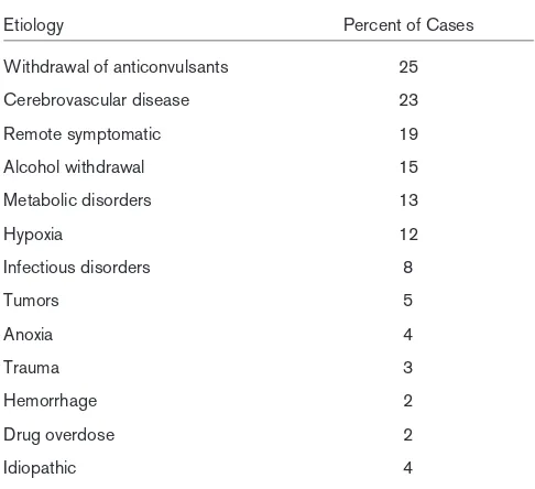
Etiologies
The causes of SE are varied. Table 1 shows the etiologies of SE in an urban hospital-based population [16]. Although the data on causes of status, reported by DeLorenzo (Table 2), show some correspondence, there are discrepancies such as the higher proportion of cerebrovascular disease, anoxia, and hypoxia [17]. These data sets include only the adult popula-tion. In children, up to 51% are secondary to infectious etiolo-gies [16].
Epidemiology, morbidity, and mortality
There have been two recent, prospective, population-based epidemiologic studies of SE. The EPISTAR (a population-based survey of SE in the French speaking part of Switzer-land) study from Switzerland showed an incidence of about 10 cases per 100,000 people annually [18]. The population in this study, however, was principally white, and cases of anoxic encephalopathies were excluded. In the prospective population-based study conducted at the Medical College of Virginia, the incidence of SE was found to be 41 cases per 100,000 people [17] (Fig. 1). This would translate to over 100,000 cases of SE within the United States annually [19].
[image:2.612.313.556.128.350.2]Reports on the mortality of SE range from a few percent to over 50% [20]. The Richmond group found a mortality rate of 22% (corresponding to 22,000–42,000 deaths per year in
Table 1
Data on the etiology of status epilepticus in an urban hospital-based practice
Etiology Percent of cases
AED non-compliance 26
Alcohol related 24
Drug toxicity 10
CNS infection 8
Cerebral tumor 6
Trauma 5
Refractory epilepsy 5
Stroke 4
Metabolic abnormalities 4
Cardiac arrest 4
Idiopathic 5
AED, anti-epileptic drugs; CNS, central nervous system. Data adapted from Neurology[16].
Table 2
Data on the etiology of status epilepticus in a hospital and community
Etiology Percent of Cases
Withdrawal of anticonvulsants 25
Cerebrovascular disease 23
Remote symptomatic 19
Alcohol withdrawal 15
Metabolic disorders 13
Hypoxia 12
Infectious disorders 8
Tumors 5
Anoxia 4
Trauma 3
Hemorrhage 2
Drug overdose 2
Idiopathic 4
[image:2.612.56.299.128.320.2]the US) with the primary predictors of poor outcome being anoxia, prolonged seizure (greater than one hour in duration), and advanced age (Fig. 1) [21]. Mortality in the Swiss group was lower (7.6%), which is most likely to be secondary to the exclusion of anoxic encephalopathies [18].
Pathophysiology of SE
As a result of sympathetic overdrive, the body responds to GCSE with both systemic and cerebral effects, whereas, to NCSE, systemic effects are more limited. Systemic effects of
GCSE are summarized in Table 3. In the initial phase of GCSE, blood pressure, glucose, and lactate increase, and pH decreases. After this initial phase, at approximately 30 minutes, a second phase begins. During this second phase, blood pressure and glucose normalize (sometimes decreas-ing even further), lactate normalizes, and respiratory compro-mise and hyperthermia ensue [22].
In both animal models and humans, GCSE has been shown to result in damage to the brain [3,22,23]. The reasons for this damage have not been completely elucidated; however, several contributing factors are believed to exist. Some of the systemic manifestations described in Table 3 can play a con-tributory role. Meldrum et al. showed that hyperthermia causes damage to the hippocampus and cortex [24]. Addi-tionally, it has also been shown that reduction of temperature results in shorter duration of SE and less damage [25]. Ini-tially during SE, there is increased cerebral blood flow to meet the elevated demands; however, as the cerebral blood flow falls, there is a mismatch between blood supply and the utilization of oxygen and glucose, resulting in a lower energy state and metabolic–substrate mismatch [22]. It is important to note that in experiments where systemic factors are con-trolled, there is still damage to the brain [2,24].
[image:3.612.59.299.94.226.2]An area of expanding research is excitotoxic-induced cell injury, an important cause of damage to neurons [26]. The damage to neurons is based on a complex interplay of multiple
Figure 1
A graphical representation of mortality and incidence for four population groups. These data are adapted from Neurology [19] and
Epilepsia[21].
100
80
60
40
20
0
All Pediatrics Adult Elderly
Survivors per 100,000 people
Deaths per 100,000 people
Table 3
An overview of the systemic effects of status epilepticus
System Effects
Lungs Due to both metabolic and respiratory acidosis, the pH of arterial blood gases (ABG) is often found to be below normal in SE. Aminoff and Simon found that 59/80 (84%) of the patients with useable ABG information had pHs below 7.35 [43]. They suggest that the aberrations in pH may not be of clinical significance or of prognostic value.
In animal studies, pulmonary vascular pressure has been found to be elevated in SE [44] and may contribute to pulmonary edema [45].
Heart The sympathetic overdrive can cause tachycardia. In a study by Boggs, potentially fatal arrhythmias were reported in 58% of the patients [46].
In a recent study, Boggs et alcompleted hemodynamic monitoring in SE patients 24 hours prior to their death, and found that there were two distinct groups with disparate cardiac manifestations [47]. In the group with a lower proportion of previously discovered atherosclerotic disease (ASHD), there was acute cardiac decline without a significant drop in mean arterial pressure (MAP) or heart rate (HR). The second group, 90% of whom had a history of multiple risk factors for ASHD, showed a gradual decrease in MAP and HR prior to death. Muscle As a result of continued seizure activity, conversion to anaerobic metabolism contributes to lactic acidosis [3]. Blood chemistries De-margination of neutrophils occurs with the stress of seizing. In patients without underlying infection, elevated white
blood cell counts (above the upper laboratory limit) were present in as many as 63% in one study [43]. Vital signs
Blood pressure The initial phase of SE results in an increased systemic blood pressure with an increase in peripheral vascular resistance [43]. As the status becomes prolonged, the blood pressure will normalize or even begin to fall with resultant hypotension.
Temperature As the seizure progresses, the body’s core temperature elevates. Aminoff and Simon looked at the temperature of 90 patients in SE. Of these, only 8 had temperatures below 98°F, only 3 had fever secondary to known infection, and over 40 had temperatures above 100.5°F, (with two of the temperatures up to 107°F) [43].
[image:3.612.53.568.452.741.2]factors. Significant to this process is the inhibition of γ -aminobutyric acid (a principle inhibitory neurotransmitter), and the excessive action of glutamate (an excitatory neurotransmit-ter). Additionally, apoptosis is likely to play a role in cell death during SE. Given the probability of cerebral injury, it is impera-tive for the clinician to recognize and treat SE expeditiously.
Treatment of acute status epilepticus
Basic life supportAirway, breathing, and circulation should be established immediately. Intravenous access, while sometimes difficult to achieve in an actively seizing patient, should be obtained. Glucose should be given empirically along with thiamine in alcoholic or otherwise malnourished patients. Laboratory studies should include basic chemistry, blood glucose, anti-convulsant levels, blood and urine toxicologic screen, com-plete blood count, and urine analysis for evidence of infection. The treatment of SE, however, should not be delayed while these tests are pending.
Benzodiazepines
Benzodiazepines, usually either diazepam or lorazepam, remain the first-line control for acute SE (ASE). No significant difference has been found between the rate of seizure control when lorazepam has been compared with either diazepam alone [27] or diazepam plus phenytoin [8]. However, lorazepam is believed to bind more tightly to receptors in the brain and therefore has a longer duration of action and less risk of recurrent seizures [28]. All benzodiazepines carry the risk of respiratory depression and hypotension, and therefore the clinician should be prepared to intubate or give pressors if necessary. Diazepam is given at a dose of 10–20 mg; lorazepam is given in 2 mg increments at approximately three-minutes intervals. If seizures have not terminated after 8 mg of lorazepam, another agent should be started.
Midazolam is a newer benzodiazepine that is associated with a very favorable hemodynamic and pharmacokinetic profile [29]. Its major advantage over lorazepam in the acute setting is that its high water solubility and rapid onset of action when given intramuscularly make this an attractive choice when secure intravenous access is unobtainable [28]. Its use in refractory SE will be discussed below.
Fosphenytoin
Traditionally, phenytoin alone at 20 mg/kg intravenous infu-sion has been considered an appropriate first-line option in the treatment of ASE. We recommend it be only used as a second-line agent after a faster acting benzodiazepine has been tried. Infusion rates are limited to 50 mg/min secondary to the potential side effects of arrhythmia and hypotension.
Conversely, fosphenytoin (a water-soluble phosphate ester prodrug of phenytoin with a neutral pH) can be delivered at 150 phenytoin-equivalent mg/min with essentially no risk of reaction at the infusion site. Because it is water soluble, it
does not need a propylene glycol vehicle. It is 100% bioavail-able when given either intravenously or intramuscularly, and it is quickly converted by phosphatases to phenytoin once it is in the vascular compartment [30].
In one small study of GCSE, fosphenytoin was associated with a success rate of 93.8% in terminating seizures [31]. Studies have shown that therapeutic plasma concentrations of phenytoin can be achieved more rapidly with fosphenytoin that phenytoin when given at 15–20 mg/kg [31]. This is prob-ably related to three factors: faster rate of infusion; displace-ment of protein-bound phenytoin; and rapid conversion of prodrug to phenytoin (half-life of 8.1 min) [32]. Although fos-phenytoin is a more expensive alternative [33], the cost of treating complications from the use of intravenous phenytoin can be substantially higher [34].
Barbiturates
The Veterans Administration Cooperative study showed no significant difference in efficacy between lorazepam (0.1 mg/kg) and phenobarbital (15 mg/kg) [8]. However, it also showed that if a patient did not respond to lorazepam or phenytoin, the response rate to phenobarbital was only 2.1% (unpublished data). We therefore recommend a more defini-tive treatment strategy for patients that have not responded to one, or at most two, first-line agents.
Treatment of refractory status epilepticus
Refractory SE is defined as ongoing seizures despite the use of two first-line agents, usually a benzodiazepine plus either phenytoin or phenobarbital. Definitive therapy often requires doses of medications that cause respiratory suppression and hypotension, so patients should be intubated and transferred to an intensive care unit if this has not already been done. Treiman et al.showed that respiratory suppression requiring mechanical ventilation occurred in 18.9%, and pressor support was needed in 32.6% of patients treated for SE [8]. Patients at this stage of SE require EEG monitoring, since the physical exam is clouded by the use of paralytics for intuba-tion and the potential for electromechanical dissociaintuba-tion.The traditional goal of therapy has a burst suppression pattern on EEG for 12–24 hours before attempting to taper medica-tions. There are no convincing prospective data to suggest that a burst suppression pattern is required to control or to prevent recurrent seizures [35], thus we recommend seizure suppression as a goal, regardless of the EEG background.
Pentobarbital
poikilothermia. Therefore, the clinician must be vigilant in detecting infection and weaning the medication as soon as safely possible. Although the half-life of pentobarbital is 90 hours, awakening begins within a few hours after the plasma concentration begins to fall.
Midazolam
Numerous clinical studies have shown that midazolam bolus (0.1–0.3 mg/kg) followed by continuous infusion (0.05–2.0 mg/kg/hour) rapidly controls seizures that have not responded to traditional first, second, and even third-line agents [36,37]. Reports of clinically significant hypotension are rare, and sedation is rapidly reversed after infusion is stopped. Prolonged use of midazolam is limited by tachyphy-laxis requiring increasing doses to maintain the desired EEG tracing. Additionally, midazolam accumulates in critically ill patients; with prolonged usage, the half-life of the terminal phase can be three to eight times the reported half-life of two to six hours [38]. In a small retrospective study, Prasad et al. found that for patients with an APACHE II (Acute Physiology and Chronic Health Evaluation II) score greater than or equal to 20, treating refractory SE with midazolam may have a lower mortality than that associated with propofol [39].
Propofol
Several studies and case reports document the efficacy of propofol in the treatment of refractory SE, GCSE, NCSE, and complex partial SE [40–42]. Propofol is fast-acting, highly lipid soluble, and has little propensity to accumulate. An initial dose of 3–5 mg/kg is followed by a maintenance dose of 1–15 mg/kg/hour, as required for seizure control. Abrupt dis-continuation of infusion has been associated with recurrent seizures. Hypotension, hypertriglyceridemia, and worsening of anemia have also been reported with this agent. Two small studies have shown an interesting, but insignificant, increase in mortality among SE patients treated with propofol versus midazolam or high dose barbiturates. Future studies are needed to better elucidate this issue.
Conclusion
As the issue of SE is often faced by practitioners of critical care, it is imperative that SE be recognized and treated as a medical emergency. Given the complex pathophysiology, being able to abort SE will contribute to a decrease in the systemic effects and neurologic injury. First line treatment with benzodiazepines, followed by the other agents dis-cussed above, provide the maximal potential for successful management of SE.
Competing interests
None declared.References
1. Commission on Classification and Terminology of the International League Against Epilepsy: Proposal for revised clinical and elec-trographic classification of epileptic seizures.Epilepsia1981, 22:489-501.
2. Nevander G, Ingvar M, Auer R, Siesjo BK: Status epilepticus in well-oxygenated rats causes neuronal necrosis. Ann Neurol
1985, 18:281-290.
3. Meldrum BS, Horton RW: Physiology of status epilepticus in primates.Arch Neurol1973, 28:1-9.
4. Lothman EW: The biochemical basis and pathophysiology of status epilepticus.Neurology1990, 40(suppl):13-23.
5. DeLorenzo RJ, Garnett LK, Towne AR, Waterhouse EJ, Boggs JG, Morton L, Choudhry MA, Barnes T, Ko D: Comparison of status epilepticus with prolonged seizure episodes lasting from 10 to 29 minutes.Epilepsia1999, 40:164-169.
6. Theodore WH, Porter RJ, Albert P, Kelley K, Bromfield E, Devinsky O, Sato S: The secondarily generalized tonic-clonic seizure: a videotape analysis.Neurology1994, 44:1403-1407.
7. Lowenstein DH, Bleck T, Macdonald RL: It’s time to revise the definition of status epilepticus.Epilepsia1999, 40:120-122. 8. Treiman DM, Meyers PD, Walton NY, Collins JF, Colling C, Rowan
AJ, Handforth A, Faught E, Calabrese VP, Uthman BM, Ramsay RE, Mamdani MB: A comparison of four treatments for gener-alized convulsive status epilepticus. N Engl J Med 1998, 339:792-798.
9. Alldredge BK, Gelb AM, Isaacs SM, Corry MD, Allen F, Ulrich S, Gottwald MD, O’Neil N, Neuhaus JM, Segal MR, Lowenstein DH: A comparison of lorazepam, diazepam, and placebo for the treatment of out-of-hospital status epilepticus.N Engl J Med
2001, 345:631-637.
10. Shinnar S, Berg AT, Moshe SL, Shinnar R: How long do new-onset seizures in children last.Ann Neurol2001, 49:659-664. 11. Treiman DM: Generalized convulsive status epilepticus in the
adult.Epilepsia1993, 34(suppl):S2-S11.
12. Treiman DM: Effective treatment for status epilepticus. In
Epilepsy Problem Solving in Clinical Practice. Edited by Schmidt D, Schachter SC. United Kingdom: Martin Dunitz Ltd; 2000:253-265.
13. Krumholz A: Epidemiology and evidence for morbidity of non-convulsive status epilepticus. J Clin Neurophysiol 1999, 16: 314-322.
14. Kaplan PW: Nonconvulsive status epilepticus. Sem Neurol
1996, 16:33-40.
15. Towne AR, Waterhouse EJ, Boggs JG, Garnett LK, Brown AJ, Smith JR Jr, DeLorenzo RJ: Prevalence of nonconvulsive status epilepticus in comatose patients. Neurology 2000, 54 :340-345.
16. Lowenstein DH, Alldredge BK: Status epilepticus at an urban public hospital in the 1980s.Neurology1993, 43:483-488. 17. DeLorenzo RJ: Clinical syndromes and epidemiology of status
epilepticus.In Epileptic Seizures: Pathophysiology and Clinical Semiology. Edited by Luders HO, Noachtar S. Philadelphia: Churchill Livingston; 2000:697-710.
18. Coeytaux A, Jallon P, Galobardes B, Morabia A: Incidence of status epilepticus in French-speaking Switzerland.Neurology
2000, 55:693-697.
19. DeLorenzo RJ, Hauser WA, Towne AR, Boggs JG, Pellock JM, Penberthy L, Garnett L, Fortner CA, Ko D: A prospective, popu-lation-based epidemiologic study of status epilepticus in Richmond, Virginia.Neurology1996, 46:1029-1035.
20. Hauser WA: Status epilepticus: epidemiologic considerations.
Neurology1990, 40(suppl):9-13.
21. Towne AR, Pellock JM, Ko D, DeLorenzo RJ: Determinants of mortality on status epilepticus.Epilepsia1994, 35:27-34. 22. Lothman EW: The biochemical basis and pathophysiology of
status epilepticus.Neurology1990, 40(suppl):13-23.
23. DeGiorgio CM, Tomiyasu U, Gott PS, Treiman DM: Hippocampal pyramidal cell loss in human status epilepticus. Epilepsia
1992, 33:23-27.
24. Meldrum BS, Brierly JB: Prolonged epileptic seizures in pri-mates.Arch Neurol1973, 28:10-17.
25. Lui Z, Gatt A, Mikati M, Holmes GL: Effect of temperature on kainic acid-induced seizures.Brain Res1993, 631:51-58. 26. Fountain NB, Lothman EW: Pathophysiology of status
epilepti-cus.J Clin Neurophysiol1995, 12:326-342.
27. Leppik I, Derivan A, Homan R, Walker J, Ramsay R, Patrick B: Double-blind study of lorazepam and diazepam in status epilepticus.JAMA1983, 249:1452-1454.
29. Bebin M, Bleck T: New anticonvulsant drugs.Drugs1994, 48: 153-171.
30. Browne TR, Davoudi H, Donn KH, Dougherty CL, Dukes GE, Evans B, Evans JE, Jamerson B, Kres J, McEntegart CM Bioavail-ability of ACC-9653 (phenytoin prodrug). Epilepsia 1989, 30(suppl 2):S27-S32.
31. Knapp L, Kugler A: Clinical experience with fosphenytoin in adults: pharmacokinetics, safety, and efficacy.J Child Neurol
1998, 13(suppl 1):S15-S18.
32. Leppik IE, Boucher R, Wilder BJ, Murthy VS, Rask CA, Watridge C, Graves NM, Rangel RJ, Turlapaty P: Phenytoin prodrug: pre-clinical and pre-clinical studies.Epilepsia1989, 30(suppl 2) :S22-S26.
33. Bleck TP: Management approaches to prolonged seizures and status epilepticus.Epilepsia1999, 40(suppl 1):S49-S53. 34. O’Brien TJ, Cascino GD, So EL, Hanna DR: Incidence and
clini-cal consequence of the purple glove syndrome in patients receiving intravenous phenytoin. Neurology 1998, 51 :1034-1039.
35. Krishnamurthy KB, Drislane FW: Depth of EEG suppression and outcome in barbiturate anesthetic treatment for refractory status epilepticus. Epilepsia 1999, 40:759-762.
36. Kumar A, Bleck T: Intravenous midazolam for the treatment of refractory status epilepticus.Crit Care Med1992, 20:483-488. 37. Igartua J, Silver P, Maytal J, Sagy M: Midazolam coma for
refrac-tory status epilepticus in children.Crit Care Med 1999, 27: 1982-1985.
38. Naritoku D, Sinha S: Prolongation of midazolam half-life after sustained infusion for status epilepticus.Neurology2000, 54: 1366-1368.
39. Prasad A, Worrall B, Bertram E, Bleck T: Propofol and midazo-lam in the treatment of refractory status epilepticus.Epilepsia
2001, 42:380-386.
40. Stecker M, Kramer T, Raps E, O’Meeghan R, Dulaney E, Skaar D: Treatment of refractory status epilepticus with propofol: clini-cal and pharmacokinetic findings.Epilepsia1998, 39:18-26. 41. Begemann M, Rowan A, Tuhrim S: Treatment of refractory
complex partial status epilepticus with propofol: case report.
Epilepsia2000, 41:105-109.
42. Mackenzie SJ, Kapadia F, Grant IS: Propofol infusion for control of status epilepticus.Anesthesia1990, 45:1043-1045. 43. Aminoff MJ, Simon RP: Status epilepticus: causes, clinical
fea-tures and consequences in 98 patients.Am J Med1980, 69: 657-666.
44. Bayne LL, Simon RP: Systemic and pulmonary vascular pres-sures during generalized seizures in sheep.Ann Neurol1981, 10:566-569.
45. White PT, Grant P, Mosier J, Craig A: Changes in cerebral dynamics associated with seizures.Neurology1961, 11 :354-361.
46. Boggs JG, Painter JA, DeLorenzo RJ: Analysis of electrocardio-graphic changes in status epilepticus.Epilepsy Res1993, 14: 87-94.