R E S E A R C H
Open Access
Immune derangement occurs in patients with
H7N9 avian influenza
Wei Wu
†, Yu Shi
†, Hainv Gao, Weifeng Liang, Jifang Sheng and Lanjuan Li
*Abstract
Introduction:Currently, little is known about the immunological characteristics of patients with avian influenza A (H7N9) virus infection.
Methods:The numbers and percentages of peripheral blood immune cells were measured in 27 patients with laboratory-confirmed H7N9 virus infection and 30 healthy controls (HCs). The functional phenotypes of T cells and monocytes, as well as serum cytokine levels, were analyzed by flow cytometry.
Results:There were 19 patients (70.4%) with acute respiratory distress syndrome, 13 (48.1%) with secondary respiratory infection, 20 (74%) with systemic inflammatory response syndrome (SIRS; defined as having at least two concurrent SIRS components), 18 (66.7%) with lymphocytopenia and 11 (40.7%) with reduced numbers of monocytes. In comparison with levels in the HCs, the levels of serum interleukin 6 (IL-6), IL-8 and IL-10 and the percentages of CD38+ or Tim-3+ T cells were significantly increased. However, the percentages of human leukocyte antigen-DR + and Tim-3+ monocytes were significantly decreased in patients compared with HCs.
Conclusions:Patients with avian H7N9 virus infection display profound SIRS concomitantly with an anti-inflammatory response, which may be associated with the rapid progression of and high mortality associated with this novel viral disease.
Introduction
Recently in China, an outbreak of influenza occurred that was caused by a novel influenza A (H7N9) viral infection of avian origin. According to published reports, patients with H7N9 virus infection present with rapid, progressive pneumonia commonly leading to the development of acute respiratory distress syndrome (ARDS), respiratory failure and even multiorgan dysfunction syndrome [1]. More importantly, patients with H7N9 virus–mediated influ-enza have a high mortality rate [2]. Previous studies have revealed the clinical characteristics [1], epidemi-ology [3-5] and virepidemi-ology [6,7], laboratory diagnosis, and treatment of patients with H7N9 virus infection [8,9]. However, little is known about the impact of H7N9 virus infection on the immune system.
In this paper, we describe the cytokine profiles and functional phenotypes of immunocompetent cells in 27 pa-tients with H7N9 virus–mediated influenza and 30 healthy controls (HCs). We determined the functional phenotypes of immunocompetent cells and serum cytokine profiles of the participants. We describe the cytokine profiles and functional phenotypes of immunocompetent cells in 27 patients.
Materials and methods
Patients
We recruited 27 patients with H7N9 virus–mediated
influenza and 30 healthy controls (HCs). The patients with avian influenza were recruited from the Inpatient Service at The First Affiliated Hospital of Zhejiang University School of Medicine between 10 and 22 April 2013. Indi-vidual patients with H7N9 were diagnosed on the basis of clinical symptoms and laboratory-confirmed H7N9 virus infection. Patients with ARDS were diagnosed according to standard criteria [10]. Thirty age- and gender-matched HCs were recruited at the Physical Examination Center of * Correspondence:ljli@zju.edu.cn
†Equal contributors
Collaborative Innovation Center for Diagnosis and Treatment of Infectious Diseases, State Key Laboratory of Diagnostic and Treatment of Infectious Diseases, The First Affiliated Hospital, Zhejiang University School of Medicine, Qingchun Road No 79, Hangzhou 310003, China
© Wu et al.; licensee BioMed Central Ltd. This is an Open Access article distributed under the terms of the Creative Commons Attribution License (http://creativecommons.org/licenses/by/2.0), which permits unrestricted use, distribution, and reproduction in any medium, provided the original work is properly cited.
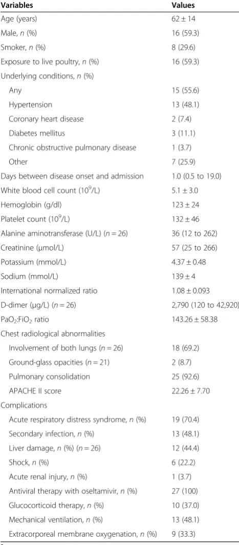
the hospital during the same period. The mean age of the HCs was 57 ± 12 years, and 56.7% of them were male (17 participants). The exclusion criteria were (1) coinfection with hepatitis B virus (HBV), hepatitis C virus (HCV) or HIV; (2) the presence of a common autoimmune disease and tumor; and/or (3) a recent history of chemotherapy, radiotherapy or use of immunosuppressants. Written in-formed consent was obtained from each participant. The experimental protocol was established in accordance with the Declaration of Helsinki and approved by the Ethics Committee of The First Affiliated Hospital of Zhejiang University School of Medicine. The patients’demographic and clinical characteristics are shown in Table 1.
Laboratory examination of H7N9 virus patients
Sputum samples were collected from individual patients immediately after hospitalization, and the presence of H7N9 virus in the collected sputum samples was deter-mined by real-time RT-PCR as previously described [8]. Briefly, the presence of the M, H7 and N9 genes of the H7N9 virus was detected by TaqMan real-time RT-PCR assays (Applied Biosystems, Foster City, CA, USA) using specific primers. The sequences of primers and probes
used were as follows: M forward: 5′-GAGTGGCTAAA
GACAAGACCAATC-3′), M reverse: 5′-TTGGACAAA
GCGTCTACGC-3′ and M probe: 6-carboxyfluorescein
(6-FAM)-TCACCGTGCCCAGTGAGCGAG-black hole quencher 1 (BHQ1); H7 forward: AGAGTCATTRCARA ATAGAATACAGAT, H7 reverse: CACYGCATGTTTC CATTCTT and H7 probe: 6-FAM-AAACATGATGCCC CGAAGCTAAAC-BHQ1; and N9 forward: GTTCTAT GCTCTCAGCCAAGG, N9 reverse: CTTGACCACCCA ATGCATTC and N9 probe: hexachlorofluorescein-TAA GCTRGCCACTATCATCACCRCC-BHQ1. The sensi-tivity of these RT-PCR assays was approximately 100 copies/ml RNA.
Flow cytometry
Venous blood samples were collected from individual patients immediately after hospitalization and from HCs when they visited the hospital. To characterize the fre-quency of T, natural killer (NK) and B cells, individual blood samples (0.5 ml) were stained with the following antibodies: phycoerythrin (PE) cyanine 5–conjugated (Pcy5) anti-CD3, fluorescein isothiocyanate (FITC)-con-jugated anti-CD4, PE-con(FITC)-con-jugated anti-CD8 (BD Biosci-ences, San Jose, CA, USA), PE-conjugated anti-CD16/ anti-CD56 or FITC-conjugated anti-CD19 (Beckman Coulter, Brea, CA, USA). Furthermore, to characterize T-cell
im-munoglobulin mucin 3–positive (Tim-3+) or CD38+ T
cells, blood samples were stained with the following antibodies: Pcy5-conjugated anti-CD3, FITC-conjugated CD4, FITC-conjugated CD8, PE-conjugated anti-Tim-3 (R&D Systems, Minneapolis, MN, USA) or
[image:2.595.304.538.113.646.2]PE-conjugated anti-CD38 (Beckman Coulter). In addition, to characterize Tim-3+ and HLA-DR + monocytes, whole-blood samples were stained with antibodies against FITC-conjugated anti-CD14 and allophycocyanin-FITC-conjugated
Table 1 Demographic, epidemiologic and clinical characteristics of 27 patients with H7N9 virus infectiona
Variables Values
Age (years) 62 ± 14
Male,n(%) 16 (59.3)
Smoker,n(%) 8 (29.6)
Exposure to live poultry,n(%) 16 (59.3)
Underlying conditions,n(%)
Any 15 (55.6)
Hypertension 13 (48.1)
Coronary heart disease 2 (7.4)
Diabetes mellitus 3 (11.1)
Chronic obstructive pulmonary disease 1 (3.7)
Other 7 (25.9)
Days between disease onset and admission 1.0 (0.5 to 19.0)
White blood cell count (109/L) 5.1 ± 3.0
Hemoglobin (g/dl) 123 ± 24
Platelet count (109/L) 132 ± 46
Alanine aminotransferase (U/L) (n= 26) 36 (12 to 262)
Creatinine (μmol/L) 57 (25 to 266)
Potassium (mmol/L) 4.37 ± 0.48
Sodium (mmol/L) 139 ± 4
International normalized ratio 1.08 ± 0.093
D-dimer (μg/L) (n= 26) 2,790 (120 to 42,920)
PaO2:FiO2ratio 143.26 ± 58.38
Chest radiological abnormalities
Involvement of both lungs (n= 26) 18 (69.2)
Ground-glass opacities (n= 21) 2 (8.7)
Pulmonary consolidation 25 (92.6)
APACHE II score 22.26 ± 7.70
Complications
Acute respiratory distress syndrome,n(%) 19 (70.4)
Secondary infection,n(%) 13 (48.1)
Liver damage,n(%) (n= 26) 12 (44.4)
Shock,n(%) 6 (22.2)
Acute renal injury,n(%) 1 (3.7)
Antiviral therapy with oseltamivir,n(%) 27 (100)
Glucocorticoid therapy,n(%) 10 (37.0)
Mechanical ventilation,n(%) 13 (48.1)
Extracorporeal membrane oxygenation,n(%) 9 (33.3)
a
APACHE II, Acute Physiology and Chronic Health Evaluation II;PaO2: FiO2, ratio
anti-Tim-3 (R&D Systems) or PE antibodies against major histocompatibility complex class II cell surface receptor encoded by the human leukocyte antigen (anti-HLA-DR) (BD Biosciences). The isotype-matched immunoglobulins were used as controls. The frequency of different types of immunocompetent cells was characterized by flow cytom-etry, and at least 1 × 10⁵ events were analyzed using an FC500 MPL flow cytometer (Beckman Coulter) or an Accuri C6 cytometer (BD Biosciences).
Measurement of serum cytokines
Individual serum samples were obtained from patients immediately after hospitalization and from HCs when they visited the hospital. The concentrations of serum interferon γ (IFN-γ), tumor necrosis factor α (TNF-α), TNF-β, interleukin 1β(IL-1β), 2, 4, 5, 6, IL-8, IL-10 and IL-12P70 in individual participants were determined by cytometric bead array (CBA) using a Flow-Cytomix Simplex Kit (Bender MedSystems/eBioscience, San Diego, CA, USA), according to the manufacturer’s instructions, which were described previously [11]. The concentrations of individual serum cytokines were deter-mined using standard curves established with the individ-ual recombinant cytokines provided. The limitation of detection was 1.6 pg/ml for IFN-γ, 3.2 pg/ml for TNF-α, 2.4 pg/ml for TNF-β, 4.2 pg/ml for IL-1β, 16.4 pg/ml for IL-2, 20.8 pg/ml for IL-4, 1.6 pg/ml for IL-5, 1.2 pg/ml for IL-6, 0.5 pg/ml for IL-8, 1.9 pg/ml for IL-10 and 1.5 pg/ml for IL-12P70.
Statistical analysis
Continuous data are expressed as mean ± SD or me-dian (range), and categorical data are given as per-centages. Comparison of the data between the two groups was analyzed by Student’s t-test, Mann–Whitney Unonparametric test and χ2 test using SPSS 16.0 for Windows software (SPSS, Chicago, IL, USA). A two-sided P-value less than 0.05 was considered statistically significant.
Results
Demographic, epidemiological and clinical characteristics of the study population
A total of 27 patients with confirmed avian-origin influ-enza A (H7N9) virus infection and 30 HCs were recruited for participation in this study. Their demographic and clinical characteristics are presented in Table 1. There were no significant differences between the patients and HCs in our sample population with regard to age or gender. After hospital admission, sputum samples were collected from individual patients and subjected to characterization of H7N9 virus genes. The sputum samples from all patients were positive for the M, H7 and N9 genes as determined by RT-PCR, confirming
that all the patients had H7N9 avian-origin influenza virus infection. Of the 27 patients, 8 (29.6%) were smokers and 16 (59.3%) had a definitive history of poultry exposure. Furthermore, 55.6% of the patients had preexisting chronic diseases, such as hypertension.
Chest radiographs showed that all patients displayed significant changes, and 92.6% of them had bilateral con-solidation in the lungs. Most patients were in critical condition, with an average Acute Physiology and Chronic Health Evaluation II score of 22.26 ± 7.70, 70.4% devel-oped ARDS and 48.1% had secondary infections in the respiratory tract. In addition, many patients developed se-vere complications, including liver damage (44.4%), renal injury (3.7%) and shock (22.2%). All patients were given oral antiviral therapy with oseltamivir, and 10 (37.0%) re-ceived glucocorticoid treatment. Approximately one-half of the patients (13 (48.1%) of 27) required mechanical ventilation, and 9 of them received extracorporeal mem-brane oxygenation.
Patients with H7N9 avian influenza developed systemic inflammatory response syndrome at admission
[image:3.595.305.539.554.718.2]Many patients with clinical presentation of H7N9 avian influenza developed systemic inflammatory response syndrome (SIRS)–related clinical symptoms and signs, such as abnormalities in body temperature, heart rate, respiratory rate and leukocyte count [12], suggesting that a hyperactivated inflammatory response may play a pivotal role in disease progression. We found that 70% patients had at least two concurrent SIRS components. In addition, 92.6% of patients had increased levels of serum C-reactive protein, 33.3% had elevated levels of serum procalcitonin and 77.8% had abnormally high erythrocyte sedimentation rates (Table 2).
Table 2 Systemic inflammatory response syndrome components and clinical inflammatory markers in 27 patients with H7N9 virus infectiona
Variables Frequency
Body temperature <36°C or >38°C,n(%) 24 (88.9)
Heart rate >90 beats/min,n(%) 7 (25.9)
Respiratory rate >20 breaths/min or PaCO2< 32 mmHg,n(%)
13 (48.1)
WBC count <4 × 10⁹/L or >12 × 109/L,n(%) 13 (48.1)
Maximum two concurrent components,n(%) 11 (40.7)
Maximum three concurrent components,n(%) 7 (25.9)
Maximum four concurrent components,n(%) 2 (7.4)
C-reactive protein >10 mg/dl,n(%) 25 (92.6)
Erythrocyte sedimentation rate >20 mm/h (n= 18) 14 (77.8)
Procalcitonin >0.5 ng/ml,n/N(%) (N= 24) 8 (33.3)
a
PaCO2, partial pressure of arterial carbon dioxide; WBC, white blood cell.
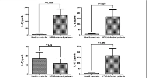
Significantly higher levels of serum cytokines in patients with H7N9 avian influenza
SIRS is a consequence of cytokine storm, so we evalu-ated the levels of serum cytokines by CBA in HCs and in the patients with H7N9 influenza at the time of admission. We detected IL-2, IL-6, IL-8 and IL-10, but we did not detect IFN-γ, TNF-α, TNF-β, IL-1β, IL-4, IL-5 or IL-12p70. We found that the levels of serum IL-6 (P= 0.0098), IL-8 (P= 0.0010) and IL-10 (P= 0.015) in the patients were significantly higher than those in the HCs (Figure 1). However, there was no significant differ-ence in the levels of serum IL-2 between these two groups (P= 0.19).
Alteration in number of peripheral blood
immunocompetent cells in patients with H7N9 avian influenza
Next, we examined the numbers of peripheral blood im-munocompetent cells. We found that 66.7% of patients developed lymphocytopenia (lymphocytes <0.8 × 10⁹/L) and 40.7% had abnormally low monocyte counts (mono-cytes <0.12 × 10⁹/L). However, only 25.9% of patients had neutropenia (neutrophils <2.0 × 10⁹/L). Further flow cyto-metric analysis indicated that the percentages of CD3+ T cells (58.88 ± 16.34% vs. 67.22 ± 9.22%; P= 0.020) and
CD8+ T cells (20.18 ± 8.58% vs. 24.19 ± 6.06%; P= 0.045) were significantly lower in the patients than in the HCs. In addition, we found no significant differences in the frequency of peripheral blood CD3 + CD4+ T cells and CD3−CD56+ NK cells between the patients and the HCs (37.46 ± 13.50% vs. 38.61 ± 8.84%; P = 0.70 for CD4+ T cells; and 15.10 ± 11.00% vs. 19.73 ± 9.55%;P= 0.095 for NK cells) (Table 3).
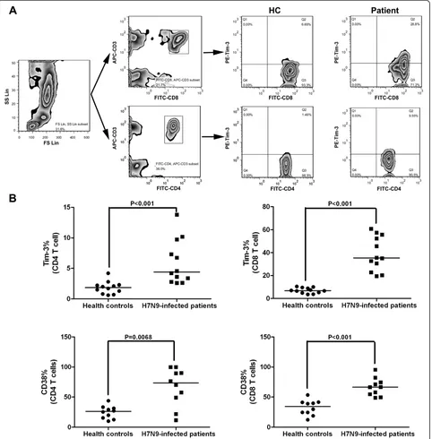
CD38 is a marker of T-cell activation, and Tim-3 is a surface marker of T-cell exhaustion [13]. Further charac-terization of functional T cells revealed that the percent-ages of peripheral blood CD38 + CD4+ and CD38 + CD8+ T cells in the patients were significantly higher than those in the HCs (P= 0.0068 and P< 0.001, respectively) (Figure 2). Concomitantly, the percentages of periph-eral blood Tim-3 + CD4+ and Tim-3 + CD8+ T cells in the patients were significantly higher than those in the HCs (P< 0.001 for both).
[image:4.595.58.543.418.676.2]We next assessed the functional phenotypes of peri-pheral blood monocytes and found that the percentages of HLA-DR + CD14+ and Tim-3 + CD14+ monocytes in total CD14+ cells in patients were significantly lower than those in the HCs (P= 0.043 and P= 0.0029, respectively) (Figure 3). Collectively, these data suggest that patients with H7N9 avian influenza had immunodysfunction.
Discussion
In the present study, we examined the immune system alterations in patients with H7N9 avian influenza. We found that a high percentage of patients developed SIRS accompanied by a high percentage of activated T cells and increased levels of serum cytokines. Concomitantly, many patients displayed lymphocytopenia, abnormally low monocyte counts, T-cell exhaustion and monocyte dys-function, which are characteristic of immune paralysis.
The presence of SIRS is predictive of organ dysfunction and mortality [14,15]. We found that 70% of patients had at least two concurrent SIRS components and detected significantly higher levels of serum IL-6 and IL-8 in the patients than in HCs, which may explain the high morbid-ity and mortalmorbid-ity associated with this disease. However, we did not detect significant alterations in the levels of serum IFN-γor TNF-αin these patients. This cytokine profile is analogous to that of patients with severe acute respiratory syndrome (SARS) [16,17]. Furthermore, it has been reported that patients with either H7N9 virus influenza or SARS coronavirus-related illness shared striking similarities with regard to their clinical presentation and disease pro-gression. Therefore, it is possible that the pathogenesis of H7N9 virus infection is similar to that of SARS coronavirus-related infection [18]. Although patients with severe influ-enza induced by the H1N1 and H5N1 viruses develop a “cytokine storm,” including high levels of serum IFN-γand TNF-α, which are commonly associated with rapid progres-sion and poor prognosis [11,19,20], we did not detect abnor-mal levels of serum IFN-γor TNF-αin patients with H7N9 virus infection. These findings suggest that different immune responses may occur in patients with varying types of influ-enza virus infection. The investigators in one recent study reported that a high frequency of programmed death recep-tor 1 (PD-1), and its ligand 1 (PD-L1), that expressed T cells impaired T-cell responses to H1N1 infection in patients with influenza [21]. It is possible that similar mechanisms may underlie the failure to detect abnormal levels of serum IFN-γ and TNF-α in patients with H7N9 virus infection.
Al-though proinflammatory IFN-γ and TNF-α responses
usually occur at early stages of immune responses, it is also possible that the failure to detect abnormal levels of serum IFN-γor TNF-αmay stem from missing the very early time point in our study. Therefore, further studies are needed to clarify the mechanisms underlying the pathogenesis of H7N9 virus infection and host immune responses.
Our results show that many patients with severe avian H7N9 influenza developed T-cell lymphocytopenia. Such a phenomenon is commonly reported in patients with SARS. However, pneumonia caused by other common respiratory vi-ruses are usually associated with a normal or elevated lympho-cyte count [22]. The lymphocytopenia in patients with H7N9 avian influenza may be a key factor leading to high morbid-ity and mortalmorbid-ity, because lymphocytopenia is an inde-pendent risk factor for ARDS, which is a very dangerous condition for patients with secondary infections [1]. The lymphocytopenia in these patients likely derives from the migration of T lymphocytes into the target tissues, such as the lungs. Alternatively, the lymphocytopenia may stem from virus-stimulated, activation-induced T-cell apoptosis and virus infection–related bone marrow suppression [23].
[image:5.595.62.541.111.224.2]T lymphocytes play a pivotal role in the defense of viral infection by directly killing virus-infected cells. In this study, we found significantly higher frequencies of CD38 + CD4+, CD38 + CD8+, 3 + CD4+ and Tim-3 + CD8+ T cells in patients with H7N9 avian influenza compared with those in the HCs. It is well-known that CD38 and Tim-3 expression are associated with T-cell activation [13]. The higher frequency of CD38+ and Tim-3+ T cells in patients with H7N9 avian influenza indicated that viral infection induced significant T-cell activation. However, Tim-3 on activated T cells usually provides a negative signal for effector T-cell function and leads to T-cell functional exhaustion [13,24,25]. Fur-thermore, engagement of Tim-3 by its specific ligand of galectin-9 can trigger T-cell apoptosis [26]. Notably, IFN-γis a potent inducer of galectin-9 protein expression, thus the low IFN-γ expression in patients with severe H7N9 influenza may limit the production of galectin-9, leading to a high frequency of impotent Tim-3+ T cells. Thus, in
Table 3 Comparison of counts and percentages of lymphocytes and lymphocyte subsets between patients with H7N9 virus infection and healthy controlsa
Variables Patients (n= 27) Healthy controls (n= 30) P-value
Neutrophils <2.0 × 10⁹/L,n(%) 7 (25.9) None –
Monocytes <0.12 × 10⁹/L,n(%) 10 (40.7) None –
Lymphocytes <0.8 × 10⁹/L,n(%) 18 (66.7) None –
CD3+ T cells (%) 58.88 ± 16.34 67.22 ± 9.22 0.020
CD4+ T cells (%) 37.46 ± 13.50 38.61 ± 8.84 0.70
CD8+ T cells (%) 20.18 ± 8.58 24.19 ± 6.06 0.045
Natural killer cells (%) 15.10 ± 11.00 19.73 ± 9.55 0.095
a
Statistical analysis was performed by Student’st-test.
turn, aberration of T-cell activation in patients with H7N9 avian influenza may render T-cell exhaustion and apop-tosis or impotence, leading to poor immune responses. We are interested in further investigating the levels of galectin-9 and T-cell function to discern the precise
mechanisms underlying immune responses to H7N9 virus infection in humans.
[image:6.595.59.541.90.580.2]the immune response, and presents antigens to trigger adaptive immune responses [27]. In this study, we found a significantly reduced frequency of peripheral blood HLA-DR + CD14+ and Tim-3 + CD14+ monocytes in patients with H7N9 avian influenza, at levels similar to those in patients with septic shock [28], trauma [29] and acute liver failure [30], as well as postoperative patients [31]. In these clinical settings, downregulation of HLA-DR expression usually represents the functional impairment of monocytes and is associated with adverse outcomes. In addition, the reduced levels of Tim-3 expression on monocytes may contribute to the functional deactivation of monocytes, as it was reported previously that constitu-tive Tim-3 expression in naïve and resting immunocom-petent cells promotes inflammation [32].
In addition, we noted that nearly one-half of the patients in our study had evidence of secondary infection, which is very dangerous because secondary bacterial or fungal infection is a common factor leading to mortality in patients with H7N9 avian influenza [1]. We speculate that the increased predisposition to secondary infection of patients is related to the deranged immune response. Therefore, it is crucial for clinicians to pay special atten-tion to patients with severe influenza by modulating immunocompetent cell function to limit adverse conse-quences in the clinic.
[image:7.595.56.544.90.463.2]further studies are warranted to measure longitudinally the dynamic changes in the immunological status of patients during the whole time course of H7N9 infection and to assess the function of different types of immu-nocompetent cells using other functional markers (for example, CD25, CD69, PD-1 and LAG-3) and antigen-specific T-cell immunity byex vivoexperiments in a larger sample population. We are interested in further investigat-ing the molecular mechanisms underlyinvestigat-ing the immunode-rangement in patients with H7N9 avian influenza.
Conclusions
Overall, our findings reveal that patients with H7N9 avian influenza commonly develop SIRS accompanied by T-cell lymphocytopenia and exhaustion, as well as monocyte dysfunction. The immunoderangement may be associated with the high mortality rate associated with this disease.
Key messages
Patients with H7N9 avian influenza usually develop ARDS and secondary infection and have a high mortality rate.
A hyperactivated inflammatory response and an anti-inflammatory response occur concomitantly in patients with H7N9 avian influenza.
Such immune derangement may contribute to the rapid progression of and high mortality associated with this disease.
Abbreviations
ARDS:Acute respiratory distress syndrome; CBA: Cytometric bead array; CRP: C-reactive protein; ECMO: Extracorporeal membrane oxygenation; ESR: Erythrocyte sedimentation rate; H7N9: Avian influenza A; HBV: Hepatitis B virus; HCV: Hepatitis C virus; SARS: Severe acute respiratory syndrome; SIRS: Systemic inflammatory response syndrome.
Competing interests
The authors declare that they have no competing interests.
Authors’contributions
WW carried out the RT-PCR and flow cytometric analysis, participated in the design of the study and helped to draft the manuscript. YS carried out the cytokine measurements, participated in the design of the study and helped to draft the manuscript. HG analyzed and interpreted the results and revised the manuscript. WL and JS participated in the design of the study and revised the manuscript. LL conceived of the study, participated in its design and coordination and helped to draft the manuscript. All authors read and approved the final manuscript.
Acknowledgements
The manuscript was proofread by Medjaden Bioscience Limited. This work was supported by the 12th Five-Year Significant New Drugs Creation Plan of the Ministry of Science and Technology of China (2011ZX09302-003-03), the Technology Group Project for Infectious Disease Control of Zhejiang Province (2009R50041) and the Chinese National Natural Science Foundation (81200301), as well as by a grant from the Health Department of Zhejiang Province (2012KYA087).
Received: 18 November 2013 Accepted: 20 February 2014 Published:
References
1. Gao HN, Lu HZ, Cao B, Du B, Shang H, Gan JH, Lu SH, Yang YD, Fang Q, Shen YZ, Xi XM, Gu Q, Zhou XM, Qu HP, Yan Z, Li FM, Zhao W, Gao ZC, Wang GF, Ruan LX, Wang WH, Ye J, Cao HF, Li XW, Zhang WH, Fang XC, He J, Liang WF, Xie J, Zeng M,et al:Clinical findings in 111 cases of influenza A (H7N9) virus infection.N Engl J Med2013,368:2277–2285. A published erratum appears inN Engl J Med2013,369:1869. 2. Uyeki TM, Cox NJ:Global concerns regarding novel influenza A (H7N9)
virus infections.N Engl J Med2013,368:1862–1864.
3. Li Q, Zhou L, Zhou M, Chen Z, Li F, Wu H, Xiang N, Chen E, Tang F, Wang D, Meng L, Hong Z, Tu W, Cao Y, Li L, Ding F, Liu B, Wang M, Xie R, Gao R, Li X, Bai T, Zou S, He J, Hu J, Xu Y, Chai C, Wang S, Gao Y, Jin L,et al: Epidemiology of human infections with avian influenza A(H7N9) virus in China.N Engl J Med2014,370:520–532.
4. Gao R, Cao B, Hu Y, Feng Z, Wang D, Hu W, Chen J, Jie Z, Qiu H, Xu K, Xu X, Lu H, Zhu W, Gao Z, Xiang N, Shen Y, He Z, Gu Y, Zhang Z, Yang Y, Zhao X, Zhou L, Li X, Zou S, Zhang Y, Li X, Yang L, Guo J, Dong J, Li Q,et al:Human infection with a novel avian-origin influenza A (H7N9) virus.N Engl J Med
2013,368:1888–1897.
5. Zhang W, Wang L, Hu W, Ding F, Sun H, Li S, Huang L, Li C:Epidemiologic characteristics of cases for influenza A(H7N9) virus infections in China.
Clin Infect Dis2013,57:619–620.
6. Zhu H, Wang D, Kelvin DJ, Li L, Zheng Z, Yoon SW, Wong SS, Farooqui A, Wang J, Banner D, Chen R, Zheng R, Zhou J, Zhang Y, Hong W, Dong W, Cai Q, Roehrl MH, Huang SS, Kelvin AA, Yao T, Zhou B, Chen X, Leung GM, Poon LL, Webster RG, Webby RJ, Peiris JS, Guan Y, Shu Y:Infectivity, transmission, and pathology of human-isolated H7N9 influenza virus in ferrets and pigs.Science2013,341:183–186. A published erratum appears inScience2013,341:959.
7. Tharakaraman K, Jayaraman A, Raman R, Viswanathan K, Stebbins NW, Johnson D, Shriver Z, Sasisekharan V, Sasisekharan R:Glycan receptor binding of the influenza A virus H7N9 hemagglutinin.Cell2013, 153:1486–1493.
8. Chen Y, Liang W, Yang S, Wu N, Gao H, Sheng J, Yao H, Wo J, Fang Q, Cui D, Li Y, Yao X, Zhang Y, Wu H, Zheng S, Diao H, Xia S, Zhang Y, Chan KH, Tsoi HW, Teng JL, Song W, Wang P, Lau SY, Zheng M, Chan JF, To KK, Chen H, Li L, Yuen KY:Human infections with the emerging avian influenza A H7N9 virus from wet market poultry: clinical analysis and characterisation of viral genome.Lancet2013,381:1916–1925.
9. Hu Y, Lu S, Song Z, Wang W, Hao P, Li J, Zhang X, Yen HL, Shi B, Li T, Guan W, Xu L, Liu Y, Wang S, Zhang X, Tian D, Zhu Z, He J, Huang K, Chen H, Zheng L, Li X, Ping J, Kang B, Xi X, Zha L, Li Y, Zhang Z, Peiris M, Yuan Z: Association between adverse clinical outcome in human disease caused by novel influenza A H7N9 virus and sustained viral shedding and emergence of antiviral resistance.Lancet2013,381:2273–2279. 10. Bernard GR, Artigas A, Brigham KL, Carlet J, Falke K, Hudson L, Lamy M,
Legall JR, Morris A, Spragg R:The American-European Consensus Conference on ARDS. Definitions, mechanisms, relevant outcomes, and clinical trial coordination.Am J Respir Crit Care Med1994,149:818–824. 11. de Jong MD, Simmons CP, Thanh TT, Hien VM, Smith GJD, Chau TNB,
Hoang DM, Chau NVV, Khanh TH, Dong VC, Qui PT, Cam BV, Ha DQ, Guan Y, Peiris JSM, Chinh NT, Hien TT, Farrar J:Fatal outcome of human influenza A (H5N1) is associated with high viral load and
hypercytokinemia.Nat Med2006,12:1203–1207.
12. American College of Chest Physicians/Society of Critical Care Medicine Consensus Conference:Definitions for sepsis and organ failure and guidelines for the use of innovative therapies in sepsis.Crit Care Med
1992,20:864–874.
13. Jones RB, Ndhlovu LC, Barbour JD, Sheth PM, Jha AR, Long BR, Wong JC, Satkunarajah M, Schweneker M, Chapman JM, Gyenes G, Vali B, Hyrcza MD, Yue FY, Kovacs C, Sassi A, Loutfy M, Halpenny R, Persad D, Spotts G, Hecht FM, Chun TW, McCune JM, Kaul R, Rini JM, Nixon DF, Ostrowski MA:Tim-3 expression defines a novel population of dysfunctional T cells with highly elevated frequencies in progressive HIV-1 infection.J Exp Med
2008,205:2763–2779.
14. Talmor M, Hydo L, Barie PS:Relationship of systemic inflammatory response syndrome to organ dysfunction, length of stay, and mortality in critical surgical illness: effect of intensive care unit resuscitation.
Arch Surg1999,134:81–87.
Prognostic values of tumor necrosis factor/cachectin, interleukin-1, interferon-α, and interferon-γin the serum of patients with septic shock. Swiss-Dutch J5 immunoglobulin study group.J Infect Dis1990, 161:982–987.
16. Jiang Y, Xu J, Zhou C, Wu Z, Zhong S, Liu J, Luo W, Chen T, Qin Q, Deng P:Characterization of cytokine/chemokine profiles of severe acute respiratory syndrome.Am J Respir Crit Care Med2005, 171:850–857.
17. Tang NL, Chan PK, Wong CK, To KF, Wu AK, Sung YM, Hui DS, Sung JJ, Lam CW:Early enhanced expression of interferon-inducible protein-10 (CXCL-10) and other chemokines predicts adverse outcome in severe acute respiratory syndrome.Clin Chem2005,51:2333–2340.
18. Spiegel M, Pichlmair A, Martínez-Sobrido L, Cros J, García-Sastre A, Haller O, Weber F:Inhibition ofβinterferon induction by severe acute respiratory syndrome coronavirus suggests a two-step model for activation of interferon regulatory factor 3.J Virol2005,79:2079–2086.
19. Woo PCY, Tung ETK, Chan KH, Lau CCY, Lau SKP, Yuen KY:Cytokine profiles induced by the novel swine-origin influenza A/H1N1 virus: implications for treatment strategies.J Infect Dis2010,201:346–353.
20. Almansa R, Anton A, Ramirez P, Martin-Loeches I, Banner D, Pumarola T, Xu L, Blanco J, Ran L, Lopez-Campos G, Martin-Sanchez F, Socias L, Loza A, Andaluz D, Maravi E, Gordón M, Gallegos MC, Fernandez V, León C, Merino P, Marcos MA, Gandía F, Bobillo F, Resino S, Eiros JM, Castro C, Mateo P, Gonzalez-Rivera M, Rello J, de Lejarazu RO,et al:Direct association between pharyngeal viral secretion and host cytokine response in severe pandemic influenza.BMC Infect Dis2011,11:232.
21. Valero-Pacheco N, Arriaga-Pizano L, Ferat-Osorio E, Mora-Velandia LM, Pastelin-Palacios R, Villasís-Keever MÁ, Alpuche-Aranda C, Sánchez-Torres LE, Isibasi A, Bonifaz L, López-Macías C:PD-L1 expression induced by the 2009 pandemic influenza A(H1N1) virus impairs the human T cell response.Clin Dev Immunol2013,2013:989673.
22. Cui W, Fan Y, Wu W, Zhang F, Wang JY, Ni AP:Expression of lymphocytes and lymphocyte subsets in patients with severe acute respiratory syndrome.Clin Infect Dis2003,37:857–859.
23. Chen J, Subbarao K:The immunobiology of SARS.Annu Rev Immunol2007, 25:443–472.
24. Golden-Mason L, Palmer BE, Kassam N, Townshend-Bulson L, Livingston S, McMahon BJ, Castelblanco N, Kuchroo V, Gretch DR, Rosen HR:Negative immune regulator Tim-3 is overexpressed on T cells in hepatitis C virus infection and its blockade rescues dysfunctional CD4+and CD8+T cells.
J Virol2009,83:9122–9130.
25. Wu W, Shi Y, Li S, Zhang Y, Liu Y, Wu Y, Chen Z:Blockade of Tim-3 signaling restores the virus-specific CD8+T-cell response in patients with chronic hepatitis B.Eur J Immunol2012,42:1180–1191.
26. Lv K, Zhang Y, Zhang M, Zhong M, Suo Q:Galectin-9 ameliorates Con A-induced hepatitis by inducing CD4+CD25low/inteffector T-cell apoptosis and increasing regulatory T cell number.PLoS One2012, 7:e48379.
27. Gordon S, Taylor PR:Monocyte and macrophage heterogeneity.
Nat Rev Immunol2005,5:953–964.
28. Pangault C, Le Tulzo Y, Tattevin P, Guilloux V, Bescher N, Drénou B: Down-modulation of granulocyte macrophage-colony stimulating factor receptor on monocytes during human septic shock.Crit Care Med2006, 34:1193–1201.
29. Flohé S, Lendemans S, Selbach C, Waydhas C, Ackermann M, Schade FU, Kreuzfelder E:Effect of granulocyte-macrophage colony-stimulating factor on the immune response of circulating monocytes after severe trauma.
Crit Care Med2003,31:2462–2469.
30. Antoniades CG, Berry PA, Davies ET, Hussain M, Bernal W, Vergani D, Wendon J:Reduced monocyte HLA-DR expression: a novel biomarker of disease severity and outcome in acetaminophen-induced acute liver failure.Hepatology2006,44:34–43.
31. Tschaikowsky K, Hedwig-Geissing M, Schiele A, Bremer F, Schywalsky M, Schüttler J:Coincidence of pro- and anti-inflammatory responses in the early phase of severe sepsis: longitudinal study of mononuclear histocompatibility leukocyte antigen-DR expression, procalcitonin,
C-reactive protein, and changes in T-cell subsets in septic and postoperative patients.Crit Care Med2002,30:1015–1023.
32. Anderson AC, Anderson DE, Bregoli L, Hastings WD, Kassam N, Lei C, Chandwaskar R, Karman J, Su EW, Hirashima M, Bruce JN, Kane LP, Kuchroo VK, Hafler DA:Promotion of tissue inflammation by the immune receptor Tim-3 expressed on innate immune cells.
Science2007,318:1141–1143.
Cite this article as:Wuet al.:Immune derangement occurs in patients with H7N9 avian influenza.Critical Care
Submit your next manuscript to BioMed Central and take full advantage of:
• Convenient online submission
• Thorough peer review
• No space constraints or color figure charges
• Immediate publication on acceptance
• Inclusion in PubMed, CAS, Scopus and Google Scholar
• Research which is freely available for redistribution
Submit your manuscript at www.biomedcentral.com/submit 10.1186/cc13788