IN-SILICO
INHIBITORS FOR PECTIN
SCREENING TO INHIBIT THE SEED GERMINATION PROCESS
1
Prathusha S. Chegu,
2Yogesh N. Joshi,
1
Department of PG Studies and Research in Biotechnology, WC
2
Department of PG Studies and Research in Bioinformatics, WC
3
Department of PG Studies and Research in Genetics, WCBT, Solapur, Mahar
ARTICLE INFO ABSTRACT
Pectin is the one of the major component of plant cellwall which maintains structural integrity of cellwall. They can be modified by pectinases such as pectin methylesterases (PMEs) which catalyses the demethylesterification of homogalacturonans releasing
roles in growth, development and seed germination. The activity of an enzyme PME affects the cell wall porosity and elasticity by allowing water uptake. The control PME activities by several proteinaceous inhibitors
Recently it has been found that the catechin isolated from Green tea can inhibit the PME from citrus and tomato plants. In the previous work we have characterized the PME from
its 3D structure by using bioinformatics tools. Now, the present study focuses to find out the novel inhibitor of PME by screening the various catechin analogue compounds from the ZINC database. The molecular docking studies of cat
PME of
complex in the Pymol software.
Copyright©2017, Prathusha S. Chegu et al. This is an open access article distributed under the Creative Commons Att use, distribution, and reproduction in any medium, provided the original work is properly cited.
INTRODUCTION
The plant cell wall is composed of polysaccharides along with proteins & aromatic substances. The wall polysaccharides are often classified into cellulose, hemicelluloses, and pectin and these three types are represented in almost all cell walls in
varying proportions (Jesper Harholt et al., 2010
consists of four different homogalacturonan (HG),
xylogalacturonan (XGA), rhamnogalacturonan I (RG rhamnogalacturonan II (RG- II) (Jesper Harholt
This is one of the principal of cellwall components involved in temporal and spatial regulation of the growt
pectin homogalacturonan composed of a linear chain of 1, 4
linked a-D-galacturonic acid (GalUA) residues, the
homogalacturonans can be methylesterified at the C carboxylic acid groups of the GalUA residues
et al., 2009). Through biochemical changes to pectin
composition and biochemical configuration, the properties of this material can be altered to trigger specific developmental
processes (Ridley et al., 2001). It has been indicated that
enzyme pectinmethylesterases plays important role in seed
*Corresponding author: Nitin N. Bolabattin,
Department of PG Studies and Research in Biotechnology, WCBT, Solapur, Maharashtra, India.
ISSN: 0975-833X
Article History:
Received 17th November, 2016
Received in revised form 10th December, 2016
Accepted 27th January, 2017
Published online 28th February,2017
Key words:
In-silico, PME, Catechin, Seed germination, Virtual screening, Docking, Hex software, ZINC database etc.
Citation: Prathusha S. Chegu, Yogesh N. Joshi,
A. thaliana by virtual screening to inhibit the seed germination process”,
RESEARCH ARTICLE
INHIBITORS FOR PECTIN- METHYLESTERASE OF
A. THALIANA
SCREENING TO INHIBIT THE SEED GERMINATION PROCESS
Yogesh N. Joshi, *
,1Nitin N. Bolabattin and
3Chetan H. Godale
Department of PG Studies and Research in Biotechnology, WCBT, Solapur, Maharashtra, India
Department of PG Studies and Research in Bioinformatics, WCBT, Solapur,
Department of PG Studies and Research in Genetics, WCBT, Solapur, Mahar
ABSTRACT
Pectin is the one of the major component of plant cellwall which maintains structural integrity of cellwall. They can be modified by pectinases such as pectin methylesterases (PMEs) which catalyses the demethylesterification of homogalacturonans releasing pectate and methanol, playing important roles in growth, development and seed germination. The activity of an enzyme PME affects the cell wall porosity and elasticity by allowing water uptake. The control PME activities by several proteinaceous inhibitors have a direct effect on the regulation of various processes in plant physiology. Recently it has been found that the catechin isolated from Green tea can inhibit the PME from citrus and tomato plants. In the previous work we have characterized the PME from
its 3D structure by using bioinformatics tools. Now, the present study focuses to find out the novel inhibitor of PME by screening the various catechin analogue compounds from the ZINC database. The molecular docking studies of catechin analogue were carried out by using the Hex software against the PME of A. thaliana which give the detailed insight of interacting residue with the ligand and target complex in the Pymol software.
open access article distributed under the Creative Commons Attribution License, which use, distribution, and reproduction in any medium, provided the original work is properly cited.
The plant cell wall is composed of polysaccharides along with aromatic substances. The wall polysaccharides are often classified into cellulose, hemicelluloses, and pectin and these three types are represented in almost all cell walls in ., 2010). Pectin is
ifferent homogalacturonan (HG),
xylogalacturonan (XGA), rhamnogalacturonan I (RG- I),
Jesper Harholt et al., 2010).
This is one of the principal of cellwall components involved in temporal and spatial regulation of the growth process. The pectin homogalacturonan composed of a linear chain of 1,
4-galacturonic acid (GalUA) residues, the
homogalacturonans can be methylesterified at the C-6 carboxylic acid groups of the GalUA residues (Fry, 2000; Wolf . Through biochemical changes to pectin composition and biochemical configuration, the properties of this material can be altered to trigger specific developmental . It has been indicated that
ys important role in seed
Department of PG Studies and Research in Biotechnology, WCBT,
germination process. During the process of seed germination the cell wall of the radical and of the tissues around it must expand (Robert Palin and Anja Geitmann
methylesterases (PMEs) (EC 3.1.1.11) remove some of the methyl ester groups from HG, releasing methanol and protons. Pectinmethylesterase (PME) catalyzes reactions accor the double-displacement mechanisms, de
transferring the C6 carboxyl groups in the pectin complexes to water molecules altering the degree and pattern of methyl esterification and trans acylation through transferring the C6 carboxyl groups to the hydroxyl groups of another pectin molecules and resulting in the formation of high molecular weight pectins with new non
which facilitate plant cell wall modification and subsequent breakdown allowing water uptake
activity thus alters cell walls and, hence, mediates various physiological and biochemical processes in plants, including elongation, growth, fruit ripening, seed germination
al., 2001) and resistance to diseases
control of methylesterification levels by PMEs has been recently reported to have a direct effect on the regulation of a wide range of processes in plant physiology. One aspect of PME regulation is the inhibition by specific proteinaceous
PME inhibitors (PMEIs). The degree of pectin
methylesterification of the cell walls of seed tissues influences
International Journal of Current Research
Vol. 9, Issue, 02, pp.46532-46536, February, 2017
INTERNATIONAL
Citation: Prathusha S. Chegu, Yogesh N. Joshi, Nitin N. Bolabattin and Chetan H. Godale, 2017. “In-silico inhibitors for pectin
by virtual screening to inhibit the seed germination process”, International Journal of Current Research, 09, (02),
A. THALIANA
BY VIRTUAL
SCREENING TO INHIBIT THE SEED GERMINATION PROCESS
Chetan H. Godale
BT, Solapur, Maharashtra, India
BT, Solapur, Maharashtra, India
Department of PG Studies and Research in Genetics, WCBT, Solapur, Maharashtra, India
Pectin is the one of the major component of plant cellwall which maintains structural integrity of cellwall. They can be modified by pectinases such as pectin methylesterases (PMEs) which catalyses pectate and methanol, playing important roles in growth, development and seed germination. The activity of an enzyme PME affects the cell wall porosity and elasticity by allowing water uptake. The control PME activities by several have a direct effect on the regulation of various processes in plant physiology. Recently it has been found that the catechin isolated from Green tea can inhibit the PME from citrus and tomato plants. In the previous work we have characterized the PME from A. thaliana & predicted its 3D structure by using bioinformatics tools. Now, the present study focuses to find out the novel inhibitor of PME by screening the various catechin analogue compounds from the ZINC database. The echin analogue were carried out by using the Hex software against the which give the detailed insight of interacting residue with the ligand and target
ribution License, which permits unrestricted
germination process. During the process of seed germination the cell wall of the radical and of the tissues around it must Anja Geitmann, 2012). The pectin methylesterases (PMEs) (EC 3.1.1.11) remove some of the methyl ester groups from HG, releasing methanol and protons. Pectinmethylesterase (PME) catalyzes reactions according to displacement mechanisms, de-esterification through transferring the C6 carboxyl groups in the pectin-PME complexes to water molecules altering the degree and pattern of methyl esterification and trans acylation through 6 carboxyl groups to the hydroxyl groups of another pectin molecules and resulting in the formation of high molecular weight pectins with new non-methoxy ester linkages which facilitate plant cell wall modification and subsequent
ptake (Müller et al., 2013). PME
activity thus alters cell walls and, hence, mediates various physiological and biochemical processes in plants, including
elongation, growth, fruit ripening, seed germination (Jiang et
and resistance to diseases (Micheli, 2001). The control of methylesterification levels by PMEs has been recently reported to have a direct effect on the regulation of a wide range of processes in plant physiology. One aspect of PME regulation is the inhibition by specific proteinaceous
ors (PMEIs). The degree of pectin
methylesterification of the cell walls of seed tissues influences
INTERNATIONAL JOURNAL OF CURRENT RESEARCH
the rate of germination of the seeds. Several PMEIs from
Arabidopsis (Arabidopsis thaliana) and other plant species
have been characterized (Pooja Kohli et al., 2015; Wolf et al.,
2003). However, the use of proteinaceous inhibitors is complex and hence not trivial. Small molecule inhibitors would be more tractable as applied enzyme inhibitors (Raiola
et al., 2004). Recently Lewis et al identified the green tea
catechin epigallocatechin gallate as a natural inhibitor of pectin
methyl esterases by gel assay in tomato (Solanum
lycopersicum) & citrus (Lewis et al., 2008). Green tea is a rich
source of catechins, which account for up to 30% of the leaf dry weight (Graham, 1992). The catechins are polyphenolic structure of C6–C3–C6 with two aromatic rings and several hydroxyl groups. The catechins are classified into two groups; free catechins and esterified catechins. The free catechins are catechin, gallocatechin, epicatechin (EC), epigallocatechin (EGC), whereas the esterified catechins are EGCG, epicatechin gallate (ECG), gallocatechin gallate (GCG), and catechin
gallate (CG) (Quan et al., 2010). Even though A. thaliana is a
model plant, the physicochemical & structural details of its PME are not available. In the absence of this experimental data
on the physicochemical & structural details of PME of A.
thaliana, In- silico approach with its ability to predict its
physicochemical & structural properties offers reasonable alternative. In the previous work we have characterized the
PME of A. thaliana & predicted its 3D structure using
bioinformatics tools. The PME of A. thaliana contains 595 amino acids & the physicochemical properties depict that the PME is alkaline, stable, cationic protein. The secondary structure reveals that PME consist of a helix, a sheet and random coil structure within its short stretch of residues. The 3D structure predicted by SWISS-MODEL was validated using PROCHECK, the percentage of most favorable region
was 86.1% (Yogesh N Joshi et al., 2016). In continuing with
the previous work the present study is focuses on the identification binding site in the PME of A. thaliana & screening of various catechin analogue compounds from the ZINC database (contains over 21 million compounds) using the HEX molecular docking software against the PME from
the A. thaliana. Our screening approach identified
nine molecules with ZINC ID & docking score
Catechin (Id- zinc_4114953,Dockingscore=-233.20),
2-(3,4-Dihydroxyphenyl) chroman-3,5,7-triol (Id- zinc_ 397894, Docking score: -233.20), Epicatechin (Id- zinc_14436215, Docking score: -233.20 ), Epigallocatechin (Id- zinc_4214953, Docking score: -232.38), (2S,3R)-2-(3,4,5-Trihydroxyphenyl) chroman-3,5,7-triol/Gallocatechin (Id- zinc_3870337, Docking score: -238.56), Epicatechin gallate (Id- zinc_4534390, Docking score: -285.73), 3-Galloylcatechin (Id-zinc_ 6040160,
Docking score:-281.83), Epigallocatechin gallate (Id-
zinc_3870412, Docking score: -238.73), Gallocatechin gallate
(Id- zinc_3470412, Docking score:-238.73). As the
Epicatechin gallate & 3- Galloylcatechin can binds to the PME with high docking score, the further invitro studies of effect of inhibition of the PME by the catechin analogues provide the best alternative to stop the inhibition of the seed germination process of the weeds, unwanted plants & poisonous plants.
MATERIALS AND METHODS
1. Prediction, Validation & Visualization of 3D structure of PME
The 3D structure of PME of A. thaliana was predicted by
using Swiss-model server (Marco Biasini et al., 2014; Arnold
et al., 2006; Kiefer et al., 2009; Guex et al., 2009). The
selection of template was accomplished by protein BLAST using PDB database having identity more than 30%. The evaluation and validation of generated model was performed
with PROCHECK server on PDBSum database (Laskowski et
al., 1993) and predicted model was visualized by Rasmol
visualization tool (Sayle and Milner-White, 1995).
2. Active site prediction of PME
The active site of PME protein was predicted by using CASTp (Computed Atlas of Surface Topography of proteins) server
(Sathish Kumar Paramashivam et al., 2015). The identification
of active sites is often the starting point for protein function, annotation and molecular docking.
3. Designing the ligand Library using ZINC database
The natural inhibitor catechin (CID: 73106) for PME was downloaded from PubChem chemical database in Mol2 chemical file format. PubChem is a database of chemical molecules and their activities against biological assays
(Laskowski et al., 2014). The analogues of catechin molecule
were generated by using ZINC database. ZINC, a free database for virtual screening’ contains over 21 million compounds in ready-to-dock, 3D formats, available at the URL http://zinc.docking.org (Irwin and Shoichet, 2005). The analogues were analyzed by MarvinView chemical viewer package. It allows for analysing easy scrolling of thousand of molecules either in a grid view or in a spreadsheet view. The analogues were saved in PDB format by using the Mravinsketch (McBride, Ryan, 2012).
4. Toxicity prediction
The ADMET (Absorption, Distribution, Metabolism,
Excretion and Toxicity) properties of the target chemical compounds were calculated using admetSAR which describes the deposition of chemical compounds within an organism. Blood-Brain Barrier (BBB) penetration, HIA (Human Intestinal Absorption), and AMES toxicity were calculated. The cytochrome P450 super family plays an important role in
drug metabolism and clearance in the liver (Balani et al.,
2005).
5. Molecular Docking
The molecular docking analysis of between catechin and its analogue with enzyme PME was carried by Hex 8.0 molecular docking software. Hex is an interactive protein docking and molecular superposition program. Hex package reads protein structures in PDB format and small-molecules in SDF files
(Macindoe et al., 2010). It is an interactive molecular program
for calculating and displaying feasible acids and small biomolecules with binding or docking score. The further docked complex was visualized by PyMOL visualizations tool for interaction studies.
RESULTS AND DISCUSSION
1. Prediction, Validation & Visualization of 3Dimensional structure of PME
The homology modeling of PME from A. thaliana was
A˚ structure of PME from Daucus carota (PDB Id:1gq8.1 Chain A) as a template. The evaluation and validation of generated model were executed with PROCHECK server on PDBSum database which is shown in Fig. I. Validation of the
predicted PME from A. thaliana by PROCHECK analysis
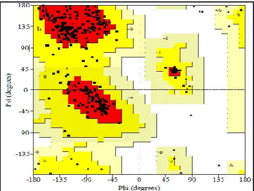
[image:3.595.309.551.181.683.2]showed that 86.1% of the residues of PL model were present in the most favoured region followed by 13.9% in the allowed region, 0% in generously allowed region and disallowed region respectively of Ramachandran plot which are shown in Table I. Further the predicted structure was visualized by Rasmol viewer which is shown in Fig. II.
Fig. I. Ramchandran plot of Predicted 3D structure of PME
[image:3.595.38.288.191.379.2]
Fig. II. Ramchandran plot of Predicted 3D structure of PME
Table I: Residue no. & its % in different regions of Ramchandran plot of PME
2. Active site prediction
Active site of PME was predicted by using CASTp server. 74 pockets were identified in PME molecule.
3. Designing ligand Library
Catechin compound was selected from PubChem as natural inhibitor for PME. The chemical compound was subjected to ZINC database to generate analogues for designing drug library. Total nine analogues were generated from ZINC database. The molecular descriptors were calculated and visualized by MarvinView package which shown in Table II.
Table II: Catechin analogues generated in ZINC database & their structures visualized in MarvinView package
S. No. ZINC ID Name of Compound Structure of the compounds
1. 4214953 Catechin/ Cianidanol
2. 3978494
2-(3,4-Dihydroxyphenyl)c hroman-3,5,7-triol
3. 14436215 Epicatechin
4. 4214953 Epigallocatechin
5. 3870337 (2S,3R)-2-(3,4,5-Trihydroxyphenyl)c hroman-3,5,7-triol/ Gallocatechin
6. 4534390 Epicatechin gallate
7. 6040160 3- Galloylcatechin
8. 3870412 Epigallocatechin gallate
9. 3870413 Gallocatechin gallate
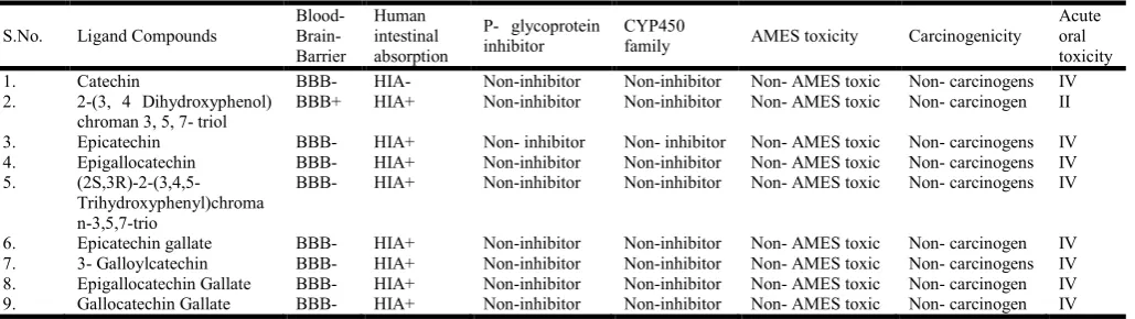
4.Toxicity prediction of ligands
The ADME and Toxicity (Absorption, Distribution,
[image:3.595.45.281.416.577.2] [image:3.595.64.264.632.743.2]5.Molecular Docking
Molecular docking studies are computational techniques for exploration of possible binding mode of a ligand (Catechin) to a given receptor (PME). Docking results of the catechin and its analogues using Hex molecular docking suite which reveals that interaction between the ligand and receptor. Eight analogues and catechin were docked with PME receptor and binding energy was calculated in Table IV. As per the table
binding energy score of 3- Galloylcatechin (Zinc_6040160 (-281.83)) & Epicatechin gallate (Zinc_4534390 (-285.53)) is
better as compared to other derivatives and Catechin molecule Zinc_4214953 was more compatible for the potential inhibitor with receptor than other ligand molecules. Further the docked
complex between the Epictechin gallate (Zinc_3870412 (-285.53)) and PME molecule were visualized and shown in
Figure 3. The below figure III reveals blocking of interacting residues Trp530, Arg503, Trp505, Asp413, Gln412, Asp465, Asp434 & Phe437 of PME with Epicatechin gallate (Zinc_4534390 (-285.53)).
[image:4.595.47.562.75.220.2]
Fig. III: Docked complex between the Epictechin gallate (Zinc_3870412 (-285.53)) and PME molecule were visualized in
Pymol
Conclusion
The tremendous growth of the weed plants & poisonous plants causes various problems to the habitat of the organisms present in that area hence their control is very essential hence we have aimed to identify the inhibitors of the enzymes playing importnat roles in their growth & developement. In the present in-silico study we have identified the novel inhibitor to the
PME enzyme of the A. thaliana to stop the seed germination
process by using the in-silico approaches. In the molecular
docking step we have found that the two analogues of the
catechin (natural inhibitor of the PME) Epicatechin gallate (Docking score= 285.53) & 3- Galloylcatechin (Docking
score= 281.83) best than the other catechin analogues. The further in- vitro inhibtion study of the PME by these analogues on the other weeds & poisonous plants can provide the new alternatives to control their growth.
REFERENCES
Arnold K, Bordoli L, Kopp J, and Schwede T. 2006. The SWISS-MODEL Workspace: A web-based environment
for protein structure homology modelling. Bioinformatics,
22:195-201.
Balani; S.K., V.S.Devishree; G.T. Miwa; L.S. Gan; J.T. Wu; F.W. Lee. 2005. "Strategy of utilizing in vitro and in vivo ADME tools for lead optimization and drug candidate
selection". Curr Top Med Chem., 5 (11): 1033–8.
Fry SC. 2000. The Growing Plant Cell Wall: Chemical and Metabolic Analysis, Blackburn Press, and Caldwell, NJ. Graham, H. N. 1992. Green tea composition, consumption, and
polyphenol chemistry. Prev. Med., 21, 334–350.
Guex N, Peitsch MC. and Schwede T. 2009. Automated comparative protein structure modeling with SWISS-MODEL and Swiss-PdbViewer: A historical perspective.
Electrophoresis, 30(S1):S162-S173.
Table III: Predicted ADMET properties of generated analogues
S.No. Ligand Compounds
Blood- Brain- Barrier
Human intestinal absorption
P- glycoprotein inhibitor
CYP450
family AMES toxicity Carcinogenicity
Acute oral toxicity 1. Catechin BBB- HIA- Non-inhibitor Non-inhibitor Non- AMES toxic Non- carcinogens IV 2. 2-(3, 4 Dihydroxyphenol)
chroman 3, 5, 7- triol
BBB+ HIA+ Non-inhibitor Non-inhibitor Non- AMES toxic Non- carcinogen II
3. Epicatechin BBB- HIA+ Non- inhibitor Non- inhibitor Non- AMES toxic Non- carcinogens IV 4. Epigallocatechin BBB- HIA+ Non-inhibitor Non-inhibitor Non- AMES toxic Non- carcinogens IV 5.
(2S,3R)-2-(3,4,5-Trihydroxyphenyl)chroma n-3,5,7-trio
BBB- HIA+ Non-inhibitor Non-inhibitor Non- AMES toxic Non- carcinogens IV
[image:4.595.153.450.254.352.2]6. Epicatechin gallate BBB- HIA+ Non-inhibitor Non-inhibitor Non- AMES toxic Non- carcinogen IV 7. 3- Galloylcatechin BBB- HIA+ Non-inhibitor Non-inhibitor Non- AMES toxic Non- carcinogens IV 8. Epigallocatechin Gallate BBB- HIA+ Non-inhibitor Non-inhibitor Non- AMES toxic Non- carcinogen IV 9. Gallocatechin Gallate BBB- HIA+ Non-inhibitor Non-inhibitor Non- AMES toxic Non- carcinogen IV
Table IV: Docking scores of each generated analogue with the PME
S.No. Compounds E –value
1. Catechin -233.20
2. Dihydroxy phenyl chromne -246.08
3. Epicatechin -233.20
4. Epigallocatechin -232.38
5. (2S- 3R)- 2- (3, 4, 5- Trihydrxy phenyl chroman) 3, 5, 7- triol -238.56
6. Epicatechin gallate -285.53
7. 3 galloylcatechin -281.83
[image:4.595.38.292.608.758.2]Irwin J. J. and Shoichet B. K. 2005. ZINC “A free database of commercially available compounds for virtual screening”
J. Chem. Inf. Model, 45, 177–182.
Jesper Harholt, Anongpat Suttangkakul, and Henrik Vibe
Scheller. June 2010. Biosynthesis of Pectin, Plant
Physiology, Vol. 153, pp. 384–395.
Jiang CM., Lai YJ., Lee BH., Chang WH., Chang HM. 2001.
De-esterification and transacylation reaction of
pectinesterase from jelly fig (Ficus awkeotsang makino)
achenes, J Food Sci., 66:810-815.
Kiefer F, Arnold K, Künzli M, Bordoli L, Schwede T. 2009. The SWISS-MODEL Repository and associated resources.
Nucleic Acids Research, 37:D387-D392.
Laskowski RA, Hutchinson EG, Michie AD, Wallace AC, Jones ML, Thornton JM. Dec 2014. "PDBsum: a Web-based database of summaries and analyses of all PDB
structures". Trends Biochem. Sci., 22 (12): 488–90.
Laskowski RA, MacArthur MW, Moss DS, Thornton JM. 1993. PROCHECK-a program to check the stereochemical
quality of protein structures. J App. Cryst, 26:283-291.
Lewis, Kristin C., Tzvia Selzer, et al. 2008. Inhibition of
pectin methyl esterase activity by green tea catechins,
Phytochemistry, 69: 2586–2592.
Macindoe, G., L. Mavridis, V. Venkatraman, M.-D. Devignes, D.W. Ritchie, 2010. “Hex Server: an FFT-based protein
docking server powered by graphics processors”. Nucleic
Acids Research, 38, W445-W449.
Marco Biasini, Stefan Bienert. et al. 2014. SWISS-MODEL:
modelling protein tertiary and quaternary structure using
evolutionary information. Nucleic Acids Research, 42(W1):
W252-W258.
McBride, Ryan1 Oct 2012. "ChemAxon opens shop in 'heart'
of Boston biotech hub". Retrieved.
Micheli F. 2001. Pectin methylesterases: cell wall enzymes
with important roles in plant physiology. Trends Plant Sci.,
6: 414–419.
Müller, K., Levesque - Tremblay, et al. 2013.
Demethylesterification of cell wallpectins in arabidopsis
plays a role in seed germination, Plant Physiol., 161,305–
316.
Pooja Kohli, Manmohit Kalia, Reena Gupta. 2015. Pectin
Methylesterases: A Review. J Bioprocess Biotech, Volume
5: 227.
Quan V, Vuong, John B, Golding, Minh Nguyen, Paul D. Roach, 2010. Extraction and isolation of catechins from
tea. J. Sep. Sci., 33, 3415–3428.
Raiola A., Camardella L., Giovane, A. et al. 2004. Two
Arabidopsis thaliana genes encode functional pectin
methylesterase inhibitors. FEBS Lett, 557: 199–203.
Ridley BL., O’Neill MA. and Mohnen D. 2001. Pectins: structure, biosynthesis, and oligogalacturonide-related
signaling, Phytochemistry, 57:929–967.
Robert Palin, Anja Geitmann. September 2012. The role of pectin in plant morphogenesis, Volume 109, Issue 3, Pages 397–402.
Sathish Kumar Paramashivam, Kalaivani Elayaperumal et al.
2015. “Insilico pharmacokinetic and molecular docking studies of small molecules derived from Indigoferaaspalat
hoidesVahl targeting receptor tyrosine kinases”
Bioinformation, 11(2): 073-084.
Sayle RA. and Milner-White EJ. 1995. RASMOL:
biomolecular graphics for all. Trends Biochem Sci., 20(9):
374.
Wolf S., Grsic-Rausch S., Rausch T., Greiner S. 2003. Identification of pollen-expressed pectin methylesterase
inhibitors in Arabidopsis. FEBS Lett, 555: 551–555.
Wolf S., Mouille G., Pelloux J. 2009. Homogalacturonan
methyl-esterification and plant development, Mol Plant, 2:
851–860.
Yogesh N Joshi, Prathusha S. Chegu, Nitin N. Bolabattin,
Chetan Godale. October 2016. In- silico Structural &
Molecular characterization of Pectin methylesterase from
Arabidopsis thaliana. EJBB, Volume 4; Issue 10; Page No.
21-24.