ANTIFUNGAL EFFICACY OF ROUTINE AND NEWER IRRIGANTS ON CANDIDA ALBICANS BIOFILM
COLONIZATION IN YOUNG AND OLD HUMAN ROOT CANAL DENTIN
1
Dr. Arrvind Vikram
1
Department of Conservative Dentistry and Endodontics Ragas Dental College and Hospital, Chennai
2Department of Conservative Dentistry and Endodontics Adhiprasakthi Dental College and
ARTICLE INFO ABSTRACT
The purpose of this study was to evaluate the effect of routine and newer endodontic irrigants on Candida albicans biofilm colonization in Young and Old human root canal dentin. Eighty intact mandibular premolars were u
were from young subjects (Group I) and the remaining 40 from old subjects (Group II). Dentin disc samples of 4mm were prepared from each tooth, standardized using gates glidden drill # autoclaved for sterility. The forty samples each in the young and old group were divided into 4 subgroups with various irrigation protocols. The experimental irrigants were: A) 17% EDTA +5.25% NaOCl, B) 100% Octenisept, C) 17% EDTA +5.25% NaOCl + 1%
buffer saline. After the irrigation the experimental samples were inoculated with C.albicans and incubated for 72hrs. Out of the 10 samples in each subgroup, 8 samples were analyzed by the Colony forming unit method and 2 samp
CFU method, aliquots from the experimental samples were plated on Sabouraud dextrose agar plates and the colony forming units were counted as a measure of antifungal activity. In the CLSM fungal viability was demonstrated using special dyes SYTO 9 and Propidium iodide. The results showed that Octenisept was the most effective irrigant against C.albicans followed by addition of 1% Clotrimazole to 17% EDTA+5.25% NaOCl in both age grou
effective in both age groups. The results of this study also indicates that higher amount of fungi are found in old root dentin as demonstrated by the CFU method and confirmed by the CLSM method.
Copyright©2017, Dr. Arrvind Vikram et al. This is an open access article distributed under the Creative Commons Att
use, distribution, and reproduction in any medium, provided the original work is properly cited.
INTRODUCTION
Diseases of the pulp and periradicular tissues are often associated with invasion of Microorganisms. Kakehashi in a classic study proved that bacteria caused pulpal disease endodontic infections are known to be polymicrobial in nature with preponderance toward anaerobic species. Numerous studies have revealed that Enterococcus faecalis, Actinomyces, and Candida albicans were the most prevalent microorganisms associated with failed endodontic treatment Jin Y, Zhang T. The microorganisms remaining in the root canal space after treatment or re-colonising the filled canal system are the main cause of endodontic failure Kakoli P Fungi are eukaryotic microorganisms that exhibit two basic structural forms: a ‘‘yeast form’’ (unicellular) and a ‘‘mould form’’ (multicellular). They are common opportunistic pathogens that constitute a part of the normal microbial flora of the
*Corresponding author: Dr. N. Bharath,
Department of Conservative Dentistry and Endodontics Adhiprasakthi Dental College and Hospital, Melmaruvathur
ISSN: 0975-833X
Article History:
Received 29th April, 2017 Received in revised form 11th May, 2017
Accepted 20th June, 2017
Published online 31st July, 2017
Citation: Dr. Arrvind Vikram, Dr. Rajamani Indira and Dr. N. Bharath,
biofilm colonization in young and old human root canal dentin
Key words:
Anti fungal efficacy, Candida Albicans,
Irrigants Octenisept Colony forming unit Confocal laser scanning microscope.
RESEARCH ARTICLE
ANTIFUNGAL EFFICACY OF ROUTINE AND NEWER IRRIGANTS ON CANDIDA ALBICANS BIOFILM
COLONIZATION IN YOUNG AND OLD HUMAN ROOT CANAL DENTIN
Arrvind Vikram,
1Dr. Rajamani Indira and
*,2Dr. N. Bharath
Department of Conservative Dentistry and Endodontics Ragas Dental College and Hospital, Chennai
Department of Conservative Dentistry and Endodontics Adhiprasakthi Dental College and
Melmaruvathur
ABSTRACT
purpose of this study was to evaluate the effect of routine and newer endodontic irrigants on Candida albicans biofilm colonization in Young and Old human root canal dentin. Eighty intact mandibular premolars were used in this study and divided based on Age into two groups. Forty teeth were from young subjects (Group I) and the remaining 40 from old subjects (Group II). Dentin disc samples of 4mm were prepared from each tooth, standardized using gates glidden drill # autoclaved for sterility. The forty samples each in the young and old group were divided into 4 subgroups with various irrigation protocols. The experimental irrigants were: A) 17% EDTA +5.25% NaOCl, B) 100% Octenisept, C) 17% EDTA +5.25% NaOCl + 1%
buffer saline. After the irrigation the experimental samples were inoculated with C.albicans and incubated for 72hrs. Out of the 10 samples in each subgroup, 8 samples were analyzed by the Colony forming unit method and 2 samples were analyzed by the Confocal laser scanning microscope. In the CFU method, aliquots from the experimental samples were plated on Sabouraud dextrose agar plates and the colony forming units were counted as a measure of antifungal activity. In the CLSM fungal viability was demonstrated using special dyes SYTO 9 and Propidium iodide. The results showed that Octenisept was the most effective irrigant against C.albicans followed by addition of 1% Clotrimazole to 17% EDTA+5.25% NaOCl in both age groups. The other irrigant subgroups were less effective in both age groups. The results of this study also indicates that higher amount of fungi are found in old root dentin as demonstrated by the CFU method and confirmed by the CLSM method.
is an open access article distributed under the Creative Commons Attribution License, which use, distribution, and reproduction in any medium, provided the original work is properly cited.
Diseases of the pulp and periradicular tissues are often associated with invasion of Microorganisms. Kakehashi et al
in a classic study proved that bacteria caused pulpal disease endodontic infections are known to be polymicrobial in nature with preponderance toward anaerobic species. Numerous studies have revealed that Enterococcus faecalis, Actinomyces, bicans were the most prevalent microorganisms associated with failed endodontic treatment Jin Y, Zhang T. The microorganisms remaining in the root canal space after
colonising the filled canal system are the main Kakoli P Fungi are eukaryotic microorganisms that exhibit two basic structural forms: a ‘‘yeast form’’ (unicellular) and a ‘‘mould form’’ (multicellular). They are common opportunistic pathogens that constitute a part of the normal microbial flora of the oral
Department of Conservative Dentistry and Endodontics Adhiprasakthi Dental
cavity. The most important fungi belong to genus Candida with C. albicans being the most predomin
isolated yeast from the oral cavity Waltimo TMT Candida is a versatile microorganism capable of adapting itself to a wide range of pH level and exhibits a variety of virulence factors such as adherence, hyphal formation, thigmotropism, phenotypic switching and secretes a degenerative enzyme ‘‘aspartyl protease’’ that degrades the dentinal collagen Vianna ME, Gomes B. Candida has the ability to grow on the dentinal surfaces in the absence of oral tissue fluids and penetrates into the dentinal tubules by its various growth patterns (hyphae and blastospores) and by the formation of Biofilms Tandjung L. Sen et al. suggested that Candida be considered a ‘‘dentinophilic’’ microorganism as it can invade dental hard tissues and present a reservoir f
infections many investigations have confirmed a strong association between persistent or secondary intraradicular infection with Candida albicans and posttreatment apical periodontitis. The use of antimicrobial agents in the form irrigants has been recommended as an adjunct to mechanical
International Journal of Current Research Vol. 9, Issue, 07, pp.55121-55131, July, 2017
Dr. Arrvind Vikram, Dr. Rajamani Indira and Dr. N. Bharath, 2017. “Antifungal efficacy of routine and newer irrigants on candida albicans biofilm colonization in young and old human root canal dentin”, International Journal of Current Research, 9, (07), 55121
ANTIFUNGAL EFFICACY OF ROUTINE AND NEWER IRRIGANTS ON CANDIDA ALBICANS BIOFILM
COLONIZATION IN YOUNG AND OLD HUMAN ROOT CANAL DENTIN
Bharath
Department of Conservative Dentistry and Endodontics Ragas Dental College and Hospital, Chennai
Department of Conservative Dentistry and Endodontics Adhiprasakthi Dental College and Hospital,
purpose of this study was to evaluate the effect of routine and newer endodontic irrigants on Candida albicans biofilm colonization in Young and Old human root canal dentin. Eighty intact sed in this study and divided based on Age into two groups. Forty teeth were from young subjects (Group I) and the remaining 40 from old subjects (Group II). Dentin disc samples of 4mm were prepared from each tooth, standardized using gates glidden drill #2 and autoclaved for sterility. The forty samples each in the young and old group were divided into 4 subgroups with various irrigation protocols. The experimental irrigants were: A) 17% EDTA +5.25% NaOCl, B) 100% Octenisept, C) 17% EDTA +5.25% NaOCl + 1% Clotrimazole and D) Phosphate buffer saline. After the irrigation the experimental samples were inoculated with C.albicans and incubated for 72hrs. Out of the 10 samples in each subgroup, 8 samples were analyzed by the Colony les were analyzed by the Confocal laser scanning microscope. In the CFU method, aliquots from the experimental samples were plated on Sabouraud dextrose agar plates and the colony forming units were counted as a measure of antifungal activity. In the CLSM method, fungal viability was demonstrated using special dyes SYTO 9 and Propidium iodide. The results showed that Octenisept was the most effective irrigant against C.albicans followed by addition of 1% ps. The other irrigant subgroups were less effective in both age groups. The results of this study also indicates that higher amount of fungi are found in old root dentin as demonstrated by the CFU method and confirmed by the CLSM method.
ribution License, which permits unrestricted
cavity. The most important fungi belong to genus Candida with C. albicans being the most predominant and commonly isolated yeast from the oral cavity Waltimo TMT Candida is a versatile microorganism capable of adapting itself to a wide range of pH level and exhibits a variety of virulence factors such as adherence, hyphal formation, thigmotropism, notypic switching and secretes a degenerative enzyme ‘‘aspartyl protease’’ that degrades the dentinal collagen Vianna ME, Gomes B. Candida has the ability to grow on the dentinal surfaces in the absence of oral tissue fluids and penetrates into l tubules by its various growth patterns (hyphae and blastospores) and by the formation of Biofilms Tandjung L.
. suggested that Candida be considered a ‘‘dentinophilic’’ microorganism as it can invade dental hard tissues and present a reservoir for disseminating candidal infections many investigations have confirmed a strong association between persistent or secondary intraradicular infection with Candida albicans and posttreatment apical periodontitis. The use of antimicrobial agents in the form of irrigants has been recommended as an adjunct to mechanical
INTERNATIONAL JOURNAL OF CURRENT RESEARCH
instrumentation to reduce the numbers of micro-organisms Saurabh S. Chandra Sodium hypochlorite is the most frequently used irrigant in the treatment of infected root canals because of it’s antimicrobial, sporicidal, fungicidal, tissue dissolving properties and aids in debridement of canal system. Studies have reported that C.albicans is susceptible to the action of NaOCl with increasing concentration Siqueira JF. EDTA was first introduced as a chelating agent in endodontic therapy by Nygaard-Ostby. It reacts with calcium ions in dentin and forms soluble calcium chelate. However EDTA may have an antifungal potential with its chelating property because calcium ions have a critical role in the morphogenesis and pathogenesis of Candida albicans Ozdemir H.O
Octenisept is an antiseptic for skin burns, wound disinfection and mouth rinses consisting of octenidine hydrochloride and phenoxyethanol has been suggested as a potential endodontic irrigant based on its antimicrobial effects and lower cytotoxicity It demonstrates broad spectrum antimicrobial effects covering both Gram-positive and Gram-negative bacteria, fungi and several viral species Venegas SC. Clotrimazole, a substituted imidazole, is a commonly used antifungal in both medical and dental practice. It is one of a family of azoles and is useful in treating systemic mycoses. They have a broad-spectrum antifungal activity covering the Candida species, dermatophytes, and some gram-positive and anaerobic bacteria such as Staphylococcus aureus and Streptococcus faecalis Ozdemir H.O. However the use of an antifungal agent as an adjunct in irrigation protocol has been reported only in one study in endodontic literature Jin Y. Age related histological changes occur in the pulp-dentin complex of teeth Mustafa A. Alterations in dentin tissue with age might result in different adhesion capability of bacteria and fungi. These differences in adhesion of microorganisms and its clinical significance have not been evaluated in the literature. Therefore, the aim of this study was to evaluate the effect of various irrigants on Candida albicans biofilm colonization in Young and Old human root canal dentin by using 2 different techniques: Colony Forming Unit (CFU) method and Confocal Laser Scanning Microscopic (CLSM) method. The Objectives of this study was to
1. Investigate the antifungal potential of a new irrigant – Octenisept.
2. Compare Octenisept with Clotrimazole, which is a known antifungal agent.
3. Analyze the adherence capability of C.albicans to Young and Old root canal dentin.
4. Demonstrate the viable and dead fungi in the dentinal tubules using special dyes (SYTO 9 and Propidium Iodide) with the Confocal laser scanning microscope.
Preparation of media
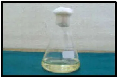
[image:2.595.339.526.52.175.2]Sabouraud dextrose broth ingredients Special peptone – 10gm/lit, Dextrose – 20gm/lit, pH - 5.6 Sabouraud dextrose broth was weighed to measure 3gm and dissolved in 100ml of distilled water in conical flask. The conical flask was plugged with cotton and sterilized in autoclave at 121°c for 15 min at 15 lbs pressure. Sabouraud dextrose agar Ingredients Peptone – 10gm/lit, Dextrose – 40gm/lit, pH - 5.6 Sabouraud dextrose agar was weighed 6.5gm and dissolved in 100ml of distilled water in conical flask. The conical flask was plugged with cotton and heated till the colour change and sterilized in autoclave at 121°c for 15 min at 15 lbs pressure.
Fig. 1. Sabouraud dextrose Broth
Fig. 2. Sabouraud dextrose Agar
Preparation of media plates
The prepared sterilized Sabouraud dextrose agar was poured into petridishes to a depth of 5mm under the laminar flow chamber. For every 100ml of the medium 6 plates were poured. The poured plates were allowed to solidify and were refrigerated. For every batch of prepared plates one plate served as a sterility check.
MATERIALS AND METHODS
[image:2.595.337.529.183.332.2]Eighty freshly extracted intact single-rooted human mandibular premolars, stored in saline solution at 4°C, were used in this study. Forty teeth were from young subjects (removal for orthodontic reasons) and the remaining forty from older subjects (removal for periodontal reasons). These served as the two groups in the study. The young subjects were in the age group of 12 – 25 years, whereas the older subjects were above the age of 50 years.
[image:2.595.313.553.601.778.2]Fig. 2. Decoronation at CEJ level
Fig. 3. Dentin Disc Samples
The teeth were cleaned to render them free from calculus, tissue tags and other debris and kept in 0.2% sodium azide solution for disinfection.
Preparation of dentin disc samples
The coronal and apical parts of the teeth were cut with a high-speed diamond disk, resulting in a 4-mm-long mid-part of the root sample per tooth and a total of 80 dentin disc samples. Standardization of each root canal was performed by enlarging the canal with #2 Gates-Glidden burs (0.7 mm diameter). Samples were washed thoroughly, sterilized by autoclave at 121°C for 15 minutes, and preincubated at 37°C in brain-heart infusion (BHI) to ensure no microbial contamination.
Grouping of Specimens
In Group I (teeth from young subjects) and Group II (teeth from older subjects), each forty dentin disc samples, were divided into four subgroups having ten dentin samples each and treated with the following irrigants.
Group I (Young group) Group II (Old group) IA - 2ml of 17% EDTA for one min
and 2 ml of 5.25% NaOCl for one min.
IIA - 2ml of 17% EDTA for one min and 2 ml of 5.25% NaOCl for one min.
IB - 2ml of Octenisept for one min. IIB - 2ml of Octenisept for one min. IC - 2ml of 17% EDTA for one min
and 2 ml of 5.25% NaOCl for one min. A 5ml flush with distilled water. 2ml of 1% Clotrimazole for one min.
IIC - 2ml of 17% EDTA for one min and 2 ml of 5.25% NaOCl for one min. A 5ml flush with distilled water. 2ml of 1% Clotrimazole for one min.
ID - 2ml of sterile phosphate buffer saline for one min.
[image:3.595.59.267.240.398.2]IID - 2ml of sterile phosphate buffer saline for one min.
Fig. 4. Experimental Irrigants
Irrigation was done with the help of a 26 gauge syringe. Following this, the set of instruments were sterilized by soaking the tip in alcohol and flaming them in Bunsen flame.
Microbiology procedures
A suspension of C.albicans was adjusted to 0.5 turbidity on the Mcfarland scale.
The sterile canals of 80 experimental dentin disc samples were inoculated with 0.3ml of the adjusted C.albicans suspension, and each sample was individually submerged in Candida albicans suspension in the glass test tube vials.
Fig. 5. Irrigating the Specimen
Fig. 6. Inoculation with C.albicans
[image:3.595.322.544.374.554.2] [image:3.595.323.542.450.714.2]Fig. 7. Samples incubated at 36ºC for 72 hrs in Incubator
Fig. 8. Collection of Samples
Fig. 9. Streaking of aliquot
At 48 hrs aliquots were taken from each sample using a syringe and plated on 4% sabouraud dextrose agar plate to verify the growth of Candida albicans in each sample tube. Out of the 10 dentin disc samples in each subgroup, 8 samples were assessed to analyze the formation of Colony Forming Units (CFU), whilst the remaining 2 samples were assessed to detect the presence of Live/Dead Fungi in the dentinal tubules using the Confocal Laser Scanning Microscope. Hence, a total of 64 dentin disc samples were subjected to the CFU method and 16 dentin disc samples were used for the CLSM method.
CFU Method
After 72hrs, the 64 samples were removed from the glass test tube vials and rinsed 3 times with 10 mL of sterile PBS. The root canal of each tooth sample was again enlarged with sterile #3 Gates-Glidden burs (0.9 mm diameter), and dentin shavings were collected into 3 mL of sterile PBS. The Gates-Glidden burs were also placed into the test tube to collect dentin shavings that adhered to the bur. All the tubes were sonicated in an ultrasonic water bath for 10 minutes to dislodge fungi from the burs and dentin shavings and to disperse fungal aggregation. A 1-µm inoculation loop was used to remove aliquots from the suspension prepared from each one of the 64 dentin disc samples and was plated individually on Sabouraud 4% dextrose agar plates. The plates were incubated at 36°c and
91% humidity for 48 hours. After the incubation period, the growth of C. albicans was assessed with light microscopy at 400X. The number of colony forming units (CFUs) of Candida served as a measure of the antifungal activity.
[image:4.595.76.245.188.476.2]
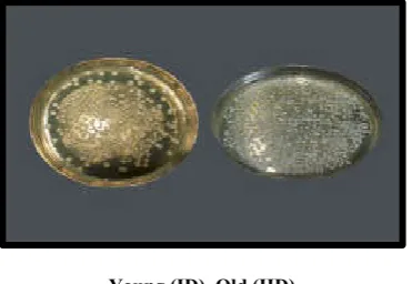
Fig. 10. Group I (Young Teeth)
Fig : Group II (Old Teeth)
Young (IA) Old (IIA)
Fig. 11. C.F.U WITH 17% EDTA + 5.25% NaOCl
Young (IB) Old (IIB)
[image:4.595.80.247.197.307.2] [image:4.595.329.538.626.755.2]Young (IC) Old (IIC)
Fig. 13. C.F.U WITH 17% EDTA + 5.25% NaOCl + 1% CLOTRIMAZOLE
[image:5.595.70.258.54.162.2]Young (ID) Old (IID)
Fig. 14. C.F.U with Phosphate Buffer Saline
Confocal Laser Scanning Microscopic method
The 16 samples to be analyzed by the Confocal Laser Scanning Microscope were removed from the glass test tube vials and rinsed 3 times with 10 mL of sterile PBS. The samples were embedded on methyl methacrylate resin blocks and four evenly distributed transverse sections (1 mm thick) were cut from each sample using the Hard tissue microtome.
Fig. 15. Hard Tissue Microtome
[image:5.595.71.257.224.352.2]Fig. 16. Teeth Samples (1mm Cross Section)
[image:5.595.314.552.258.422.2]Fig. 17. Bactec Viability Kit (INVITROGEN)
Fig. 18. Staining with SYTO 9 and Propidium Iodide Dyes
[image:5.595.83.245.494.615.2] [image:5.595.77.248.651.774.2]YOUNG (IA) OLD (IIA)
[image:6.595.36.263.53.210.2]20X 40X 63X
Fig. 19. Sub Group A: (17% EDTA + 5.25% NaOCl)
YOUNG (IB) OLD (IIB
[image:6.595.38.271.233.405.2]20X 40X 63X
Fig. 20. Sub Group B: (OCTENISEPT)
YOUNG (IC) OLD (IIC)
20X 40X 63X
Fig. 21. Sub Group C: (17% EDTA + 5.25% NaOCl + 1% CLOTRIMAZOLE)
YOUNG (ID) OLD (IID
20X 40X 63X
Fig. 22. Sub Group D: (PHOSPHATE BUFFER SALINE)
Fluorescence images were analysed with Amira 5.0 (Visage Imaging Inc., Andover, MA, USA), and image stacks were viewed with LSM Image Browser (Carl Zeiss Ltd). The initial stacks, comprising both green and red fluorescence, were split into individual component colour channels and saved as grey-scale images. For each greygrey-scale image, fluorescence was adjusted (‘thresholded’) such that signals of intensity less than 20% were regarded as background. The split greyscale images were then combined and calibrated to form a single fluorescent image which was qualitatively analyzed by three independent blinded observers to determine the proportion of green and red fluorescence denoting the presence of live and dead Candida albicans cells in the mineralized human dentinal tubules.
Statistical analysis
[image:6.595.38.281.427.591.2]The results of the present study were subjected to statistical analysis to interpret the significant differences among various irrigants used in the Young and Old groups respectively and also assessed the difference between the groups when the irrigant used was a constant. One-Way ANOVA, post hoc Tukey HSD tests and unpaired t-test were used for statistical analysis in the present study.Unpaired t-test is applied to unpaired data of independent observation made on individuals of two different or separate groups or samples drawn from two populations. In this study one way ANOVA followed by Tukey HSD test showed statistically significant difference among various subgroups concerning the discrepancy in the colony forming unit in each group. Unpaired t-test showed a significant difference in the number of colony forming unit between the groups.
Table 1. Comparison of the growth of CFUs (1 x 10³ml-1) of Candida albicans in two groups with 4 irrigants
Groups YOUNG – I (Mean±SD)
OLD – II (Mean±SD) A – 17%EDTA + 5.25%NaOCl 62.25±8.73 185.75±16.36 B - Octenisept 9.38±3.02 38.75±6.23 C – 17%EDTA + 5.25%NaOCl +
1%Clotrimazole
14.88±4.49 51.13±10.87
[image:6.595.39.289.625.782.2]D – Phosphate buffer saline 570.63±33.02 997.25±74.66 P VALUE: 0.000** 0.000**
[image:6.595.309.558.665.736.2]Table 1 demonstrates the mean CFUs of Candida albicans after a final rinse with the irrigant solution in both age groups. Octenisept was found to be effective against Candida albicans followed by 17%EDTA + 5.25%NaOCl + 1%Clotrimazole, 17%EDTA + 5.25%NaOCl and finally, phosphate buffer saline in both Age groups (P<.001). This difference was highly statistically significant
Table 2. Comparison of growth of CFUs (1 x 10³ml-1) of Candida albicans between the 4 irrigants in each group
Groups YOUNG – I (P Value) OLD – II (P Value) A × B 0.000** 0.000**
A × C 0.000** 0.000** A × D 0.000** 0.000** B × C 0.919 0.918 B × D 0.000** 0.000** C × D 0.000** 0.000**
(suggesting both irrigant combinations were equally effective). All other comparisons of irrigants within each group were highly statistically significant (P<.001).
Table 3. Comparison of growth of CFUs (1 x 10³ml-1) of Candida albicans for each irrigant sub group between the groups
Groups YOUNG – I (Mean±SD)
OLD – II
(Mean±SD) P Value A – 17%EDTA +
5.25%NaOCl
62.25±8.73 185.75±16.36 0.000**
B – Octenisept 9.38±3.02 38.75±6.23 0.000** C – 17%EDTA +
5.25%NaOCl + 1%Clotrimazole
14.88±4.49 51.13±10.87 0.000**
D – Phosphate buffer saline 570.63±33.02 997.25±74.66 0.000**
Table 3 demonstrates the comparison of mean CFUs of Candida albicans after a final rinse with:
17% EDTA + 5.25% NaOCl in both age groups. Octenisept in both age groups
17%EDTA + 5.25%NaOCl + 1% Clotrimazole in both age groups
Phosphate buffer saline in both age groups
All the irrigant solutions used in this present study were more effective in the Younger group than the older group. This difference was highly statistically significant (P<.001).
DISCUSSION
Microorganisms play a fundamental role in the etiology of pulp and periapical diseases. Therefore the most important goal of endodontic treatment is the complete debridement of the root canal system to eliminate these entire microorganisms, their by products and tissue debris from the infected root canals. Primary root canal infections are polymicrobial. Various studies in literature over the last decade have reported that the two most common organisms reported to be associated with root canal failure cases are E.faecalis and Candida albicans Douglas J L. Candida can adapt to a wide range of pH, change gene expression in response to environmental conditions, adhere to a variety of substrates, produce degenerative enzymes and change morphological form to evade the host immune system they have surface molecules that mediate adherence to the host tissue. These molecules include a receptor which binds RGD (Arginine-glycine-aspartic acid) groups on IC3b, fibrinogen, fibronectin, laminin and vitronectin. Candida species are also able to bind to Collagen type I and IV of dentin and can use dentin as a nutrient source Sen BH. Sen et al (1997) investigated the growth patterns of C.albicans in relation to human radicular dentin and observed blastospores and hyphal structures on the root canal walls of all specimens.It was proposed that the contact sensing (Thigmotropism) ability of hyphal structures of Candida made dentinal invasion inevitable. Therefore, on the basis of this invasive affinity to dentin, they considered C.albicans a dentinophilic microorganism. Sen et al (1997) & (2003) demonstrated that the presence of smear layer increased the adhesion of C.albicans to dentin. They hypothesized that this increased adhesion was attributable to the availability of the disintegrated organic structure of dentin and the availability of calcium ions as a source of growth and adhesion.
Baumgartner et al. (2000) found Candida albicans in 21% of samples taken from infected root canals while using PCR. Siqueira et al. (2004) detected Candida albicans in 2 of 22 patients in case of failed endodontic therapy by PCR. All these studies prove beyond doubt that C.albicans are the most common yeast isolates found in the root canals of endodontic failure cases.
However, investigations on Fungi in recent literature have been limited as opposed to the exhaustive analysis of the facultative anaerobe E.faecalis. Hence, Candida albicans (ATCC 90028) was the test organism chosen in this investigative study. The aim of the present study was to evaluate the effect of universally used routine endodontic irrigants - Sodium hypochlorite and EDTA, newer endodontic irrigants - Octenisept and Clotrimazole on Candida albicans biofilm colonization in Young and Old human root canal dentin by using two different techniques: Colony Forming Unit (CFU) method and Confocal Laser Scanning Microscopic (CLSM) method. There is only limited knowledge on the effect of dentin aging on microbial adhesion, although such information is clinically important. Hence, teeth from younger and older subjects were included in this study, to explore the significance of age in the clinical scenario. From each tooth, a 4mm long dentin disc sample was prepared which resulted in a total of 80 dentin disc samples. Root canals were standardized with the #2 Gates-Glidden burs (0.7 mm diameter), autoclaved and incubated overnight in BHI medium to ensure no microbial contamination and complete sterility. Following this the samples in the two groups were sub grouped under various experimental irrigants. The irrigants used in the study were 17%EDTA + 5.25%NaOCl, 100% Octenisept, 17% EDTA + 5.25%NaOCl + Clotrimazole and Phosphate buffer saline. The irrigation regimen used in group IA and IIA was 2ml of 17% EDTA for one minute followed by 2ml of 5.25% NaOCl for one minute. This combination of the universal endodontic irrigants was chosen to simulate the synergistic use of these two agents in routine endodontic regime. 2ml of 17% EDTA was taken in the present study based on the previous studies by Sen et al (2000). Mustafa et al (2005) who proved the antifungal efficacy of 17%EDTA on C.albicans. The contact time was for one min as recommended by Ruff et al (2006)24. 2ml of 5.25%NaOCl was taken based on the studies by Sen et al (1999)28 and Sena et al (2006)32 who demonstrated that 5.25% NaOCl was effective against C.albicans in the absence of smear layer. One minute of contact time was taken in accordance to the previous study done by Radcliff et al. 22.
The volume and contact time of the Octenisept irrigant was 2ml and 1 minute respectively to standardize with the irrigants used in the other groups. Till date no studies have reported the use of Octenisept in the elimination of fungi from the root canal system. Hence, Octenisept was chosen as an experimental irrigant in this study. The irrigation regimen used in groups IC and IIC was as follows: 2ml of 17% EDTA for one minute followed by 2ml of 5.25% NaOCl for one minute. A 5ml flush with distilled water to terminate the action of the irrigants and followed by 2ml of 1% Clotrimazole for one minute. It was hypothesized that the addition of an antifungal agent as an irrigant would provide a substantive action on the dentin and prevent adherence of C.albicans biofilm cells on the experimental samples. This was in accordance with the study by Saurabh et al (2010) (Ozdemir et al., 2010) who evaluated the antifungal effect of 17% EDTA, 5.25% NaOCl and 2% CHX with and without an antifungal agent (Clotrimazole). One minute of contact time and the irrigant volume of 2ml was taken from the above study. In the present investigation, 1% Clotrimazole which is regularly used in the treatment of oral candidiasis was chosen. Samples in groups ID and IID were irrigated with 2ml of Phosphate buffer saline for one minute and served as a control group. Once the dentin disc samples were irrigated with the experimental irrigants, they were subjected to the Microbiological procedures. A suspension of C.albicans was adjusted to 0.5 turbidity on the Mcfarland scale.
The canals of 64 experimental dentin disc samples were inoculated with 0.3ml of the adjusted C.albicans suspension, and each sample was individually submerged in Candida albicans suspension in the glass test tube vials. The samples were incubated at 36°c and 91% humidity for 72 hrs to form biofilms which simulates the environment inside the root canal system and resembles clinical situations. Out of the 10 dentin disc samples in each subgroup, 8 samples were assessed by the CFU method whilst the remaining 2 samples were assessed to detect the presence of Live/Dead Fungi in the dentinal tubules using the Confocal Laser Scanning Microscope. Hence, a total of 64 dentin disc samples were subjected to the CFU method and 16 dentin disc samples were used for the CLSM method. CFU is a primary microbial technique, allowing determination of the number of viable fungi per sample. The samples analyzed by the CFU method were removed from the glass test tube vials and rinsed 3 times with 10 mL of sterile PBS. The root canal of each tooth sample was again enlarged with sterile #3 Gates-Glidden burs (0.9 mm diameter), and dentin shavings were collected into 3 mL of sterile PBS. The Gates-Glidden burs were also placed into the test tube to collect dentin shavings that adhered to the bur. All the tubes were sonicated in an ultrasonic water bath for 10 minutes to dislodge fungi from the burs and dentin shavings and to disperse fungal aggregation. Most studies evaluate root canal disinfection by sampling with paper points. This technique is limited by only sampling microbes from the fluid in the canal. In the present study, dentin shavings were sampled, which allowed the detection of fungi that had penetrated inside the dentinal tubules. This method realistically replicates the clinical scenario and highlights the degree to which C.albicans adhere to dentin and invade dentinal tubules following irrigation protocols. The antifungal efficacy was evaluated after 72hrs based on the number of colony forming units of Candida, which was obtained by semiquantitative analysis. This analysis was adopted in this study because of its accuracy,
reproducibility, acceptance and feasibility in the laboratory settings.
CLSM analysis determines the viable and dead fungi immobilized in the dentinal tubules and the biomass. The samples to be analyzed by the Confocal Laser Scanning Microscope were removed from the glass test tube vials and rinsed 3 times with 10 mL of sterile PBS. The samples were embedded on methyl methacrylate resin blocks and four evenly distributed transverse sections (1 mm thick) were cut using the Hard tissue microtome. The cut sections were then stained immediately with the SYTO 9 and propidium iodide (PI) reagents which are marketed as the Live/Dead stain (Baclight; Invitrogen Corporation, Carlsbad, CA, USA) and examined under the Confocal Laser Scanning Microscope. The nucleic acid–binding fluors, SYTO 9 and propidium iodide (PI), have been widely applied in environmental studies, food microbiology and dental research including endodontic investigations (Sen et al., 1999). These reagents were introduced by Invitrogen Corporation as the Baclight – Live/Dead stain, as they differentiate between viable and non-viable bacteria. However, Jin et al (2005) evaluated the viability of candidal biofilms using combination stains, SYTO9 and propidium iodide (PI) and demonstrated that SYTO9 and PI are reliable vital stains that may be used to investigate C. albicans biofilms under the Confocal Laser Scanning Microscope. Thus, the use of the fluorescent dyes to assess the viability of Candida albicans biofilms on the root canal dentin of young and old teeth has been confirmed in this study. SYTO 9 penetrates intact biological membranes, whereas PI penetrates only fungi with compromised plasma membranes and quenches the SYTO 9 fluorescence on binding the nucleic acid (Tandjung et al., 2007). Thus, simultaneous application of the stains generates red-fluorescing dead fungi and green-fluorescing live fungi, and these can be visualized by fluorescence microscopy.
Confocal Laser Scanning Microscopy (CLSM) has become an invaluable tool for a wide range of investigations in the biological and medical sciences for imaging thin optical sections in living and fixed specimens ranging in thickness up to 100 micrometers 45. Disinfection studies in the past have most commonly used the Scanning electron microscope (SEM) in the comparative assessment of antimicrobial endodontic irrigants and medicaments. Although, SEM evaluation can show the presence of total microorganisms on intratubular and intertubular dentin, it fails to determine the viability of the immobilized organisms. The CLSM analysis on the other hand determines the viable and dead fungi immobilized in the dentinal tubules and is thus the appropriate tool of choice in this investigative study. Hence the CLSM method serves as a confirmatory guide and reflects the validity of the results obtained by the CFU method. The results of the present study indicates that among the irrigants tested in the Young group, Octenisept was found to be most effective. This was followed by the combination of 17% EDTA + 5.25% NaOCl + 1% Clotrimazole. The irrigation regimen of 17% EDTA + 5.25% NaOCl (without the addition of the antifungal agent) ranked third. The specimens irrigated with PBS showed maximum candidal adherence to the dentin substrate and was the least effective. Similar results were obtained when each of these irrigant combinations were used in the Old group of teeth.
+ 5.25% NaOCl significantly reduced the Candida albicans adhesion in the root canals of both young and old groups. Control surfaces irrigated with Phosphate buffer saline indicated the highest amount of fungal adhesion in both age groups. Application of the 17% EDTA + 5.25% NaOCl combination also reduced the adhered fungi in the root canal but it was nowhere near effective as the two test irrigant groups used in the study. However, the inter group comparison revealed that the reduction in number of Candida albicans was significantly higher in the young group compared with the old group for all the irrigants used in the experiment. The CLSM evaluation results mirrored the results obtained by the CFU method and demonstrated more C.albicans in old root dentin as compared to the young root dentin. The two test irrigant combinations which performed best by the CFU analysis contained significantly fewer viable fungi in both age groups as evidenced by the scanty green fluorescence. Most of the tubules were patent and empty with little or no fungal penetration. Control specimens treated with saline contained almost 94 – 96% of viable fungi in both the groups. The results of this study indicates that higher amount of fungi are found in old root dentin as demonstrated by the CFU method and confirmed by the CLSM method. With increasing age, several histological changes occur in the dentin-pulp complex. Dentin sclerosis occurs as a result of an increase in peritubular dentin. Dentinal tubules become obliterated, resulting in narrowing of the tubule to approximately 2.5µm in diameter near the pulp and 0.9 µm in diameter near the enamel/cement Douglas J L. In spite of the reduction in size with age, the tubule is still larger in diameter compared to the average C.albicans cell diameter of 1 – 1.5µm Tandjung L. On the basis of these cellular dimensions it can be probable that fungi can attach and penetrate older dentinal tubules in spite of the obliteration phenomenon.
Dentin represents the primary substratum for candidal adhesion and biofilm formation in both primary and secondary infections of root canals38. Basically, dentin consists of an inorganic phase of apatite crystals and an organic matrix primarily of collagen. Dentinal tubules contain appreciable amounts of unmineralized collagen Douglas J L. It has been demonstrated that C.albicans adheres to collagen and maintains the capability to invade dentinal tubules15. There is limited understanding of the changes in the collagen matrix in dentin with aging. Ager et al reported that the amid 1 peak intensity of dentin collagen increased, whereas Nazari et al noted that collagen fibrils lose their extensibility depending on patient age. These alterations in dentin collagen with aging might be one of the reasons for the differences of C.albicans adhesion capability to the root canal dentin observed in this study. There is evidence that Candida has a special affinity for dentinal collagen and type I collagen significantly enhances candida adherence. The adherence of C. albicans to the extracellular matrix proteins, type I collagen and fibronectin is dependent upon the presence of extracellular calcium. This extracellular calcium is found to be abundant in old root dentin Tandjung L. Venegas et al reported that the adhesion of several types of bacteria to hydroxyapatite was enhanced with increasing Calcium ion concentration apart from the dentin surface. It was concluded that the higher mineral content in age-induced sclerotic dentin increased bacterial adhesion in old root dentin. Taken together, the higher mineral content in age induced sclerotic dentin might be a contributory factor to the increased Candida albicans adhesion.
100% Octenisept was most effective against C.albicans in both age groups as per the results obtained by the CFU method and the same confirmed by the CLSM method. However, the older teeth showed a statistically significant increase in the number of fungi as compared to the young group. The efficacy of Octenisept was in accordance with the findings of Tirali et al
and Tandjung et al. The mode of action is fungicidal by interfering with cell walls and membranes. It has been shown that octenidine resists an organic challenges, i.e. maintains its antimicrobial efficacy in the presence of organic material comparably to chlorhexidine and iodine (Pitten et al. 2003). This is of interest, as, in a root canal system both organic and inorganic inhibitory factors are present that may weaken the antimicrobial efficacy (Haapasalo et al. 2000). The efficacy observed in the present study indicates the performance of octenidine was sufficient in this biologically complex environment as it exerts a substantive effect on dentin which blocks C.albicans biofilm colonization. However, the scope of the present study was only to evaluate the antifungal efficacy of Octenisept against C.albicans. Among the irrigants used in the study, the next effective regime against the adherence of Candida albicans biofilms was the combination of 17% EDTA + 5.25% NaOCl + 1% Clotrimazole in both age groups as per the results obtained by the CFU method and confirmed by the CLSM method. However, the older teeth showed a statistically significant increase in the number of fungi as compared to the young group. 5.25% NaOCl was effective against C.albicans as shown by Radcliff et al in their studies. The antimicrobial effectiveness of sodium hypochlorite is based on its high pH (hydroxyl ions action). The high pH of sodium hypochlorite interferes with cytoplasmic membrane integrity by irreversible enzymatic inhibition, biosynthetic alterations in cell metabolism and phospholipid destruction observed in lipidicperoxidation. The amino acid chloramination reaction forming chloramines interferes in cell metabolism. Oxidation promotes irreversible enzymatic inhibition of bacteria replacing hydrogen with chlorine. Enzyme inactivation can be observed in the reaction of chlorine with amino groups (NH2)
and an irreversible oxidation of sulphydryl groups (SH) of bacteria enzymes (cysteine). Thus, sodium hypochlorite presents antimicrobial activity with action on bacterial essential enzymatic sites promoting irreversible inactivation originated by hydroxyl ions and chloramination action. Hence this could be the action of NaOCl to be effective against Calbicans.
17% EDTA was effective against C.albicans as shown by Sen
Following the two best performing irrigation protocols, the combination of 17% EDTA + 5.25% NaOCl (without the antifungal agent) ranked third against C.albicans in both age groups as per the results obtained by the CFU method and confirmed by the CLSM method. However, the older teeth showed a statistically significant increase in the number of fungi as compared to the young group. The mechanism of action of this combination of irrigants is explained in detail above. The least effective irrigant in this study was the Phosphate buffer saline which served as the control group in both age groups. However, the older teeth showed a statistically significant increase in the number of fungi as compared to the young group as per the results obtained by the CFU method and confirmed by the CLSM method. Based on its lack of antimicrobial effectiveness and current understanding of Candida pathophysiology it is not surprising to find that it had poor activity against fungi.
Candida albicans has been confirmed to have a strong association to failed endodontic cases which are refractory to treatment. Therefore, disinfection of the root canal during cleaning and shaping procedures should incorporate an antifungal agent to target the fungi specifically Kakoli P. Each irrigant has a specific action and must be used in clinical endodontic practice to ensure complete disinfection. A combination of irrigants must be used to accomplish complete disinfection of the root canal space from the results of this investigation, the use of Octenisept or Clotrimazole can be recommended as a final rinse following the use of universal endodontic irrigants.
Conclusion
Under the limitations of this ex vivo study it can be concluded that
Octenisept used as a single irrigant and the addition of an antifungal agent (1% Clotrimazole) to 17% EDTA + 5.25% NaOCl, were found to perform as the two best irrigation regimes among the irrigants tested in both age groups to reduce C.albicans adherence in root canal dentin. Higher amount of fungi were found in old root dentin for all the irrigants tested as compared to the young group. Confocal laser scanning microscopic evaluation to demonstrate fungal viability has been explored and confirmed in this investigation. Further studies can be undertaken to investigate the use of Octenisept and Clotrimazole as a final rinse following routine endodontic irrigants in clinical trials against C.albicans.
REFERENCES
Barcelos R, Gleiser R and Portela MB. 2010. Inhibitory activity of root canal irrigants against Candida albicans, Enterococcus faecalis and Staphylococcus aureus. Braz Oral Res., 24(4): 406-12.
Baumgartner C, Chad M.W, BS, Tian X. 2000. Occurrence of Candida albicans in Infections of Endodontic origin.
Journal of Endodontics., 26(12): 695-698.
Brandle N, Zehnder M, Weiger R and Waltimo T. 2008. Impact of Growth Conditions on Susceptibility of Five Microbial Species to Alkaline Stress. Journal of Endodontics., 34:579 –582.
Carneiro M V, Juliana de M R and Antonio O C J. 2001. Effect of Sodium Hypochlorite and Five Intracanal Medications
on Candida albicans in Root Canals. Journal of Endodontics, 27(6):401-403.
Douglas J L. 2003. Candida biofilms and their role in infection TRENDS in Microbiology, 11(1).
Ferguson J.W, Hatton J.F and Gillespie M.J. 2002. Effectiveness of Intracanal Irrigants and Medications against the Yeast Candida albicans. Journal of Endodontics, 28(2): 68-71.
Jin Y, Zhang T, Samaranayake Y.H and Yip H.K. 2005. The use of new probes and stains for improved assessment of cell Viability and extracellular polymeric substances in Candida albicans biofilms. Mycopathologia,159: 353–360. Kakoli P, Nandakumar R, Romberg E and Arola D. 2009. The
Effect of Age on Bacterial Penetration of Radicular Dentin.
Journal of Endodontics., 35:78–81.
Kinney J.H, Nalla R.K, Pople J.A and Ritchie R.O. 2005. Age-related transparent root dentin: mineral concentration, crystallite size, and mechanical properties. Biomaterials, 26:3363–3376.
Lau H, Ballal V, Shenoy S and Acharya S. 2010. Evaluation of antifungal efficacy of 5% doxycycline hydrochloride, 2.5% sodium hypochlorite, 17% ethylenediamine tetraacetic acid and 0.2% chlorhexidine gluconate against candida albicans - An in vitro study. Journal of Endodontology, 6-13 Mustafa A, Akdeniz BG, Sen BH and Izmir, 2005. The effect
of calcium chelating or binding agents on Candida albicans. Oral Surg Oral Med Oral Pathol Oral Radiol Endod., 100(5): 626-63.
Mustafa A, Akdeniz BG, Sen BH and Izmir. 2000. The effect of calcium chelating or binding agents on Candida albicans. Oral Immunology, 15(4): 245-248.
Ozdemir H.O, Buzoglu H.D, Calt S and Steinberg D. 2010. Effect of Ethylenediaminetetraacetic Acid and Sodium Hypochlorite Irrigation on Enterococcus faecalis Biofilm Colonization in Young and Old Human Root Canal Dentin: In Vitro Study. Journal of Endodontics, 36:842–846. Radcliffe C.E, Potouridou L and Drucker D.B. 2004.
Antimicrobial activity of varying concentrations of sodium Hypochlorite on the endodontic microorganisms Actinomyces israelii, A.naeslundii, Candida albicans and Enterococcus faecalis. International Endodontic Journal, 37: 438-446.
Ruff ML, Mcclanahan SB and Britta SB. 2006. In Vitro Antifungal Efficacy of Four Irrigants as a Final Rinse.
Journal of Endodontics., 32(4): 331-333.
Saurabh S. Chandra, M.R Srinivasan and Rajamani Indira, 2010. Antifungal Efficacy of 5.25% Sodium Hypochlorite, 2% Chlorhexidine Gluconate, and 17% EDTA With and Without an Antifungal Agent. Journal of Endodontics., 36: 675–678.
Sen BH, Chugal NM, Calif and Izmir, 2003. A new method for studying the adhesion of Candida albicans to dentin in the presence or absence of smear layer. Oral Surg Oral Med Oral Pathol Oral Radiol Endod., 2: 201-206.
Sen BH, Safavi KE and Spangberg LSW. 1999. Antifungal Effects of Sodium Hypochlorite & Chlorhexidine in Root canals. Journal of Endodontics., 25(4): 235-238.
Sen BH, Safavi KE, Spangberg LSW, Ege and Farmington, 1997. Growth patterns of Candida albicans in relation to radicular dentin. Oral Surg Oral Med Oral Pathol Oral Radiol Endod., 84: 68-73.
Sena NT, Vianna ME, Berber VB and Zaia AA. 2006. In vitro antimicrobial activity of sodium hypochlorite and chlorhexidine against selected single-species biofilms.
Siqueira JF, Rocas IN, Lopes HP, Elias CN and Milton de U. 2002. Fungal Infection of the Radicular Dentin. Journal of Endodontics., 28(11): 770-773.
Siqueira JF, Sen BH, Rio de J and Izmir, 2004. Fungi in endodontic infections. Oral Surg Oral Med Oral Pathol Oral Radiol Endod., 97: 632-641.
Tandjung L, Waltimo T, Hauser I, Heide P and Weiger R. 2007. Octenidine in root canal and dentine disinfection ex vivo. International Endodontic Journal, 40: 845–851. Tirali R, Turan Y and Akal N. 2009. In vitro antimicrobial
activity of several concentrations of NaOCl and Octenisept in elimination of endodontic pathogens. Oral Surg Oral Med Oral Pathol Oral Radiol Endod., 108:117-120. Turk T, Ates M, Sen BH and Izmir, 2008. The effect of
treatment of radicular dentin on colonization patterns of C. albicans. Oral Surg Oral Med Oral Pathol Oral Radiol Endod., 106:457-62.
Venegas SC, Palacios JM, Apella MC, Morando PJ and Blesa MA. 2006. Calcium modulates interactions between bacteria and hydroxyapatite. J Dent Res., 85:1124–8. Vianna ME, Gomes B, Berber VB, Zaia AA and Randi FC.
2004. In vitro evaluation of the antimicrobial activity of chlorhexidine and sodium hypochlorite. Oral Surg Oral Med Oral Pathol Oral Radiol Endod., 97(1): 79-84. Waltimo TMT, Orstavik D, Siren EK and Haapasalo MPP.
2000. In Vitro Yeast Infection of Human Dentin. Journal of Endodontics., 26(4): 207-209.
Waltimo TMT, Sen BH, Orstavik D and Haapasalo MPP. 2003. Yeasts In Apical Periodontitis. Crit Rev Oral Biol Med., 14(2): 128-137.