APEXUM:
*
Dr. Prem Prakash, Dr. Dakshita Joy Sinha, Dr. Shashi Prabha Tyagi and Dr. Udai Pratap Singh
Department of Conservative Dentistry
ARTICLE INFO ABSTRACT
Apical periodontitis
sequel to microbial infection of the pulp space of teeth and is a remarkably widespread problem. The clinical management of apical periodontitis involves infection control by ro
often requires up to 12
debridement one step further by minimally invasive removal of the inflamed periapical tissues through root canal access, thereby enhancing the heal
procedure uses two sequential rotary devices designed to extend beyond the apex and to mince periapical tissues on rotation in a low
The PubMed datab
for periapical surgery featured 2820 articles. A forward search was undertaken on selected articles, author names and contemporary endodontic texts.
Copyright ©2016Dr. Prem Prakash et al. This is an open access article distributed under the Creative Commons Att
use, distribution, and reproduction in any medium, provided the original work is properly cited.
INTRODUCTION
Apical periodontitis is primarily a sequel to microbial infection of the pulp space of teeth and is a widespread problem. clinical management of apical periodontitis involves infection control by root canal treatment. When microrganisms persist at the time of root filling there is higher risk that the treatment will fail and continue to maintain periapical inflammation (David Figdor, 2002).Most but not all periapical lesions will heal in response to properly performed endodontic treatment. It often requires up to 24 months and even then it is achieved in only 72-87% of the cases (David Figdor, 2002;
2009). In an extensive study Orstavik concluded that (David Figdor, 2002) at 6 months only 50% of the cases that eventually healed showed clear signs of healing (advance and complete healing) and (Metzger et al., 2009) at 12
of the lesions that eventually healed showed clear signs of healing. So he concluded a case should ideally be followed for 12 months before the tooth may be considered a safe
abutment (Metzger et al., 2009).Performing apical surgery
(apicoectomy) on every case with a periapical lesion most likely enhances healing kinetics. Nevertheless, it is hardly justified because surgery has repercussions on the well being of
*Corresponding author: Dr. Prem Prakash,
Dept. of Conservative Dentistry and Endodontics, Kothiwal Dental College & Research Centre, Moradabad (U.P.)-244001.
ISSN: 0975-833X
Article History:
Received 14th December, 2015
Received in revised form
20th January, 2016
Accepted 29th February, 2016
Published online 16th March,2016
Key words:
Apexum, Periapical Surgery, Root canal Treatment.
Citation: Dr. Prem Prakash, Dr. Dakshita Joy Sinha, Dr. Shashi Prabha Tyagi and Dr. Udai Pratap Singh
endodontics”, International Journal of Current Research,
RESEARCH ARTICLE
APEXUM: THE MAGIC WAND IN ENDODONTICS
Dr. Prem Prakash, Dr. Dakshita Joy Sinha, Dr. Shashi Prabha Tyagi and Dr. Udai Pratap Singh
of Conservative Dentistry and Endodontics, Kothiwal Dental College & Research Centre,
Moradabad (U.P.)-244001
ABSTRACT
Apical periodontitis is an inflammatory process around the apex of a tooth root. It is primarily a sequel to microbial infection of the pulp space of teeth and is a remarkably widespread problem. The clinical management of apical periodontitis involves infection control by ro
often requires up to 12-24 months. The new Apexum procedure is based on advancing the debridement one step further by minimally invasive removal of the inflamed periapical tissues through root canal access, thereby enhancing the healing kinetics of periapical lesions. The Apexum procedure uses two sequential rotary devices designed to extend beyond the apex and to mince periapical tissues on rotation in a low-speed handpiece, followed by washing out the minced tissue. The PubMed database search reavealed that the reference list for Apexum featured four articles and for periapical surgery featured 2820 articles. A forward search was undertaken on selected articles, author names and contemporary endodontic texts.
is an open access article distributed under the Creative Commons Attribution License, which use, distribution, and reproduction in any medium, provided the original work is properly cited.
Apical periodontitis is primarily a sequel to microbial infection of the pulp space of teeth and is a widespread problem. The clinical management of apical periodontitis involves infection y root canal treatment. When microrganisms persist at the time of root filling there is higher risk that the treatment will fail and continue to maintain periapical inflammation .Most but not all periapical lesions will to properly performed endodontic treatment. It often requires up to 24 months and even then it is achieved in (David Figdor, 2002; Metzger et al., In an extensive study Orstavik concluded that (David , 2002) at 6 months only 50% of the cases that eventually healed showed clear signs of healing (advance and ., 2009) at 12 months 88% of the lesions that eventually healed showed clear signs of
a case should ideally be followed for 12 months before the tooth may be considered a safe
.Performing apical surgery (apicoectomy) on every case with a periapical lesion most evertheless, it is hardly justified because surgery has repercussions on the well being of
, Kothiwal Dental College
the patient; swelling, pain and discomfort are among the expected side effects. The prolonged healing process of many periapical lesions has been attributed to
macrophages in the lesion that may maintain their state of activation long after the initial cause of their activation has been eliminated by root canal treatment. The production of bone-resorbing cytokinesby these cells may persist for many months after the completion of the root canal
preventing resolution of the periapical bone defects. Apexum; a new approach in this field represents a shift from the current endodontic paradigm. Since it does not limit the endodontic intervention to the removal of the cause (bacteria), instead it enters the periapical lesion beyond the apical foramen to convert a chronic lesion into new granulation tissue and promotes tissue repair with less postoperative discomfort, pain or swelling. Therefore the purpose of this review is to increase awareness regarding this new innovative procedure in the dental fraternity.
Historical background
The nitinol used in apexum device is a titanium alloy first developed for the US Navy. I in 2008 (David Shamah, 2008)
apexum procedure in endodontic was carried out by Metzger al for enhancing healing kinetics of periapical lesions in dogs
in January 2009 (Metzger et al
International Journal of Current Research
Vol. 8, Issue, 03, pp.27527-27531, March, 2016
Prakash, Dr. Dakshita Joy Sinha, Dr. Shashi Prabha Tyagi and Dr. Udai Pratap Singh, 2016. “
International Journal of Current Research, 8, (03), 27527-27531.
Dr. Prem Prakash, Dr. Dakshita Joy Sinha, Dr. Shashi Prabha Tyagi and Dr. Udai Pratap Singh
Kothiwal Dental College & Research Centre,
is an inflammatory process around the apex of a tooth root. It is primarily a sequel to microbial infection of the pulp space of teeth and is a remarkably widespread problem. The clinical management of apical periodontitis involves infection control by root canal treatment and 24 months. The new Apexum procedure is based on advancing the debridement one step further by minimally invasive removal of the inflamed periapical tissues ing kinetics of periapical lesions. The Apexum procedure uses two sequential rotary devices designed to extend beyond the apex and to mince speed handpiece, followed by washing out the minced tissue. ase search reavealed that the reference list for Apexum featured four articles and for periapical surgery featured 2820 articles. A forward search was undertaken on selected articles,
ribution License, which permits unrestricted
the patient; swelling, pain and discomfort are among the The prolonged healing process of many periapical lesions has been attributed to the activated macrophages in the lesion that may maintain their state of the initial cause of their activation has been eliminated by root canal treatment. The production of resorbing cytokinesby these cells may persist for many months after the completion of the root canal treatment, thus preventing resolution of the periapical bone defects. Apexum; a new approach in this field represents a shift from the current endodontic paradigm. Since it does not limit the endodontic ntion to the removal of the cause (bacteria), instead it enters the periapical lesion beyond the apical foramen to convert a chronic lesion into new granulation tissue and promotes tissue repair with less postoperative discomfort, pain re the purpose of this review is to increase awareness regarding this new innovative procedure in the
The nitinol used in apexum device is a super elastic nickel – titanium alloy first developed for the US Navy. It was first used (David Shamah, 2008).The first reported application of apexum procedure in endodontic was carried out by Metzger et for enhancing healing kinetics of periapical lesions in dogs
et al., 2009).Soon after Metzger et
INTERNATIONAL JOURNAL OF CURRENT RESEARCH
al. conducted a clinical trial to investigate the healing kinetics of periapical lesions enhanced by apexum procedure in February 2009 (Metzger et al., 2009).
Microbiology of apical Periodontitis
Apical periodontitis is essentially an inflammatory disease of microbial etiology primarily caused by infection of the root canal system. Microorganism colonizing the root canal system enter in contact with the periradicular tissues via apical/lateral foramens or root perforations. As a consequence of the encounter between microbes and host defenses, inflammatory changes take place in the periradicular tissues and give rise to the development of apical periodontitis (Bhaskar, 1972).The endodontic microbiota is clearly different in the coronal, middle and apical parts of the root canal. Anaerobic bacteria accounted for more than 90% of the isolates in apical periodontitis (Kenneth, 2011)
Various treatment modalities available for treating apical Periodontitis
Conservative root canal treatment without adjunctive therapy
Bhaskar suggested that instrumentation should be carried 1 mm beyond the apical foramen when a periapical lesion was evident on a radiograph. This may cause transitory inflammation and ulceration of the epithelial lining resulting in resolution of the cyst (Bhaskar, 1972). Healing of large cysts like well-defined radiolucencies following conservative root canal treatment has been reported. Although the cystic fluid contains cholesterol crystals, weekly debridement and drying of the canals over a period of two to three weeks, followed by obturation did led to a complete resolution of lesions by 12to 15 months (Al-Kandari et al., 1994).
Decompression Technique
The decompression technique involves placement of adrain into the lesion, regular irrigation, periodic length adjustment and maintenance of the drain, for various periods of time
(Loushine et al., 1991). The drain could either be ‘I’ shaped
pieces of rubber dam, (Sommer, 1964) polyethylene tube along with a stent, (Patterson, 1964) hollow tubes, (Colquhoun, 1969;
Freedland, 1970)a polyvinyl tubing, (Loushine et al., 1991)
suction catheter (Rees, 1997)or a radiopaque latex tubing (Martin, 2007). Daily irrigation of the lesion could be carried out by the patient through the lumen of the drain using 0.12% chlorhexidine (Martin, 2007; Brondum and Jensen, 1991). The advantages of this technique are; it is a simple procedure, minimizes the risk of damaging adjacent vital structures, and is
easily tolerated by the patient (Loushine et al., 1991).
However, several disadvantages have also been noted; patient compliance is very essential, inflammation of the alveolar mucosa, persistence of the surgical defect at site, development of an acute or chronic infection, displacement or submergence of the drainage tube (Çalişkan et al., 1997; Mejia et al., 2004).
Active nonsurgical decompression technique
This technique uses the Endo-eze vacuum system (Ultradent, Salt Lake, Utah) to create a negative pressure, which results in
the decompression of large periapical lesions. Unlike the decompression technique, this technique is minimally invasive as the entire procedure is done through the root canal and causes less discomfort for the patient (Hoen et al., 1990).
Aspiration and irrigation technique
Hoen et al, suggested aspiration of the cystic fluid from the
periapical lesion using a buccal palatal approach. In this technique, an 18-gauge needle attached to a 20 ml syringeis used to penetrate the buccal mucosa and aspirate the cystic fluid. A second syringe filled with saline is thenused to rinse the bony lesion. The new needle is inserted through the buccal wound and passed out through the palatal tissue creating a pathway for the escape of the irrigant (Hoen et al., 1990). The disadvantage of this technique is the creation of buccal and palatal wounds that may cause discomfort to the patient (Fernandes and Ataide, 2010).
Aspiration through the root canal technique
In this technique, aspiration of the cystic fluid is done through the root canal by passing the aspirating needle through the apical foramen. This technique eliminates the creation of buccal and palatal wounds. Severely curved canals may limit the use of this technique as the canal anatomy prevents the aspirating needle from reaching the apical foramen. This technique may also not be favorable innarrow-rooted teeth. However, it is advisable not to use this technique where adjacent tissue spaces or sinus cavities are involved and in
infected periapical lesions (Hoen et al., 1990;Fernandes and
Ataide, 2010).
Method using calcium hydroxide
Souza et al., suggested that the action of calcium hydroxide
beyondthe apex may be four-fold: (a) anti-inflammatory,(b) neutralization of acid products, (c) activation of the alkaline phosphatase, and (d) antibacterial action (Ghose et al., 1987). Asuccess rate of 80.8 (Çalişkan and Şen, 1996) and 73.8% (Çalişkan, 2004) has been reported with calcium hydroxide, when used for endodontic treatment of teeth with periapical lesions. Some studies have reported that long-term exposure of root dentin to intracanal calcium hydroxide leads to a decrease
in the fracture resistance of teeth (Doyon et al., 2005;
Andreasen et al., 2006). A method using calcium hydroxide,
demineralized freeze-driedbone allograft, and Mineral Trioxide
Aggregate (MTA) has been described by Chhabra et al., for
apexification of an immature tooth associated with a large periapical lesion.
Lesion sterilization and repair therapy
The Cariology Research Unit of the Niigata University School of Dentistry has developed this concept of using a triple
antibiotic paste of ciprofloxacin, metronidazole and
minocycline, for disinfection of oral infectious lesions,
including dentinal, pulpal and periradicular lesions (Sato et
Apexum procedure
The new Apexum procedure represents ashift from the current endodontic paradigm. Foremost, it does not limit the endodontic intervention only to removing the cause (bacteria)and then allowing the host to heal at its own pace,and furthermore, the device enters the periapicallesion far beyond the apical foramen.
Apexum devices
The Apexum kit consists of two devices, the Apexum NiTi Ablator and Apexum PGA Ablator, designed to be used sequentially. Both instruments are for single use. The Apexum NiTi Ablator consists of a specially preshaped Nitinolwire. One end is bent and is designed to enter the periapical tissues through the root canal and apical foramen, whereas the other end has alatch-type connector to allow its operation by a low-speed contra-anglehandpiece. The bent part is initially concealed in a straight super elastic Nitinol tube that serves as a sheath allowing its introduction up to the apical foramen. When pushed, the wire emerges from its sheath and through the apical foramen and resumes its preshaped form. The special retrograde design of the bent part allows it torotate in the periapical soft tissues at 200 to 250 rpm and coarsely grind them while being deflected from the surrounding bone. The Nitinol sheath is used first to allow the introduction of the prebent Nitinol wire to the apical foramen and second to allow unobstructed rotation of the wire in the root canal without
twisting of the wire (Metzger et al., 2009; Metzger et al.,
2009). The second device is the Apexum PGA Ablator, built from a Nitinolshaft, equipped on one end with a latch-type connector to allow its operation by a low-speed contra-angle handpiece.PGA(poly glycolic acid) – synthetic absorbable suture material, is available in braided multifilament and monofilament, iolet or undyed. At the other end, a bio absorbable filament is attached, which is designed to enter the periapical bony crypt and rotate at 5,000 to 7,000 rpm for 30 seconds, turning the tissue that is initially minced with the NiTi Ablator into a thin suspension hat may be flushed through the root canal (Metzger et al., 2009; Metzger et al., 2009).
Apexum procedure
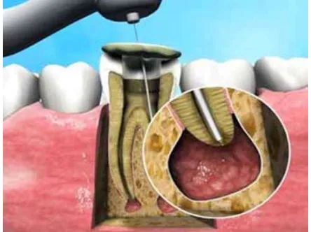
A #20 K-file is passed through the apical foramen and beyond the apex to verify patency. It is followed by a rotary #30 file that is passed 1 mm beyond the apical foramen, creating a passage with a 330µm diameter. The Apexum NiTi Ablator is then inserted, while encased in its sheath, to the working length as established at the cleaning and shaping stage. The sheath is stabilized to the occlusal surface of the tooth using glass-ionomer cement. The Nitinol filament is then pushed manually through the enlarged apical foramen and into the periapical tissues. The NiTi Ablator is then rotated in the periapical tissues for 30seconds at 200 to 250 rpm to initially mince the tissue.
The stabilizing glass-ionomer cement is then removed and the NiTi Ablator withdrawn from the root canal with its sheath to examine it for any mechanical damage or missing parts. The
root canal is then rinsed with sterile saline, and the Apexum PGA Ablator is manually inserted through the root canal and into the periapical tissues. It is then connected to a low-speed contra-angle hand piece and rotated for 30 second sat 5,000 to 7,000 rpm to turn the minced tissues into a thin suspension. Next, it is withdrawn from the root and examined for any
mechanical damage or missing parts (Metzger et al., 2009;
Metzger et al., 2009).The tissue suspension is now washed out with sterile saline solution. The cross-sectional area between the enlarged apical foramen and the outer surface of the needle is 3.4 times larger than that of the needle’s lumen. This facilitated an unobstructed backflow and prevented pressure buildup in the periapical crypt. The Apexum procedure is usually performed under local anesthesia, provided in a manner similar to that used for tooth extraction or surgical intervention. With some experience, it generally takes an additional 7 to 10minutes compared with a conventional root
canal treatment (Metzger et al., 2009; Metzger et al.,
[image:3.595.323.545.284.450.2]2009)(Fig 1).
Figure 1. Apexum procedure
Advantages
Removal and debulking of periapical lesions without using
scalpels, periosteal elevators or sutures
Minimal invasive technique.
Removes chronically inflamed periapical lesions through
root canal access.
Overcome the drawbacks of the conventional surgical
procedure.
Enhanced healing kinetics of periapical lesions (Metzger
et al., 2009; Metzger et al., 2009).
No events of severe postoperative pain or swelling
(Metzger et al., 2009).
Gentle on patient wellbeing.
The procedure does not require surgical skills.
9.The Apexumprocedure is more likely to eliminate
bacterial biofilm or at least mechanically disturb them to the extent of disrupting the host-bacteria equilibrium in favour of the host.
Disadvantages
of the apical foramen to form a passage of 330µm diameter may lead to softened guttapercha extruding beyond the apex. Management of procedural errors (instrument separation) is a matter of concern (Metzger et al., 2009; Raisingani, 2011).
Indications
Chronic apical periodontitis
As a supplement to conventional RCT for teeth with periapical lesion (David Shamah, 2008).Treat infection while the root canal is in progress (Raisingani, 2011).
Contraindications
There is no absolute contraindication in using this procedure.
Clinical applications in Endodontics
With conventional procedures treating the periapical pathology is difficult and time taking. Surgical approach on the other hand may cause lot of trauma and sometimes it may be very difficult to convince the patient for surgery. Performing apical surgery on every case with a periapical lesioncan hardly be justified because surgery has repercussions on the well-being of the patient; swelling, pain and discomfort are among the expected side effects (Kvist, 2000). Furthermore, many anatomic locations preclude apical surgery either because of inaccessibility or risk to adjacent structures. In an extensive study, Ørstavik concluded that (Ørstavik, 1996) at 6 months, only 50% of the cases that eventually healed showed clear signs of healing (advanced and complete healing), and (Friedman, 2002) at 12 months, 88% of the lesions that eventually healed showed clear signs of healing (Friedman, 2002). This may imply that a case should ideally be followed for 12 monthsbefore the tooth may be considered a safe abutment. A clinical trial conducted concluded that the Apexum procedure resulted in no events of severe postoperative pain or postoperative swelling and in only a few events of postoperative discomfort or mild pain(9%).While conventional root canal treatment resulted in some discomfort or pain for 31% of the cases. It is also important to note that when a conventional, open-flap, apical surgery is performed, many of the patients do experience pain, swelling orboth and usually need analgesics in the days after surgery.
Concern over the controversial issue of widening of the apical foramen to form a passage of 330µm diameter has also been expressed. But there is ample demonstration in the anatomic literature that the apical part of root canals is wider than 350 to 400 μm in normal adult teeth and larger when resorbing apical periodontitis has developed. The apical constriction and the apical foramen itself may harbor bacterial biofilms that may be left untouched by the limited apical preparations. The foramen may become even wider when apical resorption takes place, as is often encountered in roots with apical periodontitis. Many of the current nickel-titanium rotary file systems limit the instrumentation at this area to diameters of 250 to 300 µm while avoiding any preparation in the constriction of the apical foramen itself. The device enters the periapical lesion far beyond the apical foramen, a process expected by many operators to result in a flare-up or severe symptoms (Friedman, 2002; Siqueira, 2005). A controversy is associated with this
concept too as many authors feel that the insertion of an endodontic instrument far beyond the apical foramen should be avoided by all means because it is likely to cause a“flare-up,” a painful exacerbation of the periapical inflammatory process but it should be noted that the Apexum procedure is substantially different from simple over instrumentation during root canal treatment. The last traumatizes the tissue and may also introduce bacterial antigens into a tissue containing immunoglobulin's directed against these antigens and that is primed to respond to them. Till date two studies related to the field of endodontic have been carried out. In one the authors concluded that there was enhancement of healing kinetics of periapical lesions in dogs when the Apexum procedure was
carried out (Metzger et al., 2009).In the second study the
Apexum procedure was applied, as a supplementary step, during conventional root canal treatment in 48teeth with periapical lesions. Safety and efficacy were clinically and radiographically evaluated. The result of the study showed that no adverse events occurred in the Apexum-treated group and the healing kinetics was significantly enhanced (Metzger et al., 2009).
Uses in other fields of dentistry in Implantology
When a broken-down tooth with a largeperiapical lesion has to be extracted and replaced by an implant, the implantologist is presented with a dilemma: if there is no bone defect around the apex, an immediate implant could be successfully placed. However, when there is a large periapical lesion and no bone to engage the implant’s apical part, augmentation will be required which becomes either a long and expensive story or a compromised procedure. Suchbone augmentation is provided within a relatively short time by the Apexum procedure (Raisingani, 2011).
In oral surgery
In case of management of cysts, the decompression and aspiration irrigation techniques can be used when there is drainage of cystic fluid from the canals. These techniques act by decreasing the hydrostatic pressure within the periapical lesions. When there is no drainage of fluid from the canals, calcium hydroxide, triple antibiotic paste and apexum procedure can prove beneficial (Bansal et al., 2013).
Future prospective and Conclusion
REFERENCES
Al-Kandari, A.M., Al-Quoud, O.A. and Gnanasekhar, J.D. 1994. Healing of large periapical lesions following nonsurgical endodontic therapy: Case reports. Quintessence Int., 25:115-9.
Andreasen, J.O., Munksgaard, E.C. and Bakland, L.K. 2006. Comparisonof fracture resistance in root canals of immature sheep teethafter filling with calcium hydroxide or MTA. Dent Traumatol, 22:154-6.
Bansal, R., Khursheed, I. and Bansal, T. 2013. Endodontic
Management of a Periapical Cyst- A Review. J Adv Med
Dent Scie., 1(1);7-16.
Baumgartner, J.C., Rosenberg, P.A., Hoen, M.M. and Lin, L.M. 2008. Treatment of endodontic infections,cysts, and flare-ups. In: Ingle, JI Bakland, LK Baumgartner, eds. JC Ingle’s Endodontics. 6th ed. Hamilton, Canada: BC Decker, 690 –712
Bhaskar, S.N. 1972. Nonsurgical resolution of radicular cysts. Oral Surg Oral Med Oral Pathol., 34:458-68.
Brondum, N. and Jensen, V.J. 1991. Recurrence of keratocysts
and decompression treatment. Oral Surg Oral Med Oral
Pathol, 72:265-9.
Çalişkan, M.K. 2004. Prognosis of large cyst-like periapical lesions following nonsurgical root canal treatment: A clinical review. Int Endod J., 37:408-16.
Çalişkan, M.K. and Şen, B.H. 1996. Endodontic treatment of teeth with apical periodontitis using calcium hydroxide: A long-term study. Endod Dent Traumatol12:215-21
Çalişkan, M.K. and Türkün, M. 1997. Periapical repair and apical closure of a pulpless tooth using calcium hydroxide. Oral Surg Oral Med Oral Pathol, 84:683-7.
Colquhoun, N.K. 1969. Treatment of large periapical lesions by an indwelling tube. J. Br. Endod Soc., 3:14-6.
David Figdor. 2002. Apical periodontitis: A very prevalent problem. Oral Surg Oral Med Oral Pathol, 94:651-2. David Shamah. 2008. Apexum goes to the root of the problem.
Israel.http://israel21c.org
Doyon, G.E., Dumsha, T. and von Fraunhofer, J.A. 2005. Fracture resistance of human root dentin exposed to intracanal calcium hydroxide. J Endod., 31:895-7.
Fernandes, M. and Ataide, I. Non-surgical management of a large periapical lesion using a simple aspiration technique: A case report. Int. Endod J., 2010;43:536-42
Freedland, J.B. 1970. Conservative reduction of large
periapical lesions. Oral Surg Oral Med Oral Pathol.,
29:455-64.
Friedman, S. 2002. Prognosis of initial endodontic treatment. Endod Topics., 2:59–88.
Ghose, L.J., Baghdady, V.S. and Hikmat, B.Y. 1987. Apexification of immature apices of pulpless permanent
anterior teeth with calcium hydroxide. J Endod., 13:
285-90.
Hoen, M.M., LaBounty, G.L. and Strittmatter, E.J. 1990. Conservative treatment of persistent periradicular lesions using aspiration and irrigation. J. Endod., 16:1.82-6. Hoshino, E. and Takushige, T. 1998. LSTR 3Mix-MP method-
better and efficientclinical procedures of lesion sterilization and tissue repair (LSTR)therapy. Dent Rev., 666:57-106 Kenneth M. Hargreaves,Stephen Cohen. Microbiology and
treatment of endodontic infections.In:JOSÉ F. SIQUEIRA, JR and ISABELA N. RÔÇAS, editors. Cohen’s pathway of
the pulp 10th ed.pg 560-600.
Kvist, T. and Reit, C. 2000. Postoperative discomfort associated with surgical and nonsurgical endodontic retreatment. Dental Traumatol,16:71– 4.
Loushine, R.J., Weller, R.N., Bellizzi, R. and Kulild, J.C. 1991. A 2-day decompression: A case report of a maxillary first molar. J Endod., 17:85-7.
Martin, S.A. 2007. Conventional endodontic therapy of upper central incisor combined with cyst decompression: A case report. J Endod., 33:753-7.
Mejia, J.L., Donado, J.E. and Basrani, B. 2004. Active non-surgical decompression of large periapical lesions- 3 case reports. J Can Dent Assoc, 70:691-4.
Metzger, Z., Huber, R., Slavesscu, D., Dragomirescu, D., Tobis, I. and Better, H. 2009. Healing kinetics of periapical lesions enhanced by the Apexum procedure:A clinical trial. J Endod., 35:153-9.
Metzger, Z., Huber, R., Tobis, I. and Better, H. 2009. Enhancement of healing kinetics of periapical lesions in dogs by the Apexum procedure. J Endod., 35 :40-5. Ørstavik, D. 1996. Time course and risk analysis of the
development and healing of chronic apical periodontitis in man. Int Endod J., 29:150 –5.
Patterson, S.S. 1964. Endodontic therapy: Use of a polyethylene tube and stint for drainage. J Am Dent Assoc., 69:710-4.
Raisingani, D. 2011. Apexum: A minimum invasive procedure. Int J Clin Ped Dent., 4(3): 224-7.
Rees, J.S. 1997. Conservative management of a large maxillary cyst. Int Endod J., 30:64-7.
Sato, I., Kurihara- Ando, N., Kota, K., Iwaku, M. and Hoshino, E. 1996. Sterilizationof infected root- canal dentine by
topical application of a mixture ofciprofloxacin,
metronidazole and minocycline in situ. Int Endod J.,
29:118-24.
Siqueira, J. 2005. Reaction of periradicular tissues to root canal
treatment: benefits and drawbacks. Endod Topics;10:123–
47.
Sommer, R.F., Ostrander, F.D. and Crowley, M.C. 1964. Clinical Endodontics. 2nd, ed. Philadelphia, USA: W.B. Saunders and Co..