HISTOPATHOLOGICAL SPECTRUM OF SKIN LESIONS
Dr. Sheela L Gaikwad, *Dr. Uddhav D Kumawat
Department of Pathology, SR
ARTICLE INFO ABSTRACT
Introduction:
to a variety of disease
range of diseases comprising of inflammatory disorders to highly malignant neoplasms.
Material
oral & nasal mucosa and tongue, are studied ret Ramanand
Results:
period. Patient’s age ranged from 7 years to 80 years with maximum numbers of patients were from age group of 31 to 40 years
outnumbered non
common benign and malignant neoplastic lesions respectively. Non was most common non
Conclusion:
histopathological examination combined with awareness in mind regarding prevalence of diverse skin lesions in rural population can help in correct diagnosis.
Copyright©2016, Dr. Sheela L Gaikwad et al., This
unrestricted use, distribution, and reproduction in any medium, provided the original work is properly cited.
INTRODUCTION
The skin is the largest organ of the integumentary system in human. The integumentary system constitutes the skin (integument) together with its accessory organs (hair, glands, and nails). The skin acts as a buffer against the external environment and thus is more vulnerable to a variety of diseasecausing microorganisms and physical assaults. The skin is affected by wide range of diseases comprising of inflammatory disorders to highly malignant neoplasms
(Abubakar et al., 2016). Skin lesions can be classified as
following: Disorders of Pigmentation and Melanocytes, Benign Epithelial Tumours, Premalignant and Malignant Epidermal Tumours, Tumours of the Dermis, Tumours of Cellular Immigrants to the Skin, Disorders of Epidermal
Maturation, Acute Inflammatory Dermatoses, Chronic
Inflammatory Dermatoses, Blistering (Bullous) Diseases, Disorders of Epidermal Appendages, Panniculitis, Infection
and Infestation (Kumar et al., 2014). The WHO Classification
of skin tumours editorial and consensus confer
*Corresponding author: Dr. Uddhav D Kumawat,
Department of Pathology, SRTR, GMC, Ambajogai, Maharashtra, India.
ISSN: 0975-833X
Article History:
Received 17th May, 2016
Received in revised form 23rd June, 2016
Accepted 14th July, 2016
Published online 20th August,2016
Key words:
Histopathology, Skin lesions, Neoplastic lesions.
Citation: Dr. Sheela L Gaikwad, Dr. Uddhav D Kumawat,
lesions experience at rural based hospital”, International Journal of Current Research
RESEARCH ARTICLE
GICAL SPECTRUM OF SKIN LESIONS-EXPERIENCE AT RURAL BASED HOSPITAL
Dr. Uddhav D Kumawat, Dr. Nagsen A Sakhare and
of Pathology, SRTR, GMC, Ambajogai, Maharashtra, India
ABSTRACT
Introduction: The skin acts as a buffer against the external environment and thus is more vulnerable to a variety of diseasecausing microorganisms and physical assaults. Th
range of diseases comprising of inflammatory disorders to highly malignant neoplasms.
Material & Methods: All the skin lesion received from Jan 2016 to June 2016, excluding those of oral & nasal mucosa and tongue, are studied retrospectively in the dept. of Pathology at Swami Ramanand Teerth Rural Government Medical College, Ambajogai.
Results: Total 113 (10%) skin lesion specimens out of 1130 specimens were
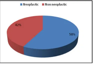
period. Patient’s age ranged from 7 years to 80 years with maximum numbers of patients were from age group of 31 to 40 years and with male predominance (M:F= 2.1:1). Neoplastic (58%) lesions outnumbered nonneoplastic ones (42%). Epidermal cysts and squamous cell carcinoma were the most common benign and malignant neoplastic lesions respectively. Non
was most common nonneoplastic lesion.
Conclusion: Diversity of skin lesions were obtained among rural pop
histopathological examination combined with awareness in mind regarding prevalence of diverse skin lesions in rural population can help in correct diagnosis.
This is an open access article distributed under the Creative Commons Att use, distribution, and reproduction in any medium, provided the original work is properly cited.
The skin is the largest organ of the integumentary system in human. The integumentary system constitutes the skin (integument) together with its accessory organs (hair, glands, and nails). The skin acts as a buffer against the external onment and thus is more vulnerable to a variety of causing microorganisms and physical assaults. The skin is affected by wide range of diseases comprising of inflammatory disorders to highly malignant neoplasms can be classified as following: Disorders of Pigmentation and Melanocytes, Benign Epithelial Tumours, Premalignant and Malignant Epidermal Tumours, Tumours of the Dermis, Tumours of Cellular Immigrants to the Skin, Disorders of Epidermal Inflammatory Dermatoses, Chronic Inflammatory Dermatoses, Blistering (Bullous) Diseases, Disorders of Epidermal Appendages, Panniculitis, Infection 2014). The WHO Classification of skin tumours editorial and consensus conference in Lyon,
Dr. Uddhav D Kumawat,
Department of Pathology, SRTR, GMC, Ambajogai, Maharashtra,
France classified skin tumours into: Keratinocytic Tumours,
Melanocytic Tumours, Appendageal Tumours,
Haematolymphoid Tumours, Soft Tissue Tumours, Neural Tumours, and Inherited Tumour Syndromes. As per WHO Classification of tumours the lifetime risk for the development of skin cancer in the USA is now 1 in 5 (LeBoit
melanoma skin cancers (NMSC) constitute maj
cancer in Caucasians with increasing incidence. Among NMSC, Basal cell carcinoma is most common (75%) followed by Squamous cell carcinoma (
2012). In Caucasians, annual increase in the incidence rate of melanoma has been approximately 3
(Erdei and Torres, 2010). Epithelial cysts of the skin form heterogenous group among which epidermal cysts are commonest (Warvi and Gates,
disorders in this research at department of pat provide essential data of skin lesions in rural area.
MATERIALS AND METHODS
This is a six month retrospective study of skin lesions carried out at the Department of Pathology, Swami Ramanand Rural Government medical college,
to June 2016. All the lesions related to skin and subcutaneous International Journal of Current Research
Vol. 8, Issue, 08, pp.36223-36227, August, 2016
INTERNATIONAL
Dr. Sheela L Gaikwad, Dr. Uddhav D Kumawat, Dr. Nagsen A Sakhare and Dr. Grace F D’costa,2016.
International Journal of Current Research, 8, (08), 3622336227.
EXPERIENCE AT RURAL BASED HOSPITAL
and Dr. Grace F D’costa
TR, GMC, Ambajogai, Maharashtra, India
The skin acts as a buffer against the external environment and thus is more vulnerable causing microorganisms and physical assaults. The skin is affected by wide range of diseases comprising of inflammatory disorders to highly malignant neoplasms.
All the skin lesion received from Jan 2016 to June 2016, excluding those of rospectively in the dept. of Pathology at Swami Teerth Rural Government Medical College, Ambajogai.
Total 113 (10%) skin lesion specimens out of 1130 specimens were received during study period. Patient’s age ranged from 7 years to 80 years with maximum numbers of patients were from and with male predominance (M:F= 2.1:1). Neoplastic (58%) lesions ermal cysts and squamous cell carcinoma were the most common benign and malignant neoplastic lesions respectively. Nonspecific chronic inflammation
Diversity of skin lesions were obtained among rural population in this study. Meticulous histopathological examination combined with awareness in mind regarding prevalence of diverse skin
access article distributed under the Creative Commons Attribution License, which permits
France classified skin tumours into: Keratinocytic Tumours,
Melanocytic Tumours, Appendageal Tumours,
Tumours, Soft Tissue Tumours, Neural Tumours, and Inherited Tumour Syndromes. As per WHO Classification of tumours the lifetime risk for the development of skin cancer in the USA is now 1 in 5 (LeBoit, 2006). Non melanoma skin cancers (NMSC) constitute major form of cancer in Caucasians with increasing incidence. Among NMSC, Basal cell carcinoma is most common (75%) followed by Squamous cell carcinoma (Samarasinghe V & Madan V, . In Caucasians, annual increase in the incidence rate of approximately 3–7% per year worldwide . Epithelial cysts of the skin form heterogenous group among which epidermal cysts are , 1943). We studied various skin disorders in this research at department of pathology aiming to provide essential data of skin lesions in rural area.
MATERIALS AND METHODS
This is a six month retrospective study of skin lesions carried out at the Department of Pathology, Swami Ramanand Teerth Rural Government medical college, Ambajogai from Jan 2016 to June 2016. All the lesions related to skin and subcutaneous INTERNATIONAL JOURNAL OF CURRENT RESEARCH
tissue are included in this study. Lesions of oral mucosa, tongue and nasal mucosa are excluded form study. Demographic data such as age, sex and site of the biopsies were obtained from patients’ histopathology request cards and histopathology registers. Stained Haematoxylin and Eosin (H&E) slides were reviewed. Whenever needed, slides were stained with special stains.
RESULTS
[image:2.595.313.554.52.165.2]Total 113 (10%) skin lesion specimens out of 1130 specimens were received in histopathology section from January 2016 to June 2016. Patient’s age ranged from 7 years to 80 years with mean and median age being 44.1 and 40 respectively. Maximum numbers of patients were from age group of 31 to 40 years.
[image:2.595.306.563.195.413.2]Figure 1. Bar diagram showing age wise distribution of skin lesions among males and females (n=113)
Figure 2. Pie diagram showing frequency distribution of neoplastic and non-neoplastic lesions (n= 113)
[image:2.595.38.290.249.392.2]Figure 3. Bar diagram showing sex wise distribution of neoplastic and non-neoplastic lesions (n=113)
Figure 4. Pie diagram showing keratinocytic malignancies
Figure 5. 5A. Basaloid squamous cell carcinoma (H&E x100). Nuclei palisade at the periphery of basaloid nests. B. Malignant melanoma (H&E x400). Showing sheets of malignant melanocytes with hyperchromatic nuclei and prominent nucleoli
[image:2.595.70.256.425.555.2] [image:2.595.308.563.475.687.2] [image:2.595.38.286.591.733.2]Figure 7. A. Congenital nevus (H&E x100). Nevus cells are deep within reticular dermis and in close association with appendages. B. Nevus sebaceous (H&E x100). There is epidermal hyperplasia, hyperplastic sebaceous glands and immature hair follicle
[image:3.595.40.285.52.237.2]Figure 8. A. Intradermal nevus (H&E x400). Nests of nevus cells are present within the dermis. B. Actinomycetoma (H&E x400). Colonies of filamentous bacteria rimmed by eosinophilic material and is surrounded by dense neutrophilic reaction
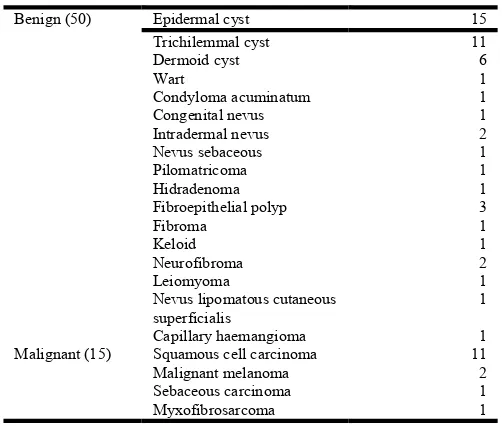
Table 1. Neoplastic lesions (n=65)
Benign (50) Epidermal cyst 15
Trichilemmal cyst 11
Dermoid cyst 6
Wart 1
Condyloma acuminatum 1
Congenital nevus 1
Intradermal nevus 2
Nevus sebaceous 1
Pilomatricoma 1
Hidradenoma 1
Fibroepithelial polyp 3
Fibroma 1
Keloid 1
Neurofibroma 2
Leiomyoma 1
Nevus lipomatous cutaneous superficialis
1
Capillary haemangioma 1
Malignant (15) Squamous cell carcinoma 11
Malignant melanoma 2
Sebaceous carcinoma 1
Myxofibrosarcoma 1
Table 2. Non neoplastic lesions (n=48)
Inflammatory (27) Nonspecific chronic inflammatory lesion 22
Granulomatous inflammation 2
Necrotising granulomatous inflammation 1
Folliculitis 1
Chronic abscess 1
Infectious (4) Actinomycosis 1
Mycetoma 2
Eumycetoma 1
Other(17) Sebaceous hyperplasia 2
Calcified cysts 3
Mucous cyst 1
Corn 3
Idiopathic calcinosis of scrotum 2
Calcinosis cutis 4
Pseudoepitheliomatous hyperplasia 1
No specific pathology 1
In that 77 specimen were from male and 36 from female with male to female ratio being 2.1:1 (Fig. 1). Neoplastic lesions of skin were the major lesions that we got in our study (Fig.2, Table 1). In that, benign lesions were maximum with Epidermal cysts contributing higher proportion. Among malignant cases squamous cell carcinoma was the predominant malignancy followed by malignant melanoma. In 11 squamous cell carcinoma 4 were its variants accordingly two Basaloid squamous cell carcinoma, one Spindle squamous cell carcinoma and one Verrucous carcinoma (Fig.4). Inflammatory lesions of epidermis and dermis were the second most lesions following neoplastic lesions (Table 2). Most of inflammatory lesions were reported as nonspecific chronic inflammation. One lesion was given as necrotising granulomatous inflammation which on ZiehlNeelsen staining was negative for acid fast bacilli. We reported 6 adnexal lesions in our study. In this 4 were of sebaceous differentiation, 1 of eccrine and 1 with follicular differentiation. We reported two lesions as Sebaceous hyperplasia and placed in nonneoplastic category.
DISCUSSION
Skin lesion prevalence in general population varies from 6.3%
to 11.16% (Grover et al., 2008; Gangadharan et al., 1976;
Dayal and Gupta, 1977; Kuruvilla et al., 2000; Rao and Kumar
2003; Das, 2002; Devi and Zamzachin, 2006; Pradeep Nair and Gopalakrishnan Nair, 1999), our result of skin lesion prevalence fall within this range. We found male predominance of skin lesions in our study (Fig.1). It is comparable to findings of Dayal and Gupta (1977), Grover
et al. (2008) and Rao and Kumar (2003) where they noticed
male predominance. This male predominance again
encountered on classifying these lesions into neoplastic and nonneoplastic category (Fig.3). Vaghela and Jha, (2016) also found male predominance among nonneoplastic lesions. In
consistent with observations of Abubakar et al. (2016), we also
[image:3.595.306.560.72.228.2] [image:3.595.42.283.292.475.2] [image:3.595.37.288.547.760.2]to released keratin following rupture of cysts. As per Park and Ko (2013), changes in wall of the cysts wall are indicative of rupture of cysts which we got in the form of foreign body reaction. In contrast to epidermal cysts, the wall of Trichilemmal cyst shows characteristic absence of granular layer of epidermis and no clearly visible intercellular bridges with abrupt keratinisation. In our study, all 11 Trichilemmal cysts showed typical histomorphology without signs of rupture. Proliferating Trichilemmal cysts can also show trichilemmal type of keratinisation, but these are usually solid with tumor like proliferation which was not present in our case
(Lever, 2005; Ramaswamy et al., 2013).
Squamous cell carcinoma was the predominant malignancy in our study (Table 1) which is comparable to findings of
Abubakar et al. (2016), Brand and Ackerman (2000) and
Wassberg et al. (2001). Among proper squamous cell
carcinoma, four were well differentiated and 3 were moderately differentiated. Degree of anaplasia in tumor and invasion into underlying dermis helped us to grade these tumors. Grading is essential as features of deep invasion, poor differentiation and associated perineural invasion give tumor more chances of recurrence or metastasis (LeBoit, 2006). Basaloid squamous cell carcinoma is a variant of squamous cell carcinoma with aggressive behaviour which was first
described by Wain et al., Its most common site is
aerodiagestive tract in head and neck region (Wain, et al.,
1986; Ereño et al., 2008). In our study, we got two basaloid
squamous cell carcinoma (Fig.5A); one at lateral canthus of eye and other at occipital region. Upon searching in literature, male predominance is evident in basaloid squamous cell
carcinoma (Ereño et al., 2008) but in our study both were
females above 60 years. Spindle squamous cell carcinoma is an unusual variant of squamous cell carcinoma. Head and neck region is the common site for this malignancy. Though male predominance is observed in literature for this lesion
(Viswanathan et al., 2010), in our case we observed it in a 72
year female (Fig.6B). Viswanathan et al., (2010) and
Thompson et al., (2002) noted median age for this lesion as 53
and 66 respectively. Intermittent exposure to ultraviolet rays (UVR) is the major environmental risk factor for melanoma, especially in combination with endogenous factors (immune deficient status, genetic predisposition). It predominantly affects adults and elderly patients, with a peak of incidence around the sixth decade of life. Sun exposed parts are commonly affected by it(LeBoit PE, 2006). In this study, two malignant melanoma lesions were present in males with age being 60 and 65. Both these lesions were present on foot (Fig.5B). Among inflammatory lesions of skin, nonspecific
chronic inflammation was predominant in which
histopathology showed epidermis with variable acanthosis, presence of lymphoplasmacytic infiltration of underlying dermis and absence of pathologic organism. One 35 year male patient presented with multiple swelling over scalp region which upon histopathologic examination turned out to be
folliculitis. In this lesion inflammatory cells are
present within the wall and ostia of the hair follicle (Lever WF, 2005). One lesion was sebaceous carcinoma (Fig.6A). It was present in a 60 year female on upper eyelid. Eyelid is commonly affected by it. Poorly differentiated sebaceous carcinoma is susceptible to misdiagnosis because of its
similarities with squamous cell carcinoma (Pereira et al.,
2005). As per literature, it commonly present in old age though
it is also reported in younger age (Pereira, 2005; Sung et al.,
2011). Sebaceous hyperplasia is characterised by enlargement of single sebaceous gland composed of numerous lobules which are arranged around wide sebaceous duct. It commonly affect facial region. It has been reported in association with MuirTorre syndrome which consist of visceral malignancies, sebaceous neoplasm and keratoacanthoma. We got two lesions as sebaceous hyperplasia; one was at lateral angle of mouth and other over nose. Still alone presence of sebaceous hyperplasia without associated neoplasms does not predispose to cancer or represent sign of Muir Torre syndrome (Lever, 2005). Intradermal nevi are one of the types of acquired melanocytic nevi without junctional activity. These usually consist of nests and cords of nevus cells in upper dermis. Two lesions of this study was intradermal nevus present in females (Fig. 8A). In our case there was no hyperkeratosis and papillomatosis, which less commonly may presents in these lesions and resemble as seborrheic keratosis of epidermis (Lever, 2005). Congenital melanocytic nevus (CMN) is benign cutaneous melanocytic neoplasm which presents since birth. These are characterised by presence of nevus cells around or in association with hair follicle, erector pili, sebaceous glands and sweat ducts (Lever, 2005). These nevi are classified according to their gross size into small (<1.5 cm), medium (1.519.9 cm) and large or giant (>20 cm). Giant CMN have increased risk of malignant transformation and need referral to specialist (Nikfarjam and Chambers, 2016). We reported one lesion as CMN in an 18 year female which was of medium size (6 cm) and present since birth (Fig. 7A). Nevus sebaceous is a hamartomatous lesion which predominantly composed of sebaceous glands. It was first described by Jadassohn in 1885.
Usually it is solitary and presents as warty growth. Simi et al.
(2008) studied 21 cases of nevus sebaceous in which they found female predominance and maximum patients were in the range of 2130 years. One female patients of 40 years age in our study presented with warty growth on nose which on microscopy came as nevus sebaceous (Fig.7B). We reported one lesion as Nevus Lipomatous Cutaneous Superficialis in a 40 years male which is a rare disease characterised by groups of ectopic fat cells in the papillary or reticular dermis. It was first described by Haffman and Zurhelle in 1921. It occurs in two clinical forms. The multiple form (classic type) is characterized by multiple soft nontender skincolored or yellow papules, nodules, or plaques that usually develop shortly after birth or during the first two decades of life. It usually develops shortly after birth or during the first two decades of life. The solitary form presents after the second
decade of life (Goldblum et al., 2013). We reported three cases
as actinomycetoma. All three were male presented with discharging sinuses over foot. In these, lesions composed of filamentous bacteria surrounded by neutrophils, plasma cells and foreign body giant cell reaction (Fig. 8B). One lesions was diagnosed as Eumycetoma in which pathologic organism was septate with hyphae. Mycetoma is a chronic infection of skin and subcutaneous tissue, characterised by discharging sinuses and presence colonial grains in the exudate. It is classified as Actinomycotic Mycetoma and eumycotic Mycetoma in which first one is caused by filamentous bacteria while fungus is
(actinomycetoma) or Gomori methenamine silver or periodic acidSchiff stains (eumycetoma) can differentiate these lesions, meticulous histopathological examination can also be
helpful in diagnosis (Alam et al., 2009). To conclude, diversity
of skin lesions we got among rural population in this study. These lesions were present predominantly in patients with age more than 30. Neoplastic skin lesions were the major entity, with epidermal cysts being the most common lesions. Though less in number, nonneoplastic lesions also were noticeable. So meticulous histopathological examination combined with awareness in mind regarding prevalence of diverse skin lesions in rural population can help in correct diagnosis.
Conflicts of interest: None
Acknowledgement: The author thanks Dr. Sheetal Khadse for
her contribution to the study. No sponsors or institutional grants were obtained for this study. This study has not been published previously in any journal.
REFERENCES
Abubakar SD, Tangaza AM, Sahabi SM, Legbo JN. 2016.
Histopathological pattern of skin lesions in
UsmanuDanfodiyo University Teaching Hospital Sokoto, Nigeria.6:1015.
Alam K, Maheshwari V, Bhargava S, Jain A, Fatima U, ulHaq E. 2009. Histological diagnosis of madura foot
(mycetoma): a must for definitive treatment. Journal of
Global Infectious Diseases. 1(1):64.
Brand D, Ackerman AB. 2000. Squamous cell carcinoma, not basal cell carcinoma, is the most common cancer in
humans. Journal of the American Academy of
Dermatology, 42(3):5236.
Das K. 2002. Pattern of dermatological diseases in Gauhati
Medical College and Hospital Guwahati. Indian Journal of
Dermatology, Venereology and Leprology., 69(1):168.
Dayal S. and Gupta G. 1977. A cross section of skin diseases
in Bundelkhand region, UP. Indian Journal of
Dermatology, Venereology, and Leprology, 43(5):258.
Devi TB, Zamzachin G. 2006. Pattern of skin diseases in
Imphal. Indian journal of Dermatology, 51(2):149
Erdei E. and Torres SM. 2010. A new understanding in the
epidemiology of melanoma. Expert Rev Anticancer Ther.,
Nov; 10(11): 181123. doi: 10.1586/era.10.170.Review. PubMed PMID: 21080806; PubMed Central PMCID: PMC3074354
Ereño C, Gaafar A, Garmendia M, Etxezarraga C, Bilbao FJ, López JI. 2008. Basaloid Squamous Cell Carcinoma of the
Head and Neck. Head and Neck Pathology, 2(2):8391.
Gangadharan C, Joseph A, Sarojini P. 1976. Pattern of skin
diseases in Kerala. Indian Journal of Dermatology,
Venereology, and Leprology, 42(1):49.
Goldblum JR, Weiss SW, Folpe AL. 2013. Enzinger and Weiss's Soft Tissue Tumors. 6th ed. Elsevier Health Sciences.
Grover S, Ranyal RK, Bedi MK. 2008. A cross section of skin
diseases in rural Allahabad. Indian Journal of
Dermatology, 53(4):179.
Kumar V, Abbas AK, Aster JC. 2014. Robbins & Cotran Pathologic Basis of Disease ElsevieronVitalSource: Elsevier Health Sciences.
Kuruvilla M, Sridhar K, Kumar P, Rao G. 2000. Pattern of
skin diseases in BantwalTaluq, Dakshina Kannada. Indian
Journal of Dermatology, Venereology, and Leprology,
66(5):247.
LeBoit PE. 2006. Pathology and Genetics of Skin Tumours: IARC Press.
Lever WF, Elder DE. 2005. Lever's Histopathology of the Skin: Lippincott Williams & Wilkins.
Nikfarjam J, Chambers E. 2016. Congenital melanocytic nevi and the risk of malignant melanoma: establishing a
guideline for primarycare physicians. Einstein Journal of
Biology and Medicine, 27(2):5966.
Park JS. and Ko DK. 2013. A histopathologic study of epidermoid cysts in Korea: comparison between ruptured
and unruptured epidermal cyst. International Journal of
Clinical and Experimental Pathology, 6(2):242.
Pereira PR, Odashiro AN, Rodrigues‐Reyes AA, Correa ZMS, De Souza Filho JP, Burnier MN. 2005. Histopathological
review of sebaceous carcinoma of the eyelid. Journal of
Cutaneous Pathology, 32(7):496501.
Pradeep Nair S, Gopalakrishnan Nair T. 1999. Pattern of
dermatological diseases in Trivandrum. Indian Journal of
Dermatology Venereology and Leprology, 65(6):2613
Ramaswamy AS, Manjunatha HK, Sunilkumar B, Arunkumar
SP. 2013. Morphological spectrum of pilar cysts. North
American Journal of Medical Sciences, 5(2):124.
Rao G, Kumar S. 2003. Pattern of skin diseases in an Indian
village. Indian Journal of Medical Sciences, 57(3):108
Samarasinghe V. and Madan V. 2012. Nonmelanoma skin
cancer. J CutanAesthet Surg., 5:3–10
Simi C, Rajalakshmi T, Correa M. 2008. Clinicopathologic analysis of 21 cases of nevus sebaceus: a retrospective
study. Indian Journal of Dermatology, Venereology, and
Leprology., 74(6):625.
Sung D, Kaltreider SA, GonzalezFernandez F. 2011. Early
onset sebaceous carcinoma. Diagnostic Pathology, 6(1):1.
Thompson LD, Wieneke JA, Miettinen M, Heffner DK. 2002. Spindle cell (sarcomatoid) carcinomas of the larynx: a
clinicopathologic study of 187 cases. The American
Journal of Surgical Pathology, 26(2):15370.
Vaghela PG, Jha BM. 2016. Histomorphological analysis of
nonneoplastic skin lesions. International Journal of
Medical Science and Public Health, 5(4):63841.
Viswanathan S, Rahman K, Pallavi S, Sachin J, Patil A,
Chaturvedi P, et al. 2010. Sarcomatoid (spindle cell)
carcinoma of the head and neck mucosal region: a clinicopathologic review of 103 cases from a tertiary
referral cancer centre. Head and Neck Pathology, 4(4):265
75
Wain SL, Kier R, Vollmer RT, Bossen EH. 1986. Basaloid squamous carcinoma of the tongue, hypopharynx, and
larynx:: Report of 10 cases. Human Pathology,
17(11):115866.
Warvi WN, Gates O. 1943. Epithelial cysts and cystic tumors
of the skin. Am J Pathol., 19:765–83
Wassberg C, Thorn M, Johansson AM, Bergstrom R, Berne B, Ringborg U. 2001. Increasing incidence rates of squamous
cell carcinoma of the skin in Sweden. Acta Derm
Venereol., 81: 268272.