Beneficial effect of reduced oxygen concentration with
transfer of blastocysts in IVF patients older than 40
years old
Javier I. García*, Soledad Sepúlveda, Luis Noriega-Hoces
Laboratory of Assisted Reproduction, Concebir Clinic, Lima, Peru; *Corresponding Author: jgarciaf@hotmail.com Received 6 February 2010; revised 1 March 2010; accepted 5 March 2010.
ABSTRACT
The aim of the present study was to determine the impact of oxygen concentration on implan-tation, pregnancy and delivery rates in IVF pa-tients older than 40 year old with transfer of blastocysts. Included were 558 women aged 23-45 years old undergoing IVF/ICSI procedures whose embryos were cultured at blastocyst stage under two different oxygen environments (a bi-gas system: 5.6% CO2 in air and a tri-gas system: 5.6% CO2, 5% de O2 and 89.4% N2). The main outcome measures of this study are im-plantation, pregnancy and delivery rates. Im-plantation, pregnancy and delivery rates are found to be reduced in women older than 40 years old. The implantation and pregnancy rates are significantly higher in women older than 40 years old from the 5% of O2 group, in compari-son to the 20% group (25.00% versus 2.70% and 41.38% versus 5.56%; P < 0.05). The deliveries rates were 13.79% and 5.56% in the 5% and 20% oxygen groups respectively (P: NS). The birth-weight was similar in both study groups (P: NS). Gestational age was significantly longer in wo- men from the 5% of O2 group, in comparison to the 20% (36.87 versus 35.87 weeks, P < 0.05). Results indicated that the embryonic culture with 5% of oxygen and transfer of blastocysts in women older than 40 years old improve the re-sults in the in Vitro fertilization/intracytoplasmic injection procedures (IVF/ICSI).
Keywords: ART; Blastocyst; IVF; ICSI; Oxygen
1. INTRODUCTION
Embryos from several mammal species, including hu-man, were exposed in vivo to low oxygen concentrations,
ranging from 2 to 8% observed in atmospheric air [1-3]. This probably corresponds to an adaptation mechanism, as it is proven that higher oxygen concentrations may be harmful to the embryo [4] by generating reactive oxygen species (ROS) [5-7].
In Vitro fertilization studies (IVF) in mice [8,9]; cattle
[5]; sheep [10]; rabbits [11]; hamsters [12]; rats [13]; cows [14] and pigs [15] have demonstrated that when cultured in oxygen concentrations of 5% present a higher viability and a better development to the blastocyst stage. However, a pioneer study in human embryos showed that cultures in Vitro in atmospheric concentration (20%) or reduced (5%) resulted in similar fecundation and preimplantational embryo development processes [16]. Therefore, in several laboratories of assisted reproduc-tion, the culture of human embryos using oxygen con-centration of 20% [17] is now a common practice.
Furthermore, a study in which the effect of oxygen over the 2nd and 3rd day of human embryo development was evaluated was unable to find any differences in the pregnancy and implantation rates when 5% or 20% of O2 was used [17]. This absence of differences in the results obtained might be because the beneficial effect of low O2 concentrations happen during the late stages of pre-implantational embryo development (day 4-6) [18]; how- ever, the addition of antioxidants to the culture media had as a result better rates of implantation and pregnancy when embryos cultured in 5% of O2 are transferred in the 2nd and 3rd day [5].
The importance of the transfer in blastocyst stage and the concentration of oxygen have been recently recog-nized [19,20] These studies reported significant in-creases in the pregnancy and implantation rates when transfers were done in blastocyst stage and when the cultures were made with 5% of O2 compared to the cul-ture effect in conditions of 20% of O2.
cause homeostatic stress of the embryo and a reduction in its implantatory potential [21]. Consequently, the tr- ansfer in blastocyst stage would allow a better synchro-nization with rhythm of uterine contractions and the em-bryo [22,23].
However, there are contradictory results in studies where the human embryos cultured in reduced (5%) or atmospheric oxygen concentrations (20%) are compared. Thus, no improvements in terms of pregnancy and im-plantation rates were observed if the transfer was per-formed in the 3rd day of the development in terms of pregnancy and implantation rates [17,20,24]. Similar results have been observed if transfer was done into blastocyst stage (day 5) [24-27].
The maternal age is an important factor to be taken into account in studies on. In fact, there has been a de-crease in the women’s fertility from 35 years old [28,29], being this reduction significant from the 40 year olds and over, in women attending a processes of assisted reproduction [30,31].
The Latin American Registry of Assisted Reproduc-tion (REDLARA) reported in 2006 a clinical pregnancy rate of 39.6% in patients ≤ 34 years old, 32.8% in pa-tients from 35 to 39 years old and 18.6% in women ≥ 40 years old respectively [32]. In older women there is commonly a reduction in the ovarian follicular reserve and a greater prevalence of chromosomal alterations in the oocyte, which lead to a significant reduction in the implantation rates [33,34] and high rates of miscarriages [35,36].
In the studies comparing the effect of different oxygen concentrations in the embryo cultures, it has not been taken into consideration the maternal age impact when blastocysts are transferred in the programs of assisted reproduction [19,25,27,37].
In a recent publication Kovačičet al. [26] did not find improvements in the implantation rates in older women over 40 years of age whose embryos were cultured with oxygen at 5% as compared cultures at 20% of O2 and embryo transfer in the 3rd day. It is possible that effects of low oxygen concentration may be observed if em-bryos are transferred in the 5th or 6th days.
We hypothesize that reducing the percentage of oxy-gen to 5% in the embryo culture systems would have a much more beneficial effect than the usage of oxygen at 20%.
The objective of this study was to evaluate in an IVF/ICSI program, the relationship between the preg-nancy and the implantation rates with maternal age whose embryos were cultured in 5% of O2, compared to those women whose embryos were cultured at 20% of O2. In addition, the results of pregnancies were assessed.
2. MATERIALS AND METHODS
2.1. Patients
This is a retrospective non randomized study based on secondary analysis of data obtained from 558 cycles of IVF and ICSI at the Laboratories of Assisted Reproduc-tion of Pranor Group (Lima, Peru) between January 2007 and June 2009. This study was approved by the Institutional Review Board (IRB) at the Concebir Clinic (Lima, Peru).
The study group were those gametes and embryos cultured at 37˚C in an atmosphere of 5.6% CO2, 5% of O2 and 89.4% N2 (341 cycles); and a control group of those gametes and embryos cultured at 37℃ and an at-mosphere of 5.6% CO2 in air (20% O2) (217 cycles). The same kind of incubators (Thermo Scientific, USA) was used for the bi-gas and tri-gas systems.
2.2. Ovarian Stimulation and Oocyte Collection
The patients were submitted to a controlled ovarian stimulation with Leuprolide Acetate (Lupron®, Abbott Laboratories) or Ganirelix (Orgalutran®, Organon) in combination with Recombinant FSH (Puregon®, Or-ganon Laboratories) or HMG (Humegon®, OrOr-ganon Laboratories) according to the established protocols. The follicular growth was monitored by ultrasound and the ovulation was induced by applying Human Chorionic Gonadotropin (hCG) (Ovidrel® 250 ug, Serono Labora-tories or Pregnyl® 10,000 UI, Organon LaboraLabora-tories). The follicular aspiration was made 34 to 36 hours after giving the hCG. The insemination or ICSI procedure was made-5 hours after the oocyte recovery.
2.3. Semen Samples
The semen samples were obtained by masturbation of every patient’s male in aseptic conditions. After the liq-uefaction process, the motile spermatozoa were recov-ered from the seminal plasma by centrifugation through Isolate gradients of 45% and 95% (Irvine Scientific, USA) for 10 minutes at 300 × g. the recovered sper-matozoa were washed in Sperm Washing Media (Irvine Scientific, USA). In oligospermic samples the sper-matozoa were washed in Sperm Washing Media and then placed in 10 µL drops of HTF-Hepes + 10% SSS for the ICSI.
2.4. Fertilization and Embryo Culture
was approximately 7.30 in all the culture media. The aspired oocytes were washed in a HTF-Hepes medium (IVFonline, Guelph, ON, Canada) supplement- ed with 10% vol/vol of SSS (Irvine Scientific, USA) and cultured in a 200 µL drop of HTF medium + 10% SSS under mineral oil at 37˚C for 5 hours before the insemi-nation or ICSI procedure.
The insemination was made with 50,000-100,000 mo-tile spermatozoa in 200 µL drop of HTF medium + 10% SSS, where from 1 to 5 oocytes were placed. In the cases of ICSI, the oocytes in metaphase II were injected in every patient by using methods previously described (38). After the insemination or ICSI in 0 day, all the oo-cytes were cultured up to the evaluation of the fertiliza-tion at 37˚C.
The fertilization was evaluated 16-18 hours post in-semination or ICSI by the presence of two pronuclei and two polar bodies (day 1). The zygotes with two pronu-clei were cultured individually, under mineral oil, in 10 µL drops of Global medium (IVFonline, Guelph, ON, Canada) supplemented with 10% vol/vol of SSS (Irvine Scientific, USA) from day 1 to day 3. On the 3rd day, the embryos were changed to 10 µL drops of fresh Global medium + 10% SSS and cultured 2 or 3 days more up to the transfer day in blastocyst stage. Therefore, the transfer was made in 5 or 6 days.
2.5. Embryo Transfer
The embryos were transferred in blastocyst stage, being the average of 1.96 and 2 embryos transferred in the group of 5% and 20% of O2 respectively (P < 0.05 among the evaluated groups). In the 5% of O2 group, 16 patients received 1 embryo, 324 received 2 embryos and 1 patient received 3 embryos. In the 20% of O2 group, 4 patients received 1 embryo, 209 patients received 2 em-bryos and 4 patients received 3 emem-bryos (Table 1).
The embryos that were not transferred were cryopre-served or eliminated according to their morphology. The embryo transfer was made with a Frydman Ultrasoft catheter (CCD Laboratoire, Paris, France) that was pre-viously washed with a culture medium. The catheter was completely filled with culture medium and the embryos filled in the last 10 µL of the catheter medium. All the transfers were made according to the methods previously described by Mansour [39].
The biochemical pregnancy was determined approxi-mately 12 to 14 days after the embryo transfer by meas-uring the Human Chorionic Gonadotropin beta subunit (hCG-b) in blood. The clinical pregnancy was deter-mined by the presence of the gestational sac and the heart beat which were evaluated by ultrasound at the 21st and 28th days post transfer respectively.
2.6. Statistical Analysis
Data were statistically analyzed using the χ2 test and Student’s t-test as appropriate and differences were con-sidered to be significant at P < 0.05. All statistical analy-sis was carried out using the statistic package Stata 10 (StataCorp, College Station, TX).
In this study, the cycles were organized in 3 segments according to the age of the patient: < 35 years old, 35-39 years old and ≥ 40 years old. The normal fertilization rate was calculated from the number of zygotes with two pronuclei of IVF and ICSI divided by the number of mature oocytes inseminated by 100. The rate of implan-tation was calculated dividing the number of gesimplan-tational sacs observed by ultrasound at the 21st day post transfer divided by the total number of embryos transferred by 100. The rate of clinic pregnancy was calculated from the number of patients with at least one gestational sac divided by the total embryo transfers by 100. The abor-tion rate was defined as the number of pregnancies with total loss of the gestational sacs before the 20 weeks of gestation between the numbers of pregnancies by 100.
3. RESULTS
[image:3.595.310.538.495.702.2]A total of 558 cycles in which the embryos were cultured under two different O2 environments were evaluated; embryos of 341 and 217 cycles were cultured in 5% and 20% of O2 respectively. The age of the patients was similar in both evaluated groups (P: NS). The fertiliza-tion rate was similar in the 5% of O2 group versus the 20% in each age group evaluated in this study (Table 2).
Table 1. Characteristics of the two study groups whose em-bryos were cultured in 5% or 20% of O2.
5% O2 20% O2
Cycles 341 217
Age (y)
Range 25-45 23-45
Mean ± SE 34.47 ± 0.20 34.33 ± 0.26
Indicationa
Tubal Factor 37 (11) 16 (7) Other female 161 (47) 95 (44)
Male Factor 48 (14) 29 (13) Multiple Factor 88 (26) 72 (33) Unexplained 7 (2) 5 (3)
Procedure Class a
Standard IVF 194 (57) 96 (44) ICSI 147 (43) 121 (56)
Data are Mean ± Standard Error; aValues in parentheses are percentages of
Table 2. Implantation rate, pregnancy rate, abortion rate and birth rate by age group in cycles with embryos cultured in at-mosphere of 5% and 20% of oxygen.
< 35 years 35-39 years ≥ 40 years
5% O2 20% O2 5% O2 20% O2 5% O2 20% O2
Cycles 169 108 143 91 29 18
Fertilization rate (%) 83.03 79.78 84.04 78.16 83.52 81.60
Transferred embryos 1.98 ± 0.01 1.99 ± 0.02 1.93 ± 0.02b 2.00 ± 0.02 1.93 ± 0.05a 2.06 ± 0.01
Implantation rate (%) 34.63 35.81 26.09 26.92 25.00a 2.70
Clinical Pregnancy rate (%) 50.89 51.85 42.66 42.86 41.38a 5.56
Abortion rate (%) 5.92 7.41 7.69 10.99 17.24c 00.00
Birth per transfer rate (%) 42.59 44.44 32.12 31.87 13.79 5.56d
Ongoing pregnancy 7 0 6 0 3 0
Data are Mean ± Standard Error; aP < 0.05 compared to the average in patients ≥ 40 years old from the 20% O
2 group; bP < 0.05 compared to
the average in patients of 35-39 years old from the 20% O2 group; cP < 0.05 compared to the average in patients of < 35 years old from the
5% O2 group; dP < 0.05 compared to the average in patients of < 35 years old from the 5% and 20% O2 group.
The patients ≥ 40 years old from 5% group of O2 had implantation and pregnancy rates significantly higher compared to those patients from 20% of O2 group (P < 0.05). Furthermore, these patients older than 40 years old from the 5% of O2 group received a significantly less number of embryos transferred, compared to the patients from 20% group (P < 0.05). The group of patients < 35 years old and of 35-39 years old had similar implanta-tion and pregnancy rates (P: NS).
Women ≥ 40 years old from 5% of O2 group had a higher abortion rate compared to women < 35 years old from the same study group (P < 0.05). However, in the group of patients whose embryos were cultured in 20% of O2, the older women (≥ 40 years old) had a lower delivery rates in comparison to women < 35 years old in both evaluated groups (5% and 20% of O2) (P < 0.05). In women ≥ 40 years old from 20% of O2 group there was only 1 pregnancy out of 18 transfers, which resulted in a healthy born baby. In the 5% of O2 group there were 12 pregnancies from which 5 women had an abortion before the 20th week of gestation, 4 had a normal delivery and 3 pregnancies have a normal development. The delivery rate was similar in women older than 40 years of age in the 5% of O2 group compared to the 20% of O2 group (13.79% vs. 5.56%; P > 0.05).
The pregnancy rates according to the kind of proce-dure of IVF or ICSI were similar among both proceproce-dures in the evaluated groups (5% and 20% of O2) in women < 35 years old, 35-39 years old and older than 40 years old. Less pregnancies were achieved in women ≥ 40 years old when their embryos were cultured in a 20% of O2 atmosphere, independently from the kind of procedure of IVF or ICSI, in comparison to the group of women < 35 years old and of 35-39 years old (P < 0.05) (data not
shown).
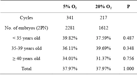
The percentage of embryos that reached the blastocyst stage in relation to the total number of fertilized oocytes is show in Table 3. There were no differences in the embryonic blastulation rate among patients from the 5% and 20% of O2 group; these percentages were equally similar in relation to the age of the patients. Furthermore, there was no difference in the pregnancy rates according to the kind of controlled ovarian stimulation in both evaluated groups in this study (Table 4).
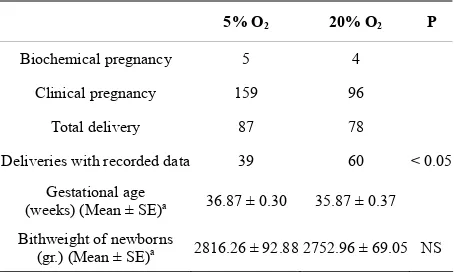
The data about the gestational age at delivery and birth weight from both groups evaluated in this study is shown in Table 5. There were 87 deliveries in the 5% of O2 group and 78 deliveries in the 20% of O2 group. However, it was only possible to register information from 39 and 60 in each group respectively. Gestational age was higher in women whose embryos were cultured in reduced concentrations of oxygen, in comparison to those women whose embryos were cultured in atmos-pheric concentrations of oxygen (P < 0.05) (Table 5).
Table 3. Blastulation rate according to age in both study groups.
5% O2 20% O2 P
Cycles 341 217
No. of embryos (2PN) 2281 1612
< 35 years old 39.82% 37.59% 0.487
35-39 years old 36.11% 39.69% 0.348
≥ 40 years old 34.01% 31.37% 0.756
Total 37.97% 37.97% 1.000
[image:4.595.310.537.582.707.2]Table 4. Pregnancy rate according to the protocol of ovarian stimulation with agonist or antagonist from the GnRH (GnRHa –GnRHant) and the recombinant FSH (rFSH) or human menopausal gonadotropin (HMG) in the study groups.
Stimulation Protocol 5% O2 20% O2
GnRHa + rFSH 60.00% 36.84%
GnRHa + HMG 50.00% 40.00%
GnRHa + rFSH + HMG 45.45% 53.33%
GnRHant + rFSH 50.43% 41.67%
GnRHant + HMG 55.00% 50.00%
GnRHant + rFSH + HMG 48.21% 60.87%
rFSH 27.27% 43.75%
HMG 57.14% 40.00%
rFSH + HMG 35.71% 38.89%
P: NS
Table 5. Gestational age and birth weight in the study groups.
5% O2 20% O2 P
Biochemical pregnancy 5 4
Clinical pregnancy 159 96
Total delivery 87 78
Deliveries with recorded data 39 60 < 0.05
Gestational age
(weeks) (Mean ± SE)a 36.87 ± 0.30 35.87 ± 0.37
Bithweight of newborns
(gr.) (Mean ± SE)a 2816.26 ± 92.88 2752.96 ± 69.05 NS aData are Mean ± Standard Error
4. DISCUSSION
Although human embryos can develop successfully in atmospheric concentrations of oxygen (20%), some au-thors have suggested that low oxygen concentrations (5%) resemble the physiological conditions of the uterus effectively, and thereby improve the quality, viability and embryo morphology [40,41].
An important result of this study was to find that cul-turing embryos at reduced concentrations of O2 (5%) is beneficial to patients ≥ 40 years old who perform as-sisted reproductive procedures with their own oocyte; these are the ones who achieve significantly higher im-plantation and pregnancy rates compared to those pa-tients of similar age whose embryos were cultured under atmospheric oxygen concentrations (20%) (25.00% ver-sus 2.70%; 41.38% verver-sus 5.56%, respectively, P < 0.05).
The implantation and pregnancy rates observed in women > 40 years old were similar to those seen in women < 35 years old and 35-39 years old (P:NS).
Meintjes et al. (20) embryos cultured in a 5% O2 en-vironment consistently resulted in higher rates of live birth implantation and live births when compared with rates among women whose embryos were cultured in an atmospheric O2 environment.
Furthermore, Nanassy et al. [27] cultured human em-bryos under oxygen atmospheric conditions (20%) until the 3rd day and then cultured the embryos in 5% and 20% of O2 from the 3rd to the 5th day of development without finding beneficial effects of 5% of O2 in the ad-vanced stages of preimplantational embryo development. These results suggest that the beneficial effect of hy-poxia on embryonic development would be along all stages of in vitro cultures even from the oocyte before fertilization up to the blastocyst stage [42], which had previously been observed in mice [8,43] cattle [44], rab-bits [45] and pigs [46].
Culturing embryos in 20% of O2, Karagenc [7] showed damage mainly in the embryonic inner cell mass (ICM). Similarly, Rhesus monkey embryo cultured in vitro in 20% of O2 showed the ICM morphologically disorgan-ized, diffuse, with few vacuolated cells, unlike the blas-tocysts with large and compact ICM cultured in low concentrations of O2 [47].
Rho et al. [48] culturing bovine embryos has shown
that low concentrations of oxygen produce higher rates of cleavage and blastocyst stages compared to embryos cultured in 20% of O2. Also in mice there is a better de-velopment to blastocyst stage, bigger number and size of ICM, and gene expression profile similar to those ob-served in embryo in vivo [49].
The discrepancies between the data obtained in ani-mals and humans could be explained based on the dif-ferences in the embryo physiology of each species and a variety of culture conditions and embryo transfer in the laboratory [47]. Beneficial effects of culture in 5% of O2 have been demonstrated in animals where embryo trans-fers routinely occur in blastocyst stage [7,50].
Dumoulin et al. [18], Pabon et al. [8], Quinn and Har-low [43] suggested that the beneficial effect of O2 in physiological concentrations should be observed in cul-tures extended to blastocyst, which are nowadays com-mon in assisted reproduction laboratories.
Several studies report increases in pregnancy and im-plantation in blastocyst transfers compared to embryo transfers on the 3rd day [51] and others report significant increases only in implantation rates [52], which consid-ering the results of this study, would have beneficial ef-fects in those patients over 40 years old.
[image:5.595.58.285.341.477.2]in a more “natural” way embryos with greater potential for development and implantation, however, this selec-tion would also depend on the O2 percentage in the sys-tems in which the embryos are cultured [47] and the oo-cyte origin associated to the patient’s age [53,54].
There are many authors who have showed the rela-tionship between chromosomal abnormalities, maternal age and embryo morphology [35,55-59]. Munné et al. [54] performed genetic diagnosis for 9 chromosomes [13,15-18,21,22, X, Y) in > 6000 embryos and found that women < 35 years old with good quality embryos had 44% of euploid embryos and that this percentage decreased to 21% in patients ≥ 41 years old. Beside, in patients with poor morphology embryos only 30% and 12% were euploid embryos in the group of women > 35 years old and ≥ 41 years old respectively. It has also been shown that chromosomal abnormalities in human oo-cytes are common and that these aneuploidies are closely related to maternal age, exceeding 60% in women over 40 years old [60].
Since older women have a higher incidence of oocytes and embryos with aneuploidy [54,60] but with similar rates of embryonic blastulation than young women (≤ 35 years old, 35-39 years old; see Table 3) as it has been demonstrated in this study, we should expect similar pregnancy and implantation rates independently from the oxygen concentration under which cultures in vitro are performed, but in this study we found that low concen-trations of oxygen (5%) are achieved significantly higher pregnancy and implantation rates in patients ≥ 40 years old, compared to the results in conditions of atmospheric concentrations of oxygen.
These results might be caused by high concentrations of oxygen that would affect the embryonic inner cell mass [7], an effect that would be even more noticeable because of the high incidence of aneuploidy observed in oocytes and embryos in older women.
In this study there were 87 births in the 5% of O2 group and 78 births in the group of 20%, being able to obtain information of their results, in terms of gesta-tional age and birthweight, in 39 and 60 cases respec-tively.
Within the data obtained, a significantly higher gesta-tional age was observed in the group of patients whose embryos were cultured under 5% O2 compared to the 20% of O2 group (36.87 ± 0.30 vs. 35.87 ± 0.37; P < 0.05) which could be a consequence of preimplantational embryo development in more physiological concentra-tions of oxygen to which the embryos were subjected during in vitro culture, but we believe that further studies are needed to determinate the possible relationship be-tween the culture in hypoxic conditions and obstetric characteristics of pregnancies.
5. ACKNOWLEDGEMENTS
The authors thank to Gustavo F. Gonzales M.D, Ph.D. for his valuable help and his review of this manuscript.
REFERENCES
[1] Bayatt-Smith, J.G., Leese, H.J. and Gosden, R.G. (1991) An investigation by mathematical modeling of whether Mouse and human preimplantation embryos in static culture can satisfy their demands for oxygen by diffusion.
Human Reproduction,6(1), 52-57.
[2] Fisher, B. and Bavister, B.D. (1993) Oxygen tension in the oviduct and uterus of rhesus monkeys, hamsters and rabbits. Journal of Reproduction and Fertility, 99(2), 673-679.
[3] Yedwab, G.A., Paz, G., Homonnai, T.Z., David, M.P. and Kraicer, P.F. (1976) Temperature, pH and partial pressure of oxygen in the cervix and uterus of women and uterus of rats during the cycle. Fertility and Sterility, 27(3), 304-309.
[4] Johnson, M.H. and Nasr-Esfahani, M.H. (1994) Radical solutions and cultural problems: Could free oxygen radi-cals be responsible for the impaired development of pre- implantation mammalian embryos in vitro? Bioassays,
16(1), 31-38.
[5] Catt, J.W. and Henman, M. (2000) Toxic effects of oxy-gen on human embryo development. Human Reproduc-tion, 15(Suppl. 2), 199-206.
[6] Bavister, B.D. (1995) Culture of preimplantation em-bryos: Facts and artifacts. Human Reproduction Update,
1(2), 91-148.
[7] Karagenc, L., Sertkaya, Z. and Ciray, N. (2004) Impact of oxygen concentration on embryonic development of mouse zygotes. Reproductive BioMedicine Online, 9(4), 409-417.
[8] Pabon, J.E., Jr, Findley, W.E. and Gibbons, W.E. (1889) The toxic effect of short exposures to the atmospheric oxygen concentration on early mouse embryonic devel-opment. Fertility and Sterility, 51(5), 896-900.
[9] Umaoka, Y., Noda, Y. and Narimoto, K. (1992) Effects of oxygen toxicity on early development of Mouse embryos.
Molecular Reproduction and Development, 31(1), 28-33. [10] Thompson, J.G., Simpson, A.C., Pugh, P.A., Donnelly,
P.E. and Tervit, H.R. (1990) Effect of oxygen concentra-tion on in-vitro development of preimplantation sheep and cattle embryos. Journal of Reproduction and Fertil-ity, 89(2), 573-578.
[11] Li, J., Foote, R.H. and Simkin, M. (1993) Development of rabbit zygotes cultured in protein-free medium with catalase, taurine or superoxide dismutase. Biology of Re-production, 49(1), 33-37.
[12] McKiernan, S.H. and Bavister, B.D. (1990) Environ-mental variables influencing in vitro development of hamster 2 cell embryos to the blastocyst stage. Biology of Reproduction, 43(3), 404-413.
[14] Fukui, Y., McGrowan, L.T. and James, R.W. (1991) Fac-tors affecting the in-vitro development to blastocysts of bovine oocytes matured and fertilized in vitro. Journal of Reproduction and Fertility, 92(1), 125-131.
[15] Kitagawa, Y., Suzuki, K. and Yoneda, A. (2004) Effects of oxygen concentration and antioxidants on the in vitro
developmental ability, production of reactive oxygen species (ROS) and DNA fragmentation in porcine em-bryos. Theriogenology, 62(7), 1186-1197.
[16] Dumoulin, J., Vanvuchelen, R. and Land, J. (1995) Effect of oxygen concentration on in-vitro fertilization and em-bryo transfer. Fertility and Sterility, 63(1), 115-119. [17] Bahçeci, M., Çiray, H.N., Karagenc, L., Uluğ, U. and
Bener, F. (2005) Effect of oxygen concentration during the incubation of embryos of women undergoing ICSI and embryo transfer: A prospective randomized study.
Reproductive BioMedicine Online, 11(4), 438-443. [18] Dumoulin, J.C.M., Meijers, C.J.J. and Bras, M. (1999)
Effect of oxygen concentration on human in-vitro fertili-zation and embryo culture. Human Reproduction, 14(2), 465-469.
[19] Waldenström, U., Engström, A., Hellberg, D., Nilsson, S. (2009) Low-oxygen compared with high-oxygen atmos-phere in blastocyst culture, a prospective randomized study. Fertility and Sterility, 91(6), 2461-2465.
[20] Meintjes, M., Chantilis, S., Douglas, J., Rodriguez, A., Guerami, A., Bookout, D., Barnett, B. and Madden, J. (2009) A controlled randomized trial evaluating the effect of lowered incubator oxygen tension on live births in a predominantly blastocyst transfer program. Human Re-production, 24(2), 300-307.
[21] Gardner, D.K., Lane, M., Calderon, I. and Leeton, J. (1996) Environment of the preimplantation human em-bryo in vivo: Metabolite analysis of oviduct and uterine fluids and metabolism of cumulus cells. Fertility and Sterility, 65(2), 349-53.
[22] Fanchin, R., Ayoubi, J.M., Righini, C., Olivennes, F., Schonauer, L.M. and Frydman, R. (2001) Uterine con-tractility decreases at the time of blastocyst transfers.
Human Reproduction, 16(6), 1115-1119.
[23] Quea, G., Romero, K., García-Velasco, J.A. (2007) Ex-tended embryo culture to increase implantation rate. Re-productive Biomedicine Online, 14(3), 375-383.
[24] Kea, B., Gebhardt, J., Watt, J., Westphal, L.M., Lathi, R.B. and Milki, A.A. (2007) Effect of reduced oxygen concentrations on the outcome of in vitro fertilization.
Fertility and Sterility, 87(1), 213-216.
[25] Kovačič, B. and Vlaisavljević, V. (2008) Influence of atmospheric versus reduced oxygen concentration on development of human blastocysts in vitro: a prospective study on sibling oocytes. Reproductive BioMedicine Online, 2(2), 229-236.
[26] Kovačič, B., Sajko, M. and Vlaisavljević, V. (2010) A prospective randomized trial on the effect of atmospheric versus reduced oxygen concentration on the outcome on intracytoplasmic sperm injection cycles. Fertility and Sterility, 94(2), 511-519.
[27] Nanassy, L., Peterson, A., Wilcox, A., Peterson, M., Hammoud, A. and Carrell, D. (2010) Comparison of 5% and ambient oxygen during days 3-5 in vitro culture of human embryos. Fertility and Sterility, 93(2), 579-585. [28] Perheentupa, A. and Huhtaniemi, I. (2009) Aging of the
human ovary and testis. Journal of Molecular Cell and Endocrinology, 299(1), 2-13.
[29] Steiner, A.Z. (2009) Clinical implications of ovarian reserve testing. Obstetrical and Gynecological Survey,
64(2), 120-128.
[30] Sauer, M.V., Paulson, R.J. and Lobo, R.A. (1992) Re-versing the natural decline in human fertility. An ex-tended clinical trial of oocytes donation to women of ad-vanced reproductive age. Journal of the American Medi-cal Association, 268(10), 1275-1279.
[31] Marcus, S.F. and Brinsden, P.R. (1996) In-vitro fertiliza-tion and embryo transfer in women aged 40 years and over. Human Reproduction Update, 2(6), 459-468. [32] Registro Latinoamericano de Reproducción Asistida
(2006) Editado por la Red Latinoamericana de Repro- ducción Asistida. Redlara, Chile, 1-95.
[33] Bar-Hava, I., Orvierto, R., Ferber, A., Ashkenazi, J., Dicker, D. and Ben-Rafael, Z. (1999) Standard in vitro
fertilization or intracytoplasmic sperm injection in ad-vanced female age, what may be expected? Gynecology and Endocrinology, 13(2), 93-97.
[34] Munné, S., Cohen, J. and Sable, D. (2002) Preimplanta-tion genetic diagnosis for advanced maternal age and other indications. Fertility and Sterility, 78(2), 234-236. [35] Munné, S., Alikani, M., Tomkin, G., Grifo, J. and Cohen,
J. (1995) Embryo morphology, developmental rates, and maternal age are correlated with chromosomal abnor-malities. Fertility and Sterility, 64(2), 382-391.
[36] Nybo Andersen, A.M., Wohlfahrt, J., Christens, P., Olsen, J. and Melbye, M. (2000) Maternal age and fetal loss: Population based register linkage study. British Medical Journal,320(7251), 1708-1712.
[37] Ciray, H.N., Aksoy, T., Yaramanci, K., Karayaka, I. and Bahçeci, M. (2009) In vitro culture under physiologic oxygen concentration improves blastocyst yield and quality: a prospective randomized survey on sibling oo-cytes. Fertility and Sterility, 91(Suppl. 1), 1459-1461. [38] García, J.I., Gonzales, G.F. and Noriega-Hoces, L. (2007)
Sperm chromatin stability and its relationship with fer-tilization rate after Intracytoplasmic Sperm Injection (ICSI) in an assisted reproduction program. Journal of Assisted Reproduction and Genetics, 24(12), 587-593. [39] Mansour, R. (2005) Minimizing embryo expulsion after
embryo transfer: A randomized controlled study. Human Reproduction, 20(1), 170-174.
[40] Gardner, D., Lane, M. (2001) Embryo culture in textbook of assisted reproductive techniques: laboratory and clini-cal perspectives. In: Gardner, D., Weissman, A., Howles, C. and Shoham, Z., Eds., Martin Dunitz Press, London, 203-222.
[41] Burton, G.J., Hempstock, J. and Jauniaux, E. (2000) Oxygen, early embryonic metabolism and free radical-mediated em-bryopathies. Reproductive Biomedicine Online, 6(1), 84- 96.
[42] Adam, A.A., Takahashi, Y., Katagiri, S. and Nagano, M. (2004) Effects of oxygen in the gas atmosphere during in vitro maturation, in vitro fertilization and in vitro culture on the efficiency of in vitro production of mouse em-bryos. Japan Journal Veterinary Research, 52(2), 77-84. [43] Quinn, P. and Harlow, G.M. (1978) The effect of oxygen
[44] Takahashi, Y. and Kanagawa, H. (1998) Effect of oxygen concentration in the gas atmosphere during in vitro in-semination of bovine oocytes on the subsequent embry-onic development in vitro. Journal of Veterinary Medical Science, 60(3), 365-367.
[45] Farrell, P.B. and Foote, R.H. (1995) Beneficial effects of culturing rabbit zygotes to blastocysts in 5% oxygen and 10% carbon dioxide. Journal of Reproduction and Fertil-ity, 103(1), 127-130.
[46] Im, G.S., Lai, L., Liu, Z., Hao, Y., Wax, D., Bonk, A. and Prather, R.S. (2004) In vitro development of preimplan-tation porcine nuclear transfer embryos cultured in dif-ferent media and gas atmospheres. Theriogenology, 61(6), 1125-1135.
[47] Bavister, B.D. (2004) The role of animal studies in sup-porting human ART. Reproduction Fertility and Devel-opment, 16(7), 719-728.
[48] Rho, G.J., Kim, D.S., Son, W.J., Cho, S.R., Kim, J.G., MK, B. and Choe, S.Y. (2007) Influence of in vitro oxy-gen concentrations on preimplantation embryo develop-ment, gene expression and production of Hanwoo calves following embryo transfer. Molecular Reproduction and Development, 74(4), 486-496.
[49] Rinaudo, P.F., Giritharan, G., Talbi, S., Dobson, A.T., Schultz, R.M. (2006) Effects of oxygen tension on gene expression in preimplantation mouse embryos. Fertility and Sterility, 86(Suppl. 4), 1252-1265.
[50] Umaoka, Y., Noda, Y. and Narimoto, K. (1991) Devel-opmental potentiality of embryos cultured under low oxygen tension with superoxide dismutase. Journal of In Vitro Fertilization and Embryo Transfer,8(5), 245-249. [51] Chang, H.J., Lee, J.R., Jee, B.C., Suh, C.S. and Kim, S.H.
(2009) Impact of blastocyst transfer on offspring sex ra-tio and the monozygotic twinning rate: A systematic re-view and meta-analisys. Fertility and Sterility, 91(6), 2381-2390.
[52] Beesley, R., Robinson, R., Propst, A., Arthur, N. and Retzloff, M. (2009) Impact of day 3 or day 5 embryo transfer on pregnancy rates and multiple gestations.
Fer-tility and Sterility, 91(5), 1717-1720.
[53] Shapiro, B., Richter, K.S., Harris, D.C. and Daneshmand, S. (2002) Influence of patient age on the growth and transfer blastocyst-stage embryos. Fertility and Sterility, 77(4), 700-705.
[54] Munné, S., Chen, S., Colls, P., Garrisi, J., Zheng, X., Cekleniak, N., Lenzi, M., Hughes, P., Fisher, J., Garrisi, M., Tomkin, G. and Cohen, J. (2007) Maternal age, mor-phology, development and chromosome abnormalities in over 6000 cleavage-stage embryos. Reproductive Bio-medicine Online, 14(5), 628-634.
[55] Munné, S., Bahçe, M., Sandalinas, M., Escudero, T., Márquez, C., Velilla, E., Colls, P., Oter, M., Alikani, M. and Cohen, J. (2004) Differences in chromosomes sus-ceptibility to aneuploidy and survival to first trimester.
Reproductive Biomedicine Online, 8(1), 81-90.
[56] Magli, M.C., Gianaroli, L. and Ferraretti, A.P. (2001) Chromosomal abnormalities in embryos. Molecular and Cellular Endocrinology, 183(Suppl. 1), S29-S34. [57] Bielanska, M., Tan, S.L. and Ao, A. (2002a)
Chromoso-mal mosaicism throughout human preimplantation de-velopment in vitro: incidence, type and relevance to em-bryo outcome. Human Reproduction, 17(2), 413-419. [58] Gianaroli, L., Magli, M.C. and Ferraretti, A.P. (2007)
Oocyte euploidy, pronuclear zygotes morphology and embryo chromosomal complement. Human Reproduction,
22(1), 241-249.
[59] Munné, S. (2006) Chromosome abnormalities and their relationship to morphology and development of human embryos. Reproductive Biomedicine Online, 12(2), 234- 253.
[60] Fragouli, E., Alfarawati, S., Katz-Jaffe, M., Alfarawati, S., Stevens, J., Colls, P., Goodall, N.N., Tormasi, S., Gutierrez- Mateo, C., Prates, R., Schoolcraft, W.B., Munné, S. and Wells, D. (2010) Comprehensive chromosome screening of polar bodies and blastocysts from couples experienc-ing repeated implantation failure. Fertility and Sterility,