Cell-PLoc 2.0: an improved package of web-servers for
predicting subcellular localization of proteins in various
organisms
Kuo-Chen Chou1,2, Hong-Bin Shen1,2
1Gordon Life Science Institute, 13784 Torrey Del Mar Drive, San Diego, CA 9213
2Institute of Image Processing & Pattern Recognition, Shanghai Jiaotong University, 800 Dongchuan Road, Shanghai, 200240, China;
Received 5 August 2010; revised 8 September 2010; accepted 12 September 2010.
ABSTRACT
Cell-PLoc 2.0 is a package of web-servers evolved from Cell-PLoc (Chou, K.C. & Shen, H.B., Nature Protocols, 2008, 2:153-162) by a top-down approach to improve the power for predicting subcellular localization of proteins in various organisms. It contains six predictors: Euk-mPLoc 2.0, Hum-mPLoc 2.0, Plant-mPLoc, Gpos-mPLoc, Gneg-mPLoc, and Virus-mPLoc, specialized for eukaryotic, human, plant, Gram- positive bacterial, Gram-negative bacterial, and virus proteins, respectively. Compared with Cell-PLoc, the predictors in the Cell-PLoc 2.0 have the following advantageous features: (1) they all have the capacity to deal with the multiplex proteins that can simultaneiously exist, or move between, two or more subcellular location sites; (2) no accession number is needed for the input of a query protein even if using the “high- level” GO (gene ontology) prediction engine; (3) the functional domain information and sequential evolution information are fused into the “ab initio” sequence-based prediction engine to enhance its accuracy. In this protocol, a step- to-step guide is provided for how to use the web server predictors in the Cell-PLoc 2.0 package, which is freely accessible to the public at
Keywords: Euk-mPLoc 2.0; Hum-mPLoc 2.0; Plant-mPLoc; Gpos-mPLoc; Gneg-mPLoc; Virus-mPLoc; Higher-level GO approach; Ab-initio approach; Functional domain; Sequential evolution; Multiplex proteins
1. INTRODUCTION
The localization of a protein in a cell is one of its
most important attributes. It can provide useful insight about the function of the protein. It is also fundamental to system biology because knowledge of the subcellu-lar locations of proteins is indispensable for in-depth understanding how the biological processes are regu-lated by the intricate pathways at the cellular level [1,2]. Particularly, the information of protein subcel-lular location is very useful for identifying and priori-tizing drug targets [3] during the process of drug de-velopment.
Given an uncharacterized protein sequence, how can we identify which subcellular location site it resides at? Does the protein stay in a single subcellular location or can it simultaneously exist in, or move between, two and more subcellular location sites? Although the answers to these questions can be determined by means of various biochemical experiments, it is time-consuming and labo-rious to acquire the desired information with experimen-tal methods alone. Particularly, in the post-genomic age, the number of newly found protein sequences has in-creased explosively. For instance, in 1986 the Swiss-Prot databank contained merely 3,939 protein sequence en-tries, but the number has since jumped to 519,348 ac-cording to the data released by the same databank on 10- Aug-20 meaning that the number of protein sequence entries now is more than 131 times the number from about 24 years ago. Facing such an avalanche of protein se-quences, it is highly desired to develop automated methods for timely identifying the subcellular locations of uncharacterized proteins based on their sequence in-formation alone.
Actually, during the past 18 years or so, various com-putational methods were developed in this regard (see, e.g., [4-59].
the development of this area. Meanwhile, they also each have their own limitations. For example, TargetP [15] is one of the popular methods in this area. Its remarkable merit is to make the prediction of the subcellular loca-tion of a protein related to its signal peptide and hence has a clearer biological meaning and basis. But TargetP [15] can only cover four subcellular location sites. For a query protein located outside its coverage scope, TargetP would either fail to predict or the predicted result thus obtained would not make any sense. The similar prob-lem also exists for PSORTb [33], one of the other popu-lar methods in this area.
The other problem for the existing methods listed above is that none of them can be used to deal with mul-tiplex proteins that may simultaneously reside at, or move between, two or more different subcellular loca-tions. Proteins with multiple location sites or dynamic feature of this kind are particularly interesting because they may have some unique biological functions worthy of our special notice [2,3]. Particularly, as pointed out by Millar et al. [60], recent evidence indicates that an in-creasing number of proteins have multiple locations in the cell.
About two years ago, a package of web-servers called Cell-PLoc was published [61] that can be used to predict subcellular localization of proteins in vari-ous organisms. It contained six web-server predictors:
Euk-mPLoc [62], Hum-mPLoc [63], Plant-PLoc
[64], Gpos-PLoc [65], Gneg-PLoc [66], and
Vi-rus-PLoc [67], specialized for eukaryotic, human,
plant, Gram-positive bacterial, Gram-negative bacte-rial, and virus proteins, respectively. As elucidated in the protocol article [61], each of the six predictors in
Cell-PLoc was established by hybridizing the
“higher-level” GO (gene ontology) [68] approach and the “ab initio” PseAAC (pseudo amino acid composi-tion) [16] approach, and hence could yield higher success rates as well as cover much wider scope. For
example, the Euk-mPLoc predictor can cover up to
22 subcellular location sites. Moreover, of the six
predictors in the Cell-PLoc package [61], Euk-
mPLoc and Hum- mPLoc can be also used to deal
with proteins with multiple-location sites. Therefore,
ever since it was published, Cell-PLoc has been
widely and increasingly used.
However, the existing version of Cell-PLoc [61] has the following shortcomings. (1) The accession number of a query protein is indispensable as an input in order to utilize the advantage of the “higher-level” GO ap-proach. Many proteins, such as hypothetical and syn-thetic proteins as well as those newly-discovered pro-teins that have not been deposited into databanks yet, do not have accession numbers, and hence cannot be
handled with the GO approach. (2) Even with their accession numbers available, many proteins cannot be meaningfully formulated in a GO space because the current GO database is far from complete yet. (3) Al-though the PseAAC approach was used as a comple-ment in Cell-PLoc [61] that could take some partial sequence order effects into account, the original Pse-AAC [16,69] did not contain the sequential evolution and functional domain information, and hence would affect the prediction quality. (4) Except Euk-mPLoc
(the predictor for eukaryotic proteins) and Hum-mPLoc
(the predictor for human proteins), all the other pre-dictors in Cell-PLoc package [61] cannot be used to deal with multiplex proteins.
To address the aforementioned four problems, a top-down approach to enhance the power of Cell-PLoc
has been implemented. The new version thus obtained is
denoted by Cell-PLoc 2.0. Compared with the old
Cell-PLoc [61], Cell-PLoc 2.0 has the following advan-tageous features.
Input Data. By means of the “homology-based GO
extraction” strategy as developed recently (see, e.g., [70]), the requirement for the accession number of a query protein is no longer needed even if using the higher-level GO approach to perform the prediction. This is especially useful for predicting the subcellular location sites of hypothetical proteins or synthetic pro-teins, as well as those new protein sequences without being deposited into data banks and hence having no accession numbers assigned yet.
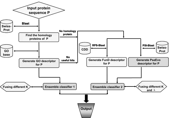
Sequence Information. For those proteins that have
no useful GO information to carry out the higher-level prediction, a hybridization approach by fusing the func-tional domain information and sequential evolution in-formation as illustrated in Figure 1 is developed to re-place the simple PseAAC approach [16] in the old
Cell-PLoc [61]. As a consequence, the success rates
have been remarkably increased for those proteins with-out useful GO numbers.
Multiplex Proteins. In the old Cell-PLoc package
[61], only two predictors, i.e., the one specialized for eukaryotic proteins and the one specialized for human proteins, can be used to treat proteins with multiple loca-tion sites. In Cell-PLoc 2.0, all the six predictors, in-cluding those specialized for plant proteins, Gram- posi-tive bacterial proteins, Gram-negaposi-tive bacterial proteins, and virus proteins, can be used to deal with the multiplex proteins.
Benchmark Datasets. With more experimental data
available in Swiss-Prot database to update the data for training the predictors, instead of version 50.7 released on 9-Sept-2006 as used in the old
RPS-Blast
Swiss-Prot
Generate FunD descriptor for P
Input protein sequence P
CDD
PSI-Blast
Generate PseEvo descriptor for P
Output
Swiss-Prot
Blast
Find the homology proteins of P
GO base
Generate GO descriptor for P
No homology protein
No useful hits
Ensemble classifier 1
Fusing different K Ensemble classifier 2 Fusing different K
and λ
RPS-Blast
Swiss-Prot
Generate FunD descriptor for P
Input protein sequence P
CDD
PSI-Blast
Generate PseEvo descriptor for P
Output
Swiss-Prot
Blast
Find the homology proteins of P
GO base
Generate GO descriptor for P
No homology protein
No useful hits
Ensemble classifier 1
Fusing different K Ensemble classifier 2 Fusing different K
[image:3.595.152.444.77.281.2]and λ
Figure 1. A flowchart to show the prediction process of the predictors in Cell-PLoc 2.0, where ensemble classifier 1 is for processing the GO descriptor samples, while ensemble classifier 2 is for the FunD (functional domain) and PseEvo (pseudo sequen-tial evolution) descriptor samples. See [70,71] for further explanation.
predictors in Cell-PLoc 2.0 were constructed based on version 55.3 released on 29-April-2008. Moreover, to make all the predictors in Cell-PLoc 2.0 have the capac-ity to deal with the multiplex proteins as well, the se-quences annotated with two or more subcellular location sites were no longer excluded even for plant proteins, Gram-positive bacterial proteins, Gram-negative bacte-rial proteins, and virus proteins as done previously in the old Cell-PLoc package [61].
Below, let us describe how to use the new Cell-PLoc 2.0 package to get the desired results.
2. EQUIPMENT AND MATERIALS
Hardware. Same as in the old Cell-PLoc [61], i.e., you need a computer that is able to access to internet.
Data. Your input protein sequences should be in
FASTA format. You can enter the sequence of a query protein by either typing or copying-and-pasting it into the input box. Spaces and line breaks will be ignored and will not affect the prediction result.
Programs. Cell-PLoc 2.0 contains the following
pro-grams: (1) Euk-mPLoc 2.0 for predicting the subcellular localization of eukaryotic proteins; (2) Hum-mPLoc 2.0
for human proteins; (3) Plant-mPLoc for plant proteins;
(4) Gpos-mPLoc for Gram-positive bacterial proteins;
(5) Gneg-mPLoc for Gram-negative bacterial proteins;
(6) Virus-mPLoc for virus proteins. The six predictors were evolved from Euk-mPLoc [62], Hum-mPLoc [63], Plant- PLoc [64], Gpos-PLoc [65], Gneg-PLoc [66], and Virus- PLoc [67] in the original Cell-PLoc package [61]
through a top-down approach to enhance their power, as elaborated in [70-75], respectively. Note that now all the six predictors in Cell-PLoc 2.0 have the capacity to deal with multiplex proteins as well, as indicated by the character “m” in front of their partial name “PLoc” that stands for the first character of “multiple”.
3. PROCEDURE
1) Go to the internetat
will see the top page of the Cell-PLoc 2.0 package on the screen of your computer, as shown in Figure 2.
2) You should use the relevant predictor to conduct the prediction: (1) if your query protein is an eukaryotic one, click the button Euk-mPLoc 2.0; (2)if it is ahuman protein, click Hum-mPLoc 2.0; (3) if it is a plant protein, click Plant-mPLoc; (4) if it is a Gram-positive bacterial protein, click Gpos-mPLoc; (5)if it is a Gram- negative bacterial protein, click Gneg-mPLoc; (6) if it is a viral protein, click Virus-mPLoc.
3) Without loss of generality, let us take Hum-mPLoc
2.0 as an example. By clicking Hum-mPLoc 2.0, you
will be prompted with the top page of the Hum-mPLoc 2.0 web-server predictor (Figure 3). To find the cover-age scope and caveat in using the predictor, click the Read Me button and you will see that the current
Hum-mPLoc 2.0 version can cover the following 14
Cell-PLoc 2.0: A package of web-servers for predicting subcellular localization of proteins in different organisms
Euk-mPLoc 2.0
Plant-mPLoc
Gneg-mPLoc
Hum-mPLoc 2.0
Gpos-mPLoc
[image:4.595.306.536.80.275.2]Virus-mPLoc
Figure 2. Illustration to show the Cell-PLoc 2.0 web-page
Hum-mPLoc 2.0: Predicting subcellular localization of human proteins including those with multiple sites
Read Me Data Citation
Enter the protein sequence(Example):
Submit Clear
Hum-mPLoc 2.0: Predicting subcellular localization of human proteins including those with multiple sites
Read Me Data Citation
Enter the protein sequence(Example):
[image:4.595.69.282.95.239.2]Submit Clear
Figure 3. A semi-screenshot to show the top page of the web- server predictor Hum-mPLoc 2.0 in the Cell-PLoc 2.0 package.
sosome, (9) microsome, (10) mitochondrion, (11) nu-cleus, (12) peroxisome, (13) plasma membrane, and (14) synapse, as schematically shown in Figure 4. You will also see the caveat from the Read Me window how to avoid meaningless prediction. To continue the prediction,
go back to the top page of the Hum-mPLoc 2.0
web-server predictor by closing the Read Me window.
4) Enter your query protein sequence into the input box as shown at the centre of Figure 3. The input se-quence should be in FASTA format. A sese-quence in FASTA format consists of a single-line description, fol-lowed by lines of sequence data. The first character of the description line is a greater-than symbol (“>”) in the first column. All lines should be shorter than 80 charac-ters. Example sequences in FASTA format can be seen by clicking on the Example button right above the input box. For more information about FASTA format, visit
5) To get the predicted result, click the Submit button.
Nucleus Nucleus Plasma membrane
Cytoplasm
Mitochondria
Endoplasmic reticulum
Cytoskeleton
Peroxisome Lysosome
Golgi apparatus
Centriole Extracell Microsome
Endosomal
Synapse
Figure 4. Schematic illustration to show the fourteen subcellu-lar location sites of human proteins that are covered by the Hum-mPLoc 2.0 predictor.
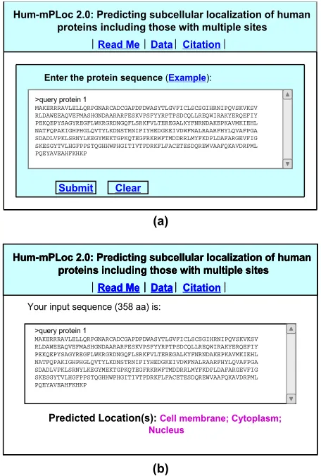
For example, if using the sequence of query protein 1 in the Example window as an input, you will see the input screen as shown in Figure 5a; after clicking the Submit
button, you will see “Cell membrane;Cytoplasm; Nu-cleus” shown on the predicted location(s) window (
Fig-ure 5b), meaning that the query protein is a multiplex
protein, which can simultaneously occur in “cell mem-brane”, “cytoplasm” and “nucleus” sites, fully consistent with experimental observations. However, if using the sequence of query protein 2 in the Example window as an input, you will instead see the input screen as shown in Figure 6a; after clicking the Submit button, you will see “Cytoplasm” shown on the predicted location(s) window (Figure 6b), meaning that the query protein is a single-location protein residing in “cytoplasm” com-partment only, also fully consistent with experimental observations.
6) By clicking the Citation button, you will find the relevant papers that document the detailed development and algorithm of Hum-mPLoc 2.0.
7) By clicking the Data button, you will find all the benchmark datasets used to train and test the Hum- mPLoc 2.0 predictor.
8) If your query protein sequence is from other organ-ism, click the relevant web-server button (Figure 2) as elaborated in Step 2, and repeat Steps 3-6.
TIMING The computational time for each prediction is within 15 seconds for most cases. The longer the query protein sequence is, the more time it is usually needed.
4. TROUBLESHOOTING
[image:4.595.56.287.288.440.2]Hum-mPLoc 2.0: Predicting subcellular localization of human proteins including those with multiple sites
Read Me Data Citation
Enter the protein sequence(Example):
Submit Clear
>query protein 1
MAKERRRAVLELLQRPGNARCADCGAPDPDWASYTLGVFICLSCSGIHRNIPQVSKVKSV RLDAWEEAQVEFMASHGNDAARARFESKVPSFYYRPTPSDCQLLREQWIRAKYERQEFIY PEKQEPYSAGYREGFLWKRGRDNGQFLSRKFVLTEREGALKYFNRNDAKEPKAVMKIEHL NATFQPAKIGHPHGLQVTYLKDNSTRNIFIYHEDGKEIVDWFNALRAARFHYLQVAFPGA SDADLVPKLSRNYLKEGYMEKTGPKQTEGFRKRWFTMDDRRLMYFKDPLDAFARGEVFIG SKESGYTVLHGFPPSTQGHHWPHGITIVTPDRKFLFACETESDQREWVAAFQKAVDRPML PQEYAVEAHFKHKP
Hum-mPLoc 2.0: Predicting subcellular localization of human proteins including those with multiple sites
Read Me Data Citation
Enter the protein sequence(Example):
Submit Clear
>query protein 1
MAKERRRAVLELLQRPGNARCADCGAPDPDWASYTLGVFICLSCSGIHRNIPQVSKVKSV RLDAWEEAQVEFMASHGNDAARARFESKVPSFYYRPTPSDCQLLREQWIRAKYERQEFIY PEKQEPYSAGYREGFLWKRGRDNGQFLSRKFVLTEREGALKYFNRNDAKEPKAVMKIEHL NATFQPAKIGHPHGLQVTYLKDNSTRNIFIYHEDGKEIVDWFNALRAARFHYLQVAFPGA SDADLVPKLSRNYLKEGYMEKTGPKQTEGFRKRWFTMDDRRLMYFKDPLDAFARGEVFIG SKESGYTVLHGFPPSTQGHHWPHGITIVTPDRKFLFACETESDQREWVAAFQKAVDRPML PQEYAVEAHFKHKP (a)
Your input sequence (358 aa) is:
Citation
Hum-mPLoc 2.0: Predicting subcellular localization of human proteins including those with multiple sites
Read Me Data
Predicted Location(s):Cell membrane; Cytoplasm; Nucleus
Your input sequence (358 aa) is:
Citation
Hum-mPLoc 2.0: Predicting subcellular localization of human proteins including those with multiple sites
Read Me Data Citation
Hum-mPLoc 2.0: Predicting subcellular localization of human proteins including those with multiple sites
Read Me Data
Hum-mPLoc 2.0: Predicting subcellular localization of human proteins including those with multiple sites
Read Me Data
Predicted Location(s):Cell membrane; Cytoplasm; Nucleus
>query protein 1
[image:5.595.309.539.77.419.2]MAKERRRAVLELLQRPGNARCADCGAPDPDWASYTLGVFICLSCSGIHRNIPQVSKVKSV RLDAWEEAQVEFMASHGNDAARARFESKVPSFYYRPTPSDCQLLREQWIRAKYERQEFIY PEKQEPYSAGYREGFLWKRGRDNGQFLSRKFVLTEREGALKYFNRNDAKEPKAVMKIEHL NATFQPAKIGHPHGLQVTYLKDNSTRNIFIYHEDGKEIVDWFNALRAARFHYLQVAFPGA SDADLVPKLSRNYLKEGYMEKTGPKQTEGFRKRWFTMDDRRLMYFKDPLDAFARGEVFIG SKESGYTVLHGFPPSTQGHHWPHGITIVTPDRKFLFACETESDQREWVAAFQKAVDRPML PQEYAVEAHFKHKP (b)
Figure 5. A semi-screenshot to show the input in the FASTA format for (a) the query protein 1 taken from the Example window, and (b) the output predicted by Hum-mPLoc 2.0 for the query protein sequence in panel (a).
your submission for prediction, consider the following points for troubleshooting.
Check the format of your input data to make sure it complies with the FASTA format as elaborated in Step 4 of the PROCEDURE.
Check the length of your input sequence to make
sure it is at least 50 amino acids long; otherwise, it might not be a real protein but its fragment.
Check the amino acid codes of your input sequence to make sure it does not contain any invalid char-acters.
You might also get meaningless result if the query protein is not among the subcellular location sites cov-ered by the web-server predictor.
5. ANTICIPATED RESULTS
In statistical prediction of subcellular localization of proteins or their any other attributes, it would be mean-ingless to simply say the success rate of a predictor
Hum-mPLoc 2.0: Predicting subcellular localization of human proteins including those with multiple sites
Read Me Data Citation
Enter the protein sequence(Example):
Submit Clear
>query protein 2
MEPSSLELPADTVQRIAAELKCHPTDERVALHLDEEDKLRHFRECFYIPKIQDLPPVDLS LVNKDENAIYFLGNSLGLQPKMVKTYLEEELDKWAKIAAYGHEVGKRPWITGDESIVGLM KDIVGANEKEIALMNALTVNLHLLMLSFFKPTPKRYKILLEAKAFPSDHYAIESQLQLHG LNIEESMRMIKPREGEETLRIEDILEVIEKEGDSIAVILFSGVHFYTGQHFNIPAITKAG QAKGCYVGFDLAHAVGNVELYLHDWGVDFACWCSYKYLNAGAGGIAGAFIHEKHAHTIKP ALVGWFGHELSTRFKMDNKLQLIPGVCGFRISNPPILLVCSLHASLEIFKQATMKALRKK SVLLTGYLEYLIKHNYGKDKAATKKPVVNIITPSHVEERGCQLTITFSVPNKDVFQELEK RGVVCDKRNPNGIRVAPVPLYNSFHDVYKFTNLLTSILDSAETKN
Hum-mPLoc 2.0: Predicting subcellular localization of human proteins including those with multiple sites
Read Me Data Citation
Enter the protein sequence(Example):
Submit Clear
>query protein 2
MEPSSLELPADTVQRIAAELKCHPTDERVALHLDEEDKLRHFRECFYIPKIQDLPPVDLS LVNKDENAIYFLGNSLGLQPKMVKTYLEEELDKWAKIAAYGHEVGKRPWITGDESIVGLM KDIVGANEKEIALMNALTVNLHLLMLSFFKPTPKRYKILLEAKAFPSDHYAIESQLQLHG LNIEESMRMIKPREGEETLRIEDILEVIEKEGDSIAVILFSGVHFYTGQHFNIPAITKAG QAKGCYVGFDLAHAVGNVELYLHDWGVDFACWCSYKYLNAGAGGIAGAFIHEKHAHTIKP ALVGWFGHELSTRFKMDNKLQLIPGVCGFRISNPPILLVCSLHASLEIFKQATMKALRKK SVLLTGYLEYLIKHNYGKDKAATKKPVVNIITPSHVEERGCQLTITFSVPNKDVFQELEK RGVVCDKRNPNGIRVAPVPLYNSFHDVYKFTNLLTSILDSAETKN (a)
Your input sequence (465 aa) is:
Citation
Hum-mPLoc 2.0: Predicting subcellular localization of human proteins including those with multiple sites
Read Me Data
Predicted Location(s):Cytoplasm >query protein 2
MEPSSLELPADTVQRIAAELKCHPTDERVALHLDEEDKLRHFRECFYIPKIQDLPPVDLS LVNKDENAIYFLGNSLGLQPKMVKTYLEEELDKWAKIAAYGHEVGKRPWITGDESIVGLM KDIVGANEKEIALMNALTVNLHLLMLSFFKPTPKRYKILLEAKAFPSDHYAIESQLQLHG LNIEESMRMIKPREGEETLRIEDILEVIEKEGDSIAVILFSGVHFYTGQHFNIPAITKAG QAKGCYVGFDLAHAVGNVELYLHDWGVDFACWCSYKYLNAGAGGIAGAFIHEKHAHTIKP ALVGWFGHELSTRFKMDNKLQLIPGVCGFRISNPPILLVCSLHASLEIFKQATMKALRKK SVLLTGYLEYLIKHNYGKDKAATKKPVVNIITPSHVEERGCQLTITFSVPNKDVFQELEK RGVVCDKRNPNGIRVAPVPLYNSFHDVYKFTNLLTSILDSAETKN
Your input sequence (465 aa) is:
Citation
Hum-mPLoc 2.0: Predicting subcellular localization of human proteins including those with multiple sites
Read Me Data
Predicted Location(s):Cytoplasm
Your input sequence (465 aa) is:
Citation
Hum-mPLoc 2.0: Predicting subcellular localization of human proteins including those with multiple sites
Read Me Data Citation
Hum-mPLoc 2.0: Predicting subcellular localization of human proteins including those with multiple sites
Read Me Data
Hum-mPLoc 2.0: Predicting subcellular localization of human proteins including those with multiple sites
Read Me Data
Predicted Location(s):Cytoplasm >query protein 2
MEPSSLELPADTVQRIAAELKCHPTDERVALHLDEEDKLRHFRECFYIPKIQDLPPVDLS LVNKDENAIYFLGNSLGLQPKMVKTYLEEELDKWAKIAAYGHEVGKRPWITGDESIVGLM KDIVGANEKEIALMNALTVNLHLLMLSFFKPTPKRYKILLEAKAFPSDHYAIESQLQLHG LNIEESMRMIKPREGEETLRIEDILEVIEKEGDSIAVILFSGVHFYTGQHFNIPAITKAG QAKGCYVGFDLAHAVGNVELYLHDWGVDFACWCSYKYLNAGAGGIAGAFIHEKHAHTIKP ALVGWFGHELSTRFKMDNKLQLIPGVCGFRISNPPILLVCSLHASLEIFKQATMKALRKK SVLLTGYLEYLIKHNYGKDKAATKKPVVNIITPSHVEERGCQLTITFSVPNKDVFQELEK RGVVCDKRNPNGIRVAPVPLYNSFHDVYKFTNLLTSILDSAETKN (b)
Figure 6. A semi-screenshot to show the input in the FASTA format for (a) the query protein 2 taken from the Example window, and (b) the output predicted by Hum-mPLoc 2.0 for the query protein sequence in panel (a).
without specifying what method and benchmark dataset were used to test its accuracy.
The following three cross-validation methods are generally used for examining the effectiveness of a sta-tistical prediction method: (1) the independent dataset test, (2) the sub-sampling (K-fold cross-validation) test, and (3) the jackknife test [76].
For the independent dataset test, although all the pro-teins to be tested are outside the training dataset used to train the predictor and hence can avoid the “memory” effect or bias, the way of how to select the independent proteins for testing could be quite arbitrary unless the number of independent proteins is sufficiently large. This kind of arbitrariness might lead to completely dif-ferent conclusions. For instance, a predictor achieving a higher success rate than the other predictor for a given independent testing dataset might fail to keep so when tested by another independent testing dataset [76].
[image:5.595.57.285.78.416.2]cross-validation. The problem with the K-fold cross- validation test as such is that the number of possible selections in dividing a benchmark dataset is an astro-nomical figure even for a very simple dataset. For ex-ample, let us consider a highly simplified dataset that consists of 300 proteins classified into five subsets, in which 60 proteins belong to subcellular location #1, 55 to location #2, 70 to location #3, 65 to location #4, and 50 to location #5. For such a simple dataset, the number of possible combinations of taking one-fifth proteins from each of the five subsets will be
1 2 3 4 5
60
60! 55! 70!
=
(60 12)!12! (55 11)!11! (70 14)!14!
65! 50!
> 5.45 10 (65 13)!13! (50 10)!10!
(1)
where 1 is the number of possible different ways of
taking 60 5 12 proteins from subset #1, 2 that of
taking 55 5 11 proteins from subset #2, 3 that of
taking 70 5 14 proteins from subset #3, 4 that of
taking 65 5 13 proteins from subset #4, and 5
that of taking 50 5 10 proteins from site-site-5. As we can see from Eq.1, even for such a simple and small dataset the number of possible ways in selecting the testing dataset for the 5-fold cross-validation would be greater than 5.45 10 60. It can be easily conceived that
for a benchmark dataset containing over a thousand pro-teins that are classified into more than five subcellular location sites, the number of the possible selections for subsampling test will be even much greater. Accordingly, in any actual subsampling cross-validation tests, only an extremely small fraction of the possible selections are taken into account. Since different selections will always lead to different results even for a same benchmark dataset and a same predictor, the subsampling test (such as 5-fold cross-validation) cannot avoid the arbitrariness either. A test method unable to yield a unique outcome cannot be deemed as an ideal one.
In the jackknife test, all the proteins in the benchmark dataset will be singled out one-by-one and tested by the predictor trained by the remaining protein samples. During the process of jackknifing, both the training dataset and testing dataset are actually open, and each protein sample will be in turn moved between the two. The jackknife test can exclude the “memory” effect. Also, the arbitrariness problem as mentioned above for the independent dataset test and subsampling test can be avoided because the outcome obtained by the jackknife cross-validation is always unique for a given benchmark dataset. As for the possible overestimation in success rate by jackknife test because of only one sample being
singled out at a time for testing, the answer is that as long as the jackknife test is performed on a stringent benchmark dataset in which none of proteins has
25%
pairwise sequence identity to any other in a
same subcellular location such as those benchmark datasets specially constructed for the six predictors in
Cell-PLoc 2.0, it is highly unlikely to yield an overesti-mated rate compared with the actual success rate in practical applications, as demonstrated in [72,74] and will be further discussed later. Besides, when the jack-knife test was used to compare two predictors, even if there was some overestimate due to using a less stringent benchmark dataset for one predictor, the same overesti-mate would exist for the other as long as they were both tested by a same dataset.
Accordingly, the jackknife test has been increasingly and widely used by investigators to examine the quality of various predictors (see, e.g., [47,51,55,58,59,77- 107]).
However, even if using the jackknife approach for cross-validation, a same predictor may still generate ob-viously different success rates when tested by different benchmark datasets. This is because the more stringent of a benchmark dataset in excluding homologous and high similarity sequences, or the more number of sub-cellular location sites it covers, the more difficult for a predictor to achieve a high overall success rate, as will be shown later.
The predictors in the old Cell-PLoc package [61] were established by hybridizing the “higher-level” GO approach with the “ab initio” sequence-correlated Pse-AAC [16] approach.Accordingly, their overall success prediction rates are generally higher than those by the best of the existing “ab initio” sequence-based proaches without combining with any higher level ap-proach, as elucidated in [61] and demonstrated in a se-ries of previous publications [62-67,108,109], and hence there is no need to repeat here.
Now, in the new version of Cell-PLoc 2.0, the same high success rates will still be achieved by the “higher-level” GO prediction engine but no require-ment for the accession number is needed for the input. And for those proteins without useful GO numbers, the corresponding success prediction rates will be further enhanced due to fusing the functional domain infor-mation and sequential evolution inforinfor-mation into the “ab initio” prediction engine in the Cell-PLoc 2.0
package as illustrated in Figure 1. Accordingly, the overall success rates by the predictors in Cell-PLoc 2.0 are not only higher than those by the other predic-tors but also those by the predicpredic-tors in the old
Cell-PLoc package [61], as can be seen from the
Table 1. Comparison between each of the six predictors in Cell-PLoc [61] and that in Cell-PLoc 2.0 by jackknife test.
Cell-PLoc Cell-PLoc 2.0
Organism lar locations covered Number of
subcellu-Predictor Overall success rateg Predictor Overall success rate
Eukaryotic 22a Euk-mPLoc 39.3% Euk-mPLoc 2.0 64.2%
Human 14b Hum-mPLoc 38.1% Hum-mPLoc 2.0 62.7%
Plant 12c Plant-PLoc 38.0% Plant-mPLoc 63.7%
Gram-positive 4d Gpos-PLoc 72.5% Gpos-mPLoc 82.2%
Gram-negative 8e Gneg-PLoc 71.5% Gneg-mPLoc 85.7%
Virus 6f Virus-PLoc 43.7% Virus-mPLoc 60.3%
aThe corresponding benchmark dataset was taken from the Supporting Information S1 of [70], in which none of protein included has 25%
pair-wise sequence identity to any other in a same subcellular location; bThe corresponding benchmark dataset was taken from the Online Supporting
Information A of [71], in which none of protein included has 25% pairwise sequence identity to any other in a same subcellular location; cThe
corresponding benchmark dataset was taken from Table S1 of [72], in which none of protein included has 25% pairwise sequence identity to any other in a same subcellular location; dThe corresponding benchmark dataset was taken from the Online Supporting Information A of [73], in which
none of protein included has 25% pairwise sequence identity to any other in a same subcellular location; eThe corresponding benchmark dataset
was taken from the Online Supporting Information A of [74], in which none of protein included has 25% pairwise sequence identity to any other in a same subcellular location; fThe corresponding benchmark dataset was taken from the Online Supporting Information A of [75], in which none of
protein included has 25% pairwise sequence identity to any other in a same subcellular location; gNote that in order to make the comparison under
exactly the same condition, only the sequences of proteins but not their accession numbers were used as inputs during the prediction.
1) Comparison with the six predictors in Cell-PLoc
[61]. Listed in Table 1 are the overall success rates by
[image:7.595.58.538.96.229.2]Cell-PLoc [61] and Cell-PLoc 2.0 using jackknife tests on six stringent benchmark datasets for eukaryotic, hu-man, plant, Gram-positive bacterial, Gram-negative bacterial, and virus proteins, respectively. For the case of eukaryotic proteins, the comparison was made between the predictor Euk-mPLoc of Cell-PLoc [61] and the predictor Euk-mPLoc 2.0 of Cell-PLoc 2.0 using the benchmark dataset classified into 22 subcellular loca-tions as given in the Supporting Information S1 of [70]. For human proteins, the comparison was made between the predictor Hum-mPLoc of Cell-PLoc [61] and the predictor Hum-mPLoc 2.0 of Cell-PLoc 2.0 using the benchmark dataset classified into 14 subcellular loca-tions as given in the Online Supporting Information A of [71]. And so forth. To avoid homology bias and redun-dancy, none of the proteins included in the six datasets has 25% pairwise sequence identity to any other in a same subcellular location. Also, to make the comparison between the two counterparts under exactly the same condition, only the sequences of proteins but not their accession numbers were used as inputs during the pre-diction. Meanwhile, the false positives (over-predictions) and false negatives (under-predictions) were also taken into account to reduce the scores for calculating the overall success rate. It is instructive to point out that it is much more complicated to count the over-predictions and under-predictions for a system containing both sin-gle-location and multiple-location proteins. For the de-tailed calculation formulation, see Eqs.43-48 as well as
Figure 4 in a comprehensive review [110]. It can be
seen from Table 1 that the overall success rates obtained by the predictors in Cell-PLoc 2.0 are about 10-25% higher than those by their counterparts in Cell-PLoc
[61].
2) Comparison with PSORTb v.2.0 [33]. The pre-dictor is widely used by biologists for predicting the subcellular locations of Gram-negative bacterial pro-teins. It is with a built-in training dataset covering the following five subcellular location sites: (1) cytoplasm, (2) extracellular, (3) inner membrane, (4) outer mem-brane, and (5) periplasm. The corresponding predictor
in Cell-PLoc 2.0 is Gneg-mPLoc that can cover eight
subcellular locations of Gram-negative proteins; i.e., in addition to the above five locations, it also covers “fimbrium”, “flagellum”, and “nucleoid”. In order to make the two predictors with different coverage scopes comparable, a degenerate testing dataset was generated by randomly picking testing proteins according to the following criteria: (1) the testing samples must be Gram-negative bacterial proteins; (2) to avoid the un-fair “memory” effect, the testing samples must be not in the training dataset of PSORTb v.2.0, nor in the training dataset of Gneg-mPLoc; (3) the experimen-tally observed subcellular locations of the testing pro-teins are known as clearly annotated in Swiss-Prot da-tabase; (4) their location sites must be within the scope covered by PSORTb v.2.0 for properly using it (for the proteins with multiple location sites, at least one of
v.2.0). For the detailed information about the testing dataset thus generated, see the Online Supporting In-formation B of [74] that contains 759 Gram-negative proteins, of which 116 are of cytoplasm, 62 of ex-tracellular, 397 of inner membrane, 89 of outer mem-brane, and 95 of periplasm. As shown in Table 2, the
overall success rates by Gneg-mPLoc and PSORTb
v.2.0 [33] in identifying the subcellular locations of
proteins in such a testing dataset were 98.0% and 79.3%, respectively, indicating the success rate by
Gneg-mPLoc of Cell-PLOc 2.0 was19% higher than
that by PSORTb v.2.0 [33]. Furthermore, some
exam-ples are given in Table 3 to show how the results
mispredicted by PSORTb v.2.0 were successfully
corrected by Greg-mPLoc. It is interesting to see from the table that the first protein with accession number
P62532 was predicted by Gneg-mPLoc belonging to
two subcellular location sites, “extracellular” and “fimbrium”, fully consistent with experimental obser-vation as annotated in Swiss-Prot database (version 55.3 released on 29- April-2008).
3) Comparison with TargetP [15]. The predictor is widely used by biologists for predicting the subcellular locations of plant proteins. It has a web-server at training dataset covering the following four items: “mi-tochondria”, “chloroplast”, “secretory pathway”, and “other”. Since the “secretory pathway” is not a final des-tination of subcellular location as annotated in Swiss- Prot databank, and should be removed from the com-parison. Also, the location of “other” is not a clear site for comparison, and should be removed too. The corre-sponding predictor in Cell-PLoc 2.0 is Plant-mPLoc
that can cover 12 subcellular locations of plant proteins; i.e., in addition to “mitochondria” and “chloroplast”, it also covers “cell membrane”, “cell wall”, “cytoplasm”, “endoplasmic reticulum”, “extracellular”, “Golgi appa-ratus”, “nucleus”, “peroxisome”, “plastid”, and “vacu-ole”. Thus, to make the two predictors with different coverage scopes comparable, a degenerate testing
data-set was generated according to the similar procedures as described in section 5.2. For the detailed information about the testing dataset thus generated, see Table S2 of [72] that contains 1,775 plant proteins of which 1,500 are of chloroplast and 275 of mitochondrion. As reported in [72], the overall success rates by Plant-mPLoc on such a testing dataset was 86%, which is more than 40% higher than that by TargetP [15] on the same testing dataset.
4) Comparison with Predotar [111]. This is another
popular predictor used by biologists for predicting the subcellular locations of plant proteins. Its web-server is at with a built-in training dataset covering the following four items: “endoplasmic reticulum”, “mitochondrion”, “plastid”, and “other”. Since the term “other” is not a clear description for subcellular location, and was re-moved from comparison. The corresponding predictor in
Cell-PLoc 2.0 is Plant-mPLoc that can cover 12
sub-cellular locations of plant proteins; i.e., in addition to “endoplasmic reticulum”, “mitochondria” and “plas-tid”, it also covers “cell membrane”, “cell wall”, “chloroplast”, “cytoplasm”, “extracellular”, “Golgi apparatus”, “nucleus”, “peroxisome”, and “vacuole”. Again, to make the two predictors with different cov-erage scopes comparable, a degenerate testing dataset was generated by following the similar procedures as described in section 5.2. For the detailed information about the testing dataset thus generated, see Table S4 of [72], where it was also reported that the overall success rates by Plant-mPLoc on such a testing data-set was 70%, which is more than 30% higher than that by Predotar [111] on the same testing dataset.
Moreover, it was also shown in [72,74] that some proteins coexisting in two or more subcellular location sites were successfully identified by Gneg-mPLoc [74]
and Plant-mPLoc [72]; cases like that are beyond the
reach ofPSORTb v.2.0 [33], TargetP [15], or Predotar
[image:8.595.76.520.607.723.2][111].
Table 2. A comparison of the predicted results by Gneg-mPLoc and PSORTb v.2.0 [33] on the testing dataset of Online Supporting Information B of [74].
Success rate Subcellular location
PSORTb v.2.0 Gneg-mPLoc
Cytoplasm 99/116=85.3% 115/116=99.1%
Extracellular 20/62=32.3% 52/62=83.9%
Inner membrane 329/397=82.9% 397/397=100%
Outer membrane 75/89=84.3% 87/89=97.8%
Periplasm 79/95=83.2% 93/95=97.9%
Table 3. Some examples to show how the subcellular location sites mispredicted by PSORTb v.2.0 were corrected by Gneg-mPLoc.
Protein accession numbera Experimental result anno-tated in Swiss-Prot
data-base
Predicted result by PSORTb v.2.0
Predicted result by Gneg-mPLoc
P62532 Extracellular;
Q8X9H8 Cytoplasm Unknown Cytoplasm P00962 Cytoplasm Unknown Cytoplasm Q83LY4 Cytoplasm Unknown Cytoplasm Q8DFR1 Cytoplasm Unknown Cytoplasm Q84H44 Cytoplasm Unknown Cytoplasm P27475 Extracellular Unknown Extracellular O50319 Extracellular Unknown Extracellular P31518 Extracellular Unknown Extracellular Q89AD4 Cytoplasm Unknown Cytoplasm Q56027 Extracellular Unknown Extracellular O52623 Extracellular Unknown Extracellular
P26219 Cell inner membrane Unknown Cell inner membrane
P77293 Cell inner membrane Unknown Cell inner membrane
P95655 Cell inner membrane Unknown Cell inner membrane.
P04123 Cell inner membrane Periplasm Cell inner membrane
Q47879 Cell outer membrane Unknown Cell outer membrane
P0A935 Cell outer membrane Unknown Cell outer membrane
P00211 Periplasm Cytoplasm Periplasm P0A182 Periplasm Unknown Periplasm Q9Z4N3 Periplasm Unknown Periplasm P31330 Periplasm Cytoplasm Periplasm
a Only the sequences but not the accession numbers were used as inputs during the prediction by Gneg-mPLoc. The accession numbers
here are just for the usage of identification.
From the above four comparisons, we can now make the following points very clear.
The more stringent a benchmark dataset is in
ex-cluding homologous and high similarity sequences, or the more subcellular location sites it covers, the more difficult for a predictor to achieve a high overall success rate. The impact of the coverage scope on the success rate can be easily understood by just considering the following cases. For a benchmark dataset only covering four subcellular locations each containing same number of proteins, the overall success rate by random assignments
would generally be 1/ 4 25% ; while for a
benchmark dataset covering 22 subcellular loca-tions, the overall success rate by random
assign-ments would be only 1/ 254.5% . This means
that the former is more than five times the latter.
Also, a predictor examined by jackknife test is very difficult to yield a high success rate when per-formed on a stringent benchmark dataset in which
none of proteins included has 25% pairwise
sequence identity to any other in a same subset (subcellular location). That is why the overall
suc-cess rate achieved by Gneg-mPLoc was 85.7%
when examined by the jackknife test on the benchmark dataset of the Online Supporting Infor-mation A of [74] but was 98.0% when examined by the independent dataset test for the proteins in the Online Supporting Information B of [74]. That is also why the overall success rate achieved by
Plant-mPLoc was only 63.7% when examined by
Table S4 of [72], respectively. However, regardless of using what test methods or test datasets, one thing is crystal clear, i.e., the overall success rates achieved by the six predictors in Cell-PLoc 2.0 are significantly higher than those by its counterparts.
Meanwhile, it has also become understandable why
the success rates as originally reported by PSORTb v.2.0 [33], TargetP [15] and Predotar [111] were over-estimated. This is because none of the success rates reported for these predictors was derived by the jackknife test. Also, the benchmark datasets used to test these predictors covered much less subcellular location sites than those used in their counterparts in Cell-PLoc 2.0. Particularly, the benchmark datasets used by PSORTb v.2.0 , Tar-getP and Predotar to estimate their success rates contained many homologous sequences. For in-stance, the cutoff threshold to reduce the homology bias for the benchmark dataset used in Predotar
[111] was set at 80%, meaning that only those
se-quences which have 80% pairwise sequence
identity to any other in a same subset were ex-cluded [111]; while for the benchmark dataset used
in TargetP [15] and PSORTb v.2.0 [33], even no
cutoff threshold was indicated to remove homolo-gous sequences. Compared with the benchmark datasets used in [70-75] where none of proteins in-cluded has 25% pairwise sequence identity to any other in a same subset, the benchmark datasets adopted by PSORTb v.2.0, TargetP, and Predotar
are much less stringent and hence cannot avoid homology bias and overestimation.
6. CONCLUDING REMARKS
Evolved from the old Cell-PLoc package [61],
Cell-PLoc 2.0 is much more flexible and powerful than
the former. In addition to yielding higher success rates than the existing prediction method, all the predictors in
Cell-PLoc 2.0 have the capacity to deal with proteins
with two or more subcellular location sites. Besides, the predictors in Cell-PLoc 2.0 cover much wider scopes than most of the existing predictors in this area. For in-stance, Hum-mPLoc 2.0 and Euk-mPLoc 2.0 can cove up to 14 sites of human proteins and 22 sites of eu-karyotic, respectively, which are about two to five times the number of subcellular location sites covered by most of the existing predictors.
However, Cell-PLoc 2.0 also has the following limi-tations and further improvements will be needed with more experimental data available in future. (1) Although
Euk-mPLoc2.0 in the Cell-PLoc 2.0 packagecan cover 22 sites of eukaryotic proteins, if a query protein is
out-side of the 22 location sites, it would still generate mea-ningless result. Therefore,we shall continuously extend the coverage scope for each of the predictors in the
Cell-PLoc series in a timely manner once more statisti-cally significant experimental data will be available in future. (2) For some subcellular locations with very small numbers of proteins, the prediction success rates are still quite low. This is because there are not sufficient location-known proteins in these sites to effectively train the prediction engine. It is anticipated that with more experimental data available for these sites in the future, this kind of situation will be improved. (3) Since the power of Cell-PLoc 2.0 is closely associated with the GO database [68,112,113] and functional domain data-base [114], with the continuous development of the GO database and functional domain database, more useful GO numbers and functional domain information will be incorporated into the prediction engine, further streng-thening its prediction power.
Once further improvements are implemented, the fu-ture version of Cell-PLoc series will be announced via a publication or a webpage.
REFERENCES
[1] Ehrlich, J.S., Hansen, M.D., Nelson, W.J. (2002) Spatio- temporal regulation of Rac1 localization and lamellipo-dia dynamics during epithelial cell-cell adhesion. Dev Cell, 3, 259-270.
[2] Glory, E., Murphy, R.F. (2007) Automated subcellular location determination and high-throughput microscopy.
Dev Cell, 12, 7-16.
[3] Smith, C. (2008) Subcellular targeting of proteins and drugs. [4] Nakai, K., Kanehisa, M. (1991) Expert system for
pre-dicting protein localization sites in Gram-negative bacte-ria. Proteins: Structure, Function and Genetics, 11, 95- 110.
[5] Nakashima, H., Nishikawa, K. (1994) Discrimination of intracellular and extracellular proteins using amino acid composition and residue-pair frequencies. Journal of Molecular Biology, 238, 54-61.
[6] Cedano, J., Aloy, P., P'erez-Pons, J.A., Querol, E. (1997) Relation between amino acid composition and cellular location of proteins. Journal of Molecular Biology, 266, 594-600.
[7] Nakai, K., Horton, P. (1999) PSORT: A program for de-tecting sorting signals in proteins and predicting their subcellular localization. Trends in Biochemical Science, 24, 34-36.
[8] Chou, K.C., Elrod, D.W. (1998) Using discriminant func-tion for predicfunc-tion of subcellular locafunc-tion of prokaryotic proteins. Biochemical and Biophysical Research Com-munications, 252, 63-68.
Nu-cleic Acids Research, 26, 2230-2236.
[10] Chou, K.C., Elrod, D.W. (1999) Protein subcellular loca-tion predicloca-tion. Protein Engineering, 12, 107-118. [11] Yuan, Z. (1999) Prediction of protein subcellular
loca-tions using Markov chain models. FEBS Letters, 451, 23-26.
[12] Nakai, K. (2000) Protein sorting signals and prediction of subcellular localization. Advances in Protein Chemistry, 54, 277-344.
[13] Murphy, R.F., Boland, M.V., Velliste, M. (2000) Towards a systematics for protein subcellular location: quantita-tive description of protein localization patterns and au-tomated analysis of fluorescence microscope images.
Proc. Int. Conf. Intell. Syst. Mol. Biol., 8, 251-259. [14] Chou, K.C. (2000) Review: Prediction of protein
struc-tural classes and subcellular locations. Current Protein and Peptide Science, 1, 171-208.
[15] Emanuelsson, O., Nielsen, H., Brunak, S., von Heijne, G. (2000) Predicting subcellular localization of proteins based on their N-terminal amino acid sequence. Journal of Molecular Biology, 300, 1005-1016.
[16] Chou, K.C. (2001) Prediction of protein cellular attrib-utes using pseudo amino acid composition. PROTEINS: Structure, Function, and Genetics (Erratum: ibid., 2001, Vol.44, 60), 43, 246-255.
[17] Feng, Z.P. (2001) Prediction of the subcellular location of prokaryotic proteins based on a new representation of the amino acid composition. Biopolymers, 58, 491-499. [18] Hua, S., Sun, Z. (2001) Support vector machine approach
for protein subcellular localization prediction. Bioinfor-matics, 17, 721-728.
[19] Feng, Z.P., Zhang, C.T. (2001) Prediction of the subcel-lular location of prokaryotic proteins based on the hy-drophobicity index of amino acids. Int. J. Biol. Macro-mol., 28, 255-261.
[20] Feng, Z.P. (2002) An overview on predicting the subcel-lular location of a protein. In. Silico. Biol., 2, 291-303. [21] Chou, K.C., Cai, Y.D. (2002) Using functional domain
composition and support vector machines for prediction of protein subcellular location. Journal of Biological Chemistry, 277, 45765-45769.
[22] Zhou, G.P., Doctor, K. (2003) Subcellular location pre-diction of apoptosis proteins. Proteins: Structure, Func-tion, and Genetics, 50, 44-48.
[23] Pan, Y.X., Zhang, Z.Z., Guo, Z.M., Feng, G.Y., Huang, Z.D., He, L. (2003) Application of pseudo amino acid composition for predicting protein subcellular location: Stochastic signal processing approach. Journal of Protein Chemistry, 22, 395-402.
[24] Park, K.J., Kanehisa, M. (2003) Prediction of protein subcellular locations by support vector machines using compositions of amino acid and amino acid pairs. Bioin-formatics, 19, 1656-1663.
[25] Gardy, J.L., Spencer, C., Wang, K., Ester, M., Tusnady, G.E., Simon, I., Hua, S., deFays, K., Lambert, C., Nakai, K., Brinkman, F.S. (2003) PSORT-B: Improving protein subcellular localization prediction for Gram-negative bacteria. Nucleic Acids Research, 31, 3613-3617.
[26] Huang, Y., Li, Y. (2004) Prediction of protein subcellular locations using fuzzy k-NN method. Bioinformatics, 20, 21-28.
[27] Xiao, X., Shao, S., Ding, Y., Huang, Z., Huang, Y., Chou,
K.C. (2005) Using complexity measure factor to predict protein subcellular location. Amino Acids, 28, 57-61. [28] Gao, Y., Shao, S.H., Xiao, X., Ding, Y.S., Huang, Y.S.,
Huang, Z.D., Chou, K.C. (2005) Using pseudo amino acid composition to predict protein subcellular location: approached with Lyapunov index, Bessel function, and Chebyshev filter. Amino Acids, 28, 373-376.
[29] Lei, Z., Dai, Y. (2005) An SVM-based system for pre-dicting protein subnuclear localizations BMC. Bioinfor-matics, 6, 291.
[30] Shen, H.B., Chou, K.C. (2005) Predicting protein subnu-clear location with optimized evidence-theoretic K-earest classifier and pseudo amino acid composition. Biochem. Biophys. Res. Comm., 337, 752-756.
[31] Garg, A., Bhasin, M., Raghava, G.P. (2005) Support vec-tor machine-based method for subcellular localization of human proteins using amino acid compositions, their or-der, and similarity search. Journal of Biological Chemis-try, 280, 14427-14432.
[32] Matsuda, S., Vert, J.P., Saigo, H., Ueda, N., Toh, H., Akutsu, T. (2005) A novel representation of protein se-quences for prediction of subcellular location using sup-port vector machines. Protein Sci., 14, 2804-2813. [33] Gardy, J.L., Laird, M.R., Chen, F., Rey, S., Walsh, C.J.,
Ester, M., Brinkman, F.S. (2005) PSORTb v.2.0: ex-panded prediction of bacterial protein subcellular local-ization and insights gained from comparative proteome analysis. Bioinformatics, 21, 617-623.
[34] Gao, Q.B., Wang, Z.Z., Yan, C., Du, Y.H. (2005) Predic-tion of protein subcellular locaPredic-tion using a combined feature of sequence. FEBS Letters, 579, 3444-3448. [35] Chou, K.C., Shen, H.B. (2006) Predicting protein
sub-cellular location by fusing multiple classifiers. Journal of Cellular Biochemistry, 99, 517-527.
[36] Guo, J., Lin, Y., Liu, X. (2006) GNBSL: A new integra-tive system to predict the subcellular location for Gram- egative bacteria proteins. Proteomics, 6, 5099-5105. [37] Xiao, X., Shao, S.H., Ding, Y.S., Huang, Z.D., Chou,
K.C. (2006) Using cellular automata images and pseudo amino acid composition to predict protein subcellular lo-cation. Amino Acids, 30, 49-54.
[38] Hoglund, A., Donnes, P., Blum, T., Adolph, H.W., Kohl-bacher, O. (2006) MultiLoc: prediction of protein sub-cellular localization using N-terminal targeting sequences, sequence motifs and amino acid composition. Bioinfor-matics, 22, 1158-1165.
[39] Lee, K., Kim, D.W., Na, D., Lee, K.H., Lee, D. (2006) PLPD: reliable protein localization prediction from im-balanced and overlapped datasets. Nucleic Acids Re-search, 34, 4655-4666.
[40] Zhang, Z.H., Wang, Z.H., Zhang, Z.R., Wang, Y.X. (2006) A novel method for apoptosis protein subcellular local-ization prediction combining encoding based on grouped weight and support vector machine. FEBS Letters, 580, 6169-6174.
[41] Shi, J.Y., Zhang, S.W., Pan, Q., Cheng, Y.-M., Xie, J. (2007) Prediction of protein subcellular localization by support vector machines using multi-scale energy and pseudo mino acid composition. Amino Acids, 33, 69-74. [42] Chen, Y.L., Li, Q.Z. (2007) Prediction of apoptosis
Theo-retical Biology, 248, 377–381.
[43] Chen, Y.L., Li, Q.Z. (2007) Prediction of the subcellular location of apoptosis proteins. Journal of Theoretical Bi-ology, 245, 775-783.
[44] Mundra, P., Kumar, M., Kumar, K.K., Jayaraman, V.K., Kulkarni, B.D. (2007) Using pseudo amino acid compo-sition to predict protein subnuclear localization: Ap-proached with PSSM. Pattern Recognition Letters, 28, 1610-1615.
[45] Emanuelsson, O., Brunak, S., von Heijne, G., Nielsen, H. (2007) Locating proteins in the cell using TargetP, Sig-nalP and related tools. Nature Protocols, 2, 953-971. [46] Lin, H., Ding, H., Feng-Biao Guo, F.B., Zhang, A.Y.,
Huang, J. (2008) Predicting subcellular localization of mycobacterial proteins by using Chou’s pseudo amino acid composition. Protein & Peptide Letters, 15, 739-744. [47] Shi, J.Y., Zhang, S.W., Pan, Q., Zhou, G.P. (2008) Using
Pseudo Amino Acid Composition to Predict Protein Subcellular Location: Approached with Amino Acid Composition Distribution. Amino Acids, 35, 321-327. [48] Li, F.M., Li, Q.Z. (2008) Predicting protein subcellular
location using Chou's pseudo amino acid composition and improved hybrid approach. Protein & Peptide Let-ters, 15, 612-616.
[49] Tantoso, E., Li, X.B. (2008) AAIndexLoc: Predicting Subcellular Localization of Proteins Based on a New Representation of Sequences Using Amino Acid Indices.
Amino Acids, 35, 345-353.
[50] Jiang, X., Wei, R., Zhang, T.L., Gu, Q. (2008) Using the concept of Chou’s pseudo amino acid composition to predict apoptosis proteins subcellular location: an ap-proach by approximate entropy. Protein & Peptide Let-ters, 15, 392-396.
[51] Zhou, X.B., Chen, C., Li, Z.C., Zou, X.Y. (2008) Im-proved prediction of subcellular location for apoptosis proteins by the dual-layer support vector machine. Amino Acids, 35, 383-388.
[52] Ding, Y.S., Zhang, T.L. (2008) Using Chou’s pseudo amino acid composition to predict subcellular localiza-tion of apoptosis proteins: an approach with immune ge-netic algorithm-based ensemble classifier. Pattern Rec-ognition Letters, 29, 1887-1892.
[53] Zhang, S.W., Zhang, Y.L., Yang, H.F., Zhao, C.H., Pan, Q. (2008) Using the concept of Chou’s pseudo amino acid composition to predict protein subcellular localization: an approach by incorporating evolutionary information and von Neumann entropies. Amino Acids, 34, 565-572. [54] Jin, Y., Niu, B., Feng, K.Y., Lu, W.C., Cai, Y.D., Li, G.Z.
(2008) Predicting subcellular localization with AdaBoost learner. Protein & Peptide Letters, 15, 286-289.
[55] Lin, H., Wang, H., Ding, H., Chen, Y.L., Li, Q.Z. (2009) Prediction of Subcellular Localization of Apoptosis Pro-tein Using Chou’s Pseudo Amino Acid Composition. Ac-ta Biotheoretica, 57, 321-330.
[56] Zhang, L., Liao, B., Li, D., Zhu, W. (2009) A novel rep-resentation for apoptosis protein subcellular localization prediction using support vector machine. Journal of Theoretical Biology, 259, 361-365.
[57] Zeng, Y.H., Guo, Y.Z., Xiao, R.Q., Yang, L., Yu, L.Z., Li, M.L. (2009) Using the augmented Chou’s pseudo amino acid composition for predicting protein submitochondria locations based on auto covariance approach. Journal of
Theoretical Biology, 259, 366-72.
[58] Du, P., Cao, S., Li, Y. (2009) SubChlo: predicting protein subchloroplast locations with pseudo-amino acid compo-sition and the evidence-theoretic K-nearest neighbor (ET-KNN) algorithm. Journal of Theoretical Biology, 261, 330-335.
[59] Cai, Y.D., He, J., Li, X., Feng, K., Lu, L., Kong, X., Lu, W. (2010) Predicting protein subcellular locations with feature selection and analysis. Protein Pept. Lett., 17, 464-472.
[60] Millar, A.H., Carrie, C., Pogson, B., Whelan, J. (2009) Exploring the function-location nexus: using multiple lines of evidence in defining the subcellular location of plant proteins. Plant Cell, 21, 1625-1631.
[61] Chou, K.C., Shen, H.B. (2008) Cell-PLoc: A package of Web servers for predicting subcellular localization of proteins in various organisms. Nature Protocols, 3, 153- 162.
[62] Chou, K.C., Shen, H.B. (2007) Euk-mPLoc: a fusion classifier for large-scale eukaryotic protein subcellular location prediction by incorporating multiple sites.
Journal of Proteome Research, 6, 1728-1734.
[63] Shen, H.B., Chou, K.C. (2007) Hum-mPLoc: An ensem-ble classifier for large-scale human protein subcellular location prediction by incorporating samples with multi-ple sites. Biochemical and Biophysical Research Com-munications, 355, 1006-1011.
[64] Chou, K.C., Shen, H.B. (2007) Large-scale plant protein subcellular location prediction. Journal of Cellular Bio-chemistry, 100, 665-678.
[65] Shen, H.B., Chou, K.C. (2007) Gpos-PLoc: An ensemble classifier for predicting subcellular localization of Gram- ositive bacterial proteins. Protein Engineering, Design, and Selection, 20, 39-46.
[66] Chou, K.C., Shen, H.B. (2006) Large-scale predictions of Gram-negative bacterial protein subcellular locations.
Journal of Proteome Research, 5, 3420-3428.
[67] Shen, H.B., Chou, K.C. (2007) Virus-PLoc: A fusion classifier for predicting the subcellular localization of vi-ral proteins within host and virus-infected cells. Bio-polymers, 85, 233-240.
[68] Ashburner, M., Ball, C.A., Blake, J.A., Botstein, D., Butler, H., Cherry, J.M., Davis, A.P., Dolinski, K., Dwight, S.S., Eppig, J.T., Harris, M.A., Hill, D.P., Issel- arver, L., Kasarskis, A., Lewis, S., Matese, J.C., Rich-ardson, J.E., Ringwald, M., Rubin, G.M., Sherlock, G. (2000) Gene ontology: Tool for the unification of biology.
Nature Genetics, 25, 25-29.
[69] Chou, K.C. (2005) Using amphiphilic pseudo amino acid composition to predict enzyme subfamily classes. Bioin-formatics, 21, 10-19.
[70] Chou, K.C., Shen, H.B. (2010) A new method for pre-dicting the subcellular localization of eukaryotic proteins with both single and multiple sites: Euk-mPLoc 2.0.
PLoS ONE, 5, e9931.
[71] Shen, H.B., Chou, K.C. (2009) A top-down approach to enhance the power of predicting human protein subcel-lular localization: Hum-mPLoc 2.0. Analytical Biochem-istry, 394, 269-74.
[73] Shen, H.B., Chou, K.C. (2009) Gpos-mPLoc: A top- down approach to improve the quality of predicting sub-cellular localization of Gram-positive bacterial proteins.
Protein & Peptide Letters, 16, 1478-1484.
[74] Shen, H.B., Chou, K.C. (2010) Gneg-mPLoc: A top- down strategy to enhance the quality of predicting sub-cellular localization of Gram-negative bacterial proteins.
Journal of Theoretical Biology, 264, 326-333.
[75] Shen, H.B., Chou, K.C. (2010) Virus-mPLoc: A fusion classifier for viral protein subcellular location prediction by incorporating multiple sites. Journal of Biomolecular Structure & Dynamics, 28, 175-186.
[76] Chou, K.C., Zhang, C.T. (1995) Review: Prediction of protein structural classes. Critical Reviews in Biochemis-try and Molecular Biology, 30, 275-349.
[77] Fang, Y., Guo, Y., Feng, Y., Li, M. (2008) Predicting DNA-binding proteins: Approached from Chou’s pseudo amino acid composition and other specific sequence fea-tures. Amino Acids, 34, 103-109.
[78] Feng, Y.E., Luo, L.F. (2008) Use of tetrapeptide signals for protein secondary-structure prediction. Amino Acids, 35, 607-614.
[79] Li, Z.C., Zhou, X.B., Dai, Z., Zou, X.Y. (2009) Predic-tion of protein structural classes by Chou’s pseudo amino acid composition: approached using continuous wavelet transform and principal component analysis. Amino Ac-ids, 37, 415-425.
[80] Nanni, L., Lumini, A. (2008) Genetic programming for creating Chou's pseudo amino acid based features for submitochondria localization. Amino Acids, 34, 653-660. [81] Wang, Y., Xue, Z., Shen, G., Xu, J. (2008) PRINTR: Pre-diction of RNA binding sites in proteins using SVM and profiles. Amino Acids, 35, 295-302.
[82] Zhao, X.M., Chen, L., Aihara, K. (2008) Protein function prediction with high-throughput data. Amino Acids, 35, 517-530.
[83] Jahandideh, S., Abdolmaleki, P., Jahandideh, M., Asada-badi, E.B. (2007) Novel two-stage hybrid neural dis-criminant model for predicting proteins structural classes.
Biophys. Chem., 128, 87-93.
[84] Chen, K., Kurgan, L.A., Ruan, J. (2008) Prediction of protein structural class using novel evolutionary colloca-tion-based sequence representation. Journal of Computa-tional Chemistry, 29, 1596-1604.
[85] Jahandideh, S., Sarvestani, A.S., Abdolmaleki, P., Jahan-dideh, M., Barfeie, M. (2007) Gamma-Turn types predic-tion in proteins using the support vector machines. Jour-nal of Theoretical Biology, 249, 785-790.
[86] Shao, X., Tian, Y., Wu, L., Wang, Y., L., J., Deng, N. (2009) Predicting DNA- and RNA-binding proteins from sequences with kernel methods. Journal of Theoretical Biology, 258, 289-293.
[87] Yang, J.Y., Peng, Z.L., Yu, Z.G., Zhang, R.J., Anh, V., Wang, D. (2009) Prediction of protein structural classes by recurrence quantification analysis based on chaos game representation. Journal of Theoretical Biology, 257, 618-626.
[88] Anand, A., Suganthan, P.N. (2009) Multiclass cancer classification by support vector machines with class-wise optimized genes and probability estimates. Journal of Theoretical Biology, 259, 533-540.
[89] Chen, C., Chen, L.X., Zou, X.Y., Cai, P.X. (2008)
Pre-dicting protein structural class based on multi-features fusion. Journal of Theoretical Biology, 253, 388-392. [90] Du, P., Li, Y. (2008) Prediction of C-to-U RNA editing
sites in plant mitochondria using both biochemical and evolutionary information. Journal of Theoretical Biology, 253, 579-589.
[91] Jahandideh, S., Hoseini, S., Jahandideh, M., Hoseini, A., Disfani, F.M. (2009) Gamma-turn types prediction in proteins using the two-stage hybrid neural discriminant model. Journal of Theoretical Biology, 259, 517-522. [92] Lin, H. (2008) The modified Mahalanobis discriminant
for predicting outer membrane proteins by using Chou’s pseudo amino acid composition. Journal of Theoretical Biology, 252, 350-356.
[93] Munteanu, C.B., Gonzalez-Diaz, H., Magalhaes, A.L. (2008) Enzymes/non-enzymes classification model com-plexity based on composition, sequence, 3D and topo-logical indices. Journal of Theoretical Biology, 254, 476- 482.
[94] Rezaei, M.A., Abdolmaleki, P., Karami, Z., Asadabadi, E.B., Sherafat, M.A., Abrishami-Moghaddam, H., Fadaie, M., Forouzanfar, M. (2008) Prediction of membrane protein types by means of wavelet analysis and cascaded neural networks. Journal of Theoretical Biology, 254, 817-820.
[95] Vilar, S., Gonzalez-Diaz, H., Santana, L., Uriarte, E. (2009) A network-QSAR model for prediction of ge-netic-component biomarkers in human colorectal cancer.
Journal of Theoretical Biology, 261, 449-458.
[96] Wang, T., Xia, T., Hu, X.M. (2010) Geometry preserving projections algorithm for predicting membrane protein types. Journal of Theoretical Biology, 262, 208-213. [97] Chen, Y., Han, K. (2009) BSFINDER: Finding Binding
Sites of HCV Proteins Using a Support Vector Machine.
Protein & Peptide Letters, 16, 373-382.
[98] Kannan, S., Hauth, A.M., Burger, G. (2008) Function prediction of hypothetical proteins without sequence si-milarity to proteins of known function. Protein & Peptide Letters, 15, 1107-1116.
[99] Nanni, L., Lumini, A. (2009) A Further Step Toward an Optimal Ensemble of Classifiers for Peptide Classifica-tion, a Case Study: HIV Protease. Protein & Peptide Let-ters, 16, 163-167.
[100] Gu, F., Chen, H. (2009) Evaluating Long-term Relation-ship of Protein Sequence by Use of d-Interval Condi-tional Probability and its Impact on Protein Structural Class Prediction. Protein Pept. Lett., 16, 1267-1276. [101] Ji, G., Wu, X., Shen, Y., Huang, J., Quinn Li, Q. (2010) A
classification-based prediction model of messenger RNA polyadenylation sites. Journal of Theoretical Biology, 265, 287-296.
[102] Yang, X.Y., Shi, X.H., Meng, X., Li, X.L., Lin, K., Qian, Z.L., Feng, K.Y., Kong, X.Y., Cai, Y.D. (2010) Classifi-cation of transcription factors using protein primary structure. Protein & Peptide Letters, 17, 899-908. [103] Gu, Q., Ding, Y.S., Zhang, T.L. (2010) Prediction of
G-Protein-Coupled Receptor Classes in Low Homology Using Chou’s Pseudo Amino Acid Composition with Approximate Entropy and Hydrophobicity Patterns. Pro-tein Pept. Lett., 17, 559-567.
affinities between the human amphiphysin SH3 domain and its peptide ligands. Protein Pept. Lett., 17, 246- 253. [105] Shi, R., Hu, X. (2010) Predicting enzyme subclasses by
using support vector machine with composite vectors.
Protein Pept. Lett., 17, 599-604.
[106] Wang, T., Yang, J. (2010) Predicting subcellular localiza-tion of gram-negative bacterial proteins by linear dimen-sionality reduction method. Protein Pept. Lett., 17, 32-37. [107] Yang, J., Jiang, X.F. (2010) A novel approach to predict
protein-protein interactions related to Alzheimer’s dis-ease based on complex network. Protein Pept. Lett., 17, 356-366.
[108] Chou, K.C., Shen, H.B. (2006) Hum-PLoc: A novel en-semble classifier for predicting human protein subcellu-lar localization. Biochemical and Biophysical Research Communications, 347, 150-157.
[109] Chou, K.C., Shen, H.B. (2006) Predicting eukaryotic protein subcellular location by fusing optimized evi-dence-theoretic K-nearest neighbor classifiers. Journal of Proteome Research, 5, 1888-1897.
[110] Chou, K.C., Shen, H.B. (2007) Review: Recent pro-gresses in protein subcellular location prediction.
Ana-lytical Biochemistry, 370, 1-16.
[111] Small, I., Peeters, N., Legeai, F., Lurin, C. (2004) Predo-tar: A tool for rapidly screening proteomes for N-terminal targeting sequences. Proteomics, 4, 1581-1590.
[112] Camon, E., Magrane, M., Barrell, D., Binns, D., Fleisch-mann, W., Kersey, P., Mulder, N., Oinn, T., Maslen, J., Cox, A., Apweiler, R. (2003) The gene ontology annotation (GOA) project: Implementation of GO in SWISS- PROT, TrEMBL, and InterPro. Genome Res., 13, 662-672. [113] Barrell, D., Dimmer, E., Huntley, R.P., Binns, D.,
O'Do-novan, C., Apweiler, R. (2009) The GOA database in 2009-an integrated Gene Ontology Annotation resource.
Nucleic Acids Research, 37, D396-403.
![Figure 4 in a comprehensive review [110]. It can be seen from Table 1 that the overall success rates obtained by the predictors in Cell-PLoc 2.0 are about 10-25% higher than those by their counterparts in Cell-PLoc [61]](https://thumb-us.123doks.com/thumbv2/123dok_us/8984441.395015/7.595.58.538.96.229/figure-comprehensive-table-overall-success-obtained-predictors-counterparts.webp)
![Table 2. A comparison of the predicted results by Gneg-mPLoc and PSORTb v.2.0 [33] on the testing dataset of Online Supporting Information B of [74]](https://thumb-us.123doks.com/thumbv2/123dok_us/8984441.395015/8.595.76.520.607.723/comparison-predicted-results-psortb-testing-dataset-supporting-information.webp)

