Hypothalamic loss of Snord116 recapitulates
the hyperphagia of Prader-Willi syndrome
Joseph Polex-Wolf, … , Stephen O’Rahilly, Giles S.H. Yeo
J Clin Invest.
2018;
128(3)
:960-969.
https://doi.org/10.1172/JCI97007
.
Profound hyperphagia is a major disabling feature of Prader-Willi syndrome (PWS).
Characterization of the mechanisms that underlie PWS-associated hyperphagia has been
slowed by the paucity of animal models with increased food intake or obesity. Mice with a
microdeletion encompassing the
Snord116
cluster of noncoding RNAs encoded within the
Prader-Willi minimal deletion critical region have previously been reported to show growth
retardation and hyperphagia. Here, consistent with previous reports, we observed growth
retardation in
Snord116
+/–Pmice with a congenital paternal
Snord116
deletion. However,
these mice neither displayed increased food intake nor had reduced hypothalamic
expression of the proprotein convertase 1 gene
PCSK1
or its upstream regulator
NHLH2
,
which have recently been suggested to be key mediators of PWS pathogenesis.
Specifically, we disrupted
Snord116
expression in the mediobasal hypothalamus in
Snord116
flmice via bilateral stereotaxic injections of a Cre-expressing adeno-associated
virus (AAV). While the Cre-injected mice had no change in measured energy expenditure,
they became hyperphagic between 9 and 10 weeks after injection, with a subset of animals
developing marked obesity. In conclusion, we show that selective disruption of
Snord116
expression in the mediobasal hypothalamus models the hyperphagia of PWS.
Research Article
Genetics
Metabolism
Find the latest version:
Introduction
Prader-Willi syndrome (PWS) causes severe obesity, with affected individuals displaying profound food-seeking behavior (1, 2). People affected by PWS progress along a complex natural history with distinct phases, commencing with an early “fail-ure to thrive” characterized by hypotonia and feeding difficul-ties. Hyperphagia, decreased energy expenditure, and obesity appear later in childhood (3). PWS is typically associated with large deletions on human chromosome 15; however, recent patient reports have shown that microdeletions encompassing the SNORD116 cluster of noncoding RNAs (ncRNAs) on pater-nal chromosome 15q11.2 result in a phenotype that overlaps substantially with PWS (4–8). These patients display most of the major diagnostic features of PWS (9, 10), thereby suggesting a PWS minimal deletion region encompassing SNORD116 and 2 neighboring ncRNAs, RNAs IPW and SNORD109A (Supplemen-tal Figure 1; supplemen(Supplemen-tal material available online with this article; https://doi.org/10.1172/JCI97007DS1).
The SNORD116 genomic locus encodes multiple species of ncRNAs, including long noncoding RNAs (lncRNAs), arrays of small nucleolar RNAs (snoRNAs), and lncRNAs with snoRNA ends (sno-lncRNAs), the function of which remains largely unknown (11–13). The Prader-Willi region on human chromo-some 15 is syntenic to mouse chromochromo-some 7, with the SNORD116 cluster in both species comprising an imprinted array of snoRNA repeats interspersed by exons of an lncRNA host gene, 116HG (12, 14). SNORD116/Snord116 is also transcribed as part of a large transcriptional unit denoted as small nucleolar host gene 14 (SNHG14, formerly LNCAT) in both humans and mice. The pro-gression of transcription through SNHG14 is different between neuronal and nonneuronal cells (15–17), and in mice, expression of Snord116 snoRNAs becomes enhanced within hypothalamic nuclei by weaning age (18).
None of the existing mouse models of PWS fully recapitulate the human phenotype (19). Many such models with gene dele-tions in the Prader-Willi region are lethal at the embryonic stage or have poor postnatal survival. Two viable mouse lines removing
Snord116 have been reported: a 150-kb deletion of the Snord116
cluster from the paternal allele yielding the Snord116+/–P mouse
(20) and a deletion of Snord116 with 2 exons of the neighboring ncRNA (21). In both models, there is no expression of any Snord116 transcripts, as the maternal allele is silenced by imprinting. Mice with a maternal allele Snord116 deletion are, unsurprisingly, indis-tinguishable from WT controls. Several additional Snord116 dele-tion models have also been reported, including Snord116 deledele-tions that are biallelic (22), specific to neuropeptide Y (NPY) neurons (22), or tamoxifen inducible (23).
Profound hyperphagia is a major disabling feature of Prader-Willi syndrome (PWS). Characterization of the mechanisms that underlie PWS-associated hyperphagia has been slowed by the paucity of animal models with increased food intake or obesity. Mice with a microdeletion encompassing the Snord116 cluster of noncoding RNAs encoded within the Prader-Willi minimal deletion critical region have previously been reported to show growth retardation and hyperphagia. Here, consistent with previous reports, we observed growth retardation in Snord116+/–P mice with a congenital paternal Snord116 deletion. However,
these mice neither displayed increased food intake nor had reduced hypothalamic expression of the proprotein convertase 1 gene PCSK1 or its upstream regulator NHLH2, which have recently been suggested to be key mediators of PWS pathogenesis. Specifically, we disrupted Snord116 expression in the mediobasal hypothalamus in Snord116fl mice via bilateral stereotaxic
injections of a Cre-expressing adeno-associated virus (AAV). While the Cre-injected mice had no change in measured energy expenditure, they became hyperphagic between 9 and 10 weeks after injection, with a subset of animals developing marked obesity. In conclusion, we show that selective disruption of Snord116 expression in the mediobasal hypothalamus models the hyperphagia of PWS.
Hypothalamic loss of Snord116 recapitulates
the hyperphagia of Prader-Willi syndrome
Joseph Polex-Wolf,1 Brian Y.H. Lam,1 Rachel Larder,1 John Tadross,1 Debra Rimmington,1 Fàtima Bosch,2
Verónica Jiménez Cenzano,2 Eduard Ayuso,2 Marcella K.L. Ma,1 Kara Rainbow,1 Anthony P. Coll,1 Stephen O’Rahilly,1 and Giles S.H. Yeo1
1Medical Research Council (MRC) Metabolic Diseases Unit, University of Cambridge Metabolic Research Laboratories, Wellcome Trust–MRC Institute of Metabolic Science, Addenbrooke’s Hospital,
Cambridge, United Kingdom. 2Center of Animal Biotechnology and Gene Therapy and Department of Biochemistry and Molecular Biology, School of Veterinary Medicine, Universitat Autònoma de Barcelona,
Bellaterra, Spain, and Centro de Investigación Biomédica en Red de Diabetes y Enfermedades Metabólicas Asociadas (CIBERDEM), Barcelona, Spain.
Related Commentary: p. 900
Authorship note: A.P. Coll, S. O’Rahilly, and G.S.H. Yeo contributed equally to this work.
Conflict of interest: S. O’Rahilly receives payment through Cambridge Enterprise for scientific advisory services to Pfizer, AstraZeneca, and MedImmune.
License: This work is licensed under the Creative Commons Attribution 4.0 Inter-national License. To view a copy of this license, visit http://creativecommons.org/ licenses/by/4.0/.
Submitted: August 21, 2017; Accepted: December 5, 2017.
ies also report that Snord116+/–P mice appear to be hyperphagic.
Recently Burnett et al. have reported a human model of PWS using human iPS cell–derived (iPSC-derived) neurons (24). They report that such neurons, with deletions encompassing Snord116, show reduced expression of PCSK1 (encod-ing prohormone covertase), which has been previously implicated in human obesity (25), and a transcription factor
NHLH2, which regulates expression of
the PCSK1 gene (26, 27).
In the present study, we assessed energy intake phenotypes in the
con-genital Snord116+/–P mouse and found
that differences in food intake are not significant when corrected appropri ately for the effect of lower body weight. Deletion of Snord116 specifically in the mediobasal hypothalamus of adult mice, however, revealed hyperphagia in the absence of early effects on growth. Of note, a subset of such animals devel-oped marked obesity.
Results
Congenital paternal Snord116 deletion (Snord116+/–P) mice have impaired growth, but do not develop hyperphagia or obesity.
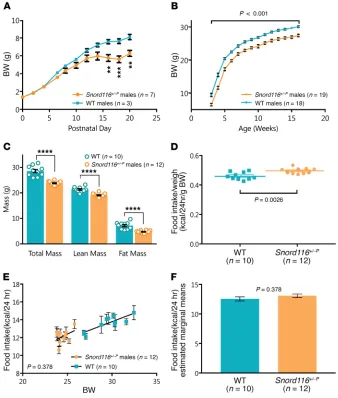
We investigated growth, body compo-sition, and food intake phenotypes in mice with a congenital paternal deletion of Snord116 (Snord116+/–P). As previously reported, Snord116+/–P mice had signifi-cantly lower body weight than WT lit-termates from early postnatal life (P16), which continued though adulthood (Figure 1, A and B). Similar results were also observed in female mice (Supple-mental Figure 2). Snord116+/–P mice thus remain small without transitioning to
developing obesity. Adult Snord116+/–P
mice had reduced lean and fat mass as assessed by dual-energy x-ray absorpti-ometry (DEXA) (Figure 1C).
We next determined whether con-genital Snord116 deficiency may affect food consumption in adult mice. However, differences in body size needed to be taken into account. Previous reports accounted for these differences by divid-ing food intake by body weight (20, 22, 28, 29). When daily food intake measurements over a 10-day period were divided by total body weight in our study, we observed, as others had previously, significantly increased food intake in the Snord116+/–P mice (Figure 1D). The raw food intake data are shown in Supplemental Figure 3.
Considering that there is an inherent difference in body weight between Snord116+/–P and WT mice, we hypothesized that it is poss-All Snord116 deletion mouse models reported thus far
remain smaller than controls through adulthood and do not transition to obesity as seen in human PWS — a summary of
Snord116 deletion model phenotypes is presented in
Supple-mental Table 1, with focus on growth and energy homeostasis. The most consistent phenotype observed in Snord116-deficient mice is low body weight and early growth retardation, remi-niscent of failure to thrive in PWS infants. Differences in body weight start emerging between P2 and P5, and body length and fat mass are also reported to be diminished. A number of
[image:3.585.43.381.60.458.2](24), we examined hypothalamic expression of both genes in
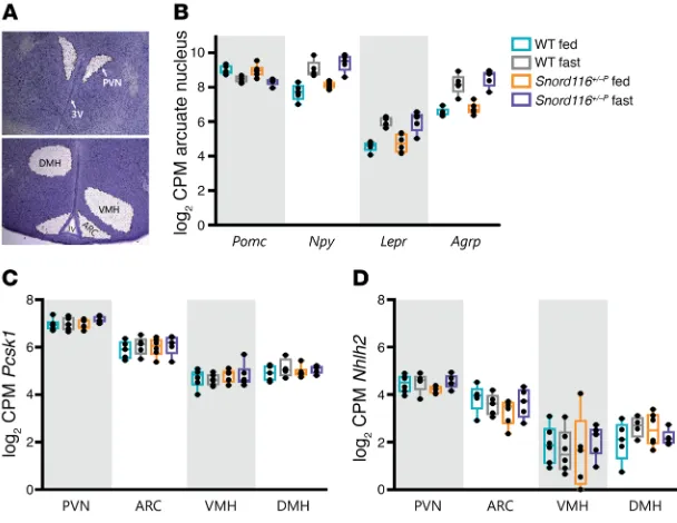
Snord116+/–P mice. Pcsk1 and Nhlh2 mRNA were detectable in all 4 hypothalamic nuclei. No differences in expression were observed by genotype or nutritional status within any of the 4 nuclei (para-ventricular nucleus [PVN], ARC, ventromedial hypothalamus [VMH], or dorsomedial hypothalamus [DMH]) (Figure 2, C and D). These data were confirmed in RNA extracts from whole hypo-thalami obtained from a different set of experimental animals, which also demonstrated that there was no evidence of
differen-tial expression of Pcsk1 and Nhlh2 between WT and Snord116+/–P
mice in either fed or fasted states (Supplemental Figure 6).
Adult-onset deletion of Snord116 in the mediobasal hypothalamus results in hyperphagia, with a subset of mice further developing obe-sity and increased fat mass. An adult-onset mediobasal
hypotha-lamic deletion of Snord116 was generated by stereotaxic injection of AAV-Cre (where AAV indicates adeno-associated virus) into
Snord116fl mice at 10 weeks of age, with AAV-GFP–injected mice used as controls. Successful bilateral targeting of Cre for each mouse was verified using Cre immunohistochemistry (Figure 3A). Of 26 AAV-Cre injections, 21 were verified as hits, with bilateral expression of Cre in the mediobasal hypothalamus, while 5 were excluded from analysis as misses, with no bilateral hypothalamic expression of Cre. Cre expression in all hits and misses is docu-mented in Supplemental Figure 7. Efficacy of the Cre-mediated deletion of Snord116 was verified in a separate set of AAV-Cre– injected (n = 10) and AAV-GFP–injected (n = 9) mice, with a reduc-tion of Snord116 expression in the mediobasal hypothalamus of Cre-injected mice (28%, P = 0.01, Student’s t test), but not in other regions, including the PVN and cerebral cortex (Figure 3B).
Food intake and energy expenditure were assessed in the post-surgery period. Food intake was measured over 2 time points: first, between 2 and 4 weeks after surgery, when no weight differences were evident between the Cre- versus GFP-injected mice, and sec-ond, between 9 and 10 weeks after surgery, when some Cre-injected mice showed increased body weight compared with controls. ible that the lower body weight of the Snord116+/–P mice may account
for the relative differences in food intake. An improved method, based on analysis of covariance (ANCOVA), has been reported to better model energy homeostasis phenotypes under these circumstances (30). This allows us to test whether a difference in food intake exists when we remove the confounding effect of body weight. Snord116+/–P mice did not have significantly different 24-hour food intake com-pared with WT mice after differences in body weight were accounted for using this model (ANCOVA, P = 0.378) (Figure 1, E and F).
We also studied food intake after a 24-hour fast in Snord116+/–P versus WT mice. Using ANCOVA to correct for body-weight dif-ferences, there were no differences in food intake after either 4 hours (P = 0.714, ANCOVA) or 24 hours (P = 0.856, ANCOVA) after reintroduction of food (Supplemental Figure 4).
Expression of leptin/melanocortin pathway genes in the hypo-thalamus of Snord116+/–P mice across fed and food-deprived states remains normal. To assess whether Snord116 deficiency affects
the expression of hypothalamic genes in appetitive pathways, we evaluated expression of leptin/melanocortin pathway genes. RNA-sequencing (RNA-seq) was performed using RNA extracted from laser-captured hypothalamic nuclei from adult (16 to 19
weeks old) WT and Snord116+/–P mice in either the ad libitum–
fed or 24-hour fasted states (Figure 2A). We verified that laser- capture microdissection yielded material specific to the dissected nucleus by verifying enriched expression of positive control genes (Supplemental Figure 5). Within the arcuate nucleus (ARC), the expression of Npy, leptin receptor (Lepr), and agouti-related pro-tein (Agrp) showed the expected increase with fasting in both WT and Snord116+/–P mice (FDR < 0.03). Proopiomelanocortin (Pomc) also displayed a trend for a decreased expression with fasting in both WT (log2 fold change [FC] = –0.52, P < 0.03, FDR = 0.53) and
Snord116+/–P (log
2 FC = –0.67, P < 0.003, FDR = 0.23) mice (Figure
[image:4.585.36.340.56.291.2]2B). Following from recent work suggesting that Snord116 defi-ciency disrupts expression of prohormone convertase 1 (Pcsk1) and its transcription activator, nescient helix-loop-helix 2 (Nhlh2)
Figure 2. Expression of hypothalamic leptin/ melanocortin pathway genes is unaltered in
congeni-tal Snord116 deletion compared with that in WT mice
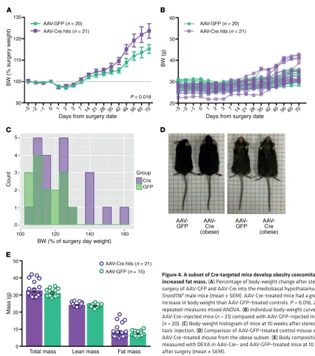
(Figure 4B), and at 10 weeks after surgery, a subset (5/21) of AAV-Cre–treated mice gained over 140% of presurgery weight (Figure 4C). Mice in this obese AAV-Cre–treated subset appeared obvi-ously larger than AAV-GFP controls (Figure 4D). Body composition was analyzed (DEXA) at 10 weeks after surgery, with increased fat mass underlying the obesity of a subset of Cre-treated mice (Figure 4E and Supplemental Figure 8).
Snord116fl Cre-injected mice have normal hypothalamic leptin-melanocortin gene expression, with obese animals displaying increased Socs3 expression. We analyzed whether the
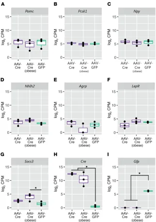
hyperpha-gic phenotype observed in the AAV-Cre–targeted mice may be driven by altered expression of hypothalamic genes in appetitive pathways. RNA-seq was performed on laser-captured hypotha-lamic regions obtained from adjacent brain sections with Cre immunohistochemistry as well as matching regions from AAV-GFP–injected mice. For the AAV-Cre mice, we analyzed gene expression in mice that were from the obese (>140% presurgery body weight) and nonobese subsets. No significant changes were detected in the expression of Pomc, Lepr, Npy, Agrp, Pcksk1, or Energy expenditure was measured at 6 weeks after surgery, when
differences in weight between the Cre- and GFP-injected mice began to appear. There were no detectable differences between Cre- and GFP-injected mice when measured between 2 and 4 weeks after surgery (P = 0.084, ANCOVA) (Figure 3C). Differences in energy expenditure as measured through indirect calorimetry were not significant when assessed at 6 weeks after surgery (P = 0.348, ANCOVA) (Figure 3D). Food intake studies performed between 9 and 10 weeks after surgery revealed consistent hyper-phagia in Cre-injected mice regardless of body weight (P = 0.001, ANCOVA) (Figure 3E).
[image:5.585.91.493.56.379.2]Body weight and composition were also assessed over the 10-week postsurgery period. AAV-Cre–injected mice gained more weight (as a percentage of presurgery weight) compared with AAV-GFP–injected controls (P = 0.016, ANOVA), with differences appearing from 6 weeks after surgery (Figure 4A). Analysis of the individual body-weight curves highlighted greater variability in the weight gain of AAV-Cre–injected mice (final weight, 29.8–42.8 g) compared with AAV-GFP–injected mice (final weight, 28.8–35.8 g)
test) in all AAV-Cre hits compared with that in GFP-injected con-trols (Supplemental Figure 9A). No significant differences were detected in levels of testosterone or IGF-1 (Supplemental Figure 9, B and C).
Discussion
Small deletions encompassing the SNORD116 cluster result in a phe-notype substantially overlapping with PWS in humans, commencing with an early failure to thrive and with a later onset of hyperphagia and obesity. Limited aspects of the PWS phenotype have, however, been recapitulated with deletions of Snord116 in mice.
Consistent with previous reports (20, 22, 28), we observed impaired growth in congenital Snord116+/–P mice from early post-Nhlh2 (for all genes, FDR > 0.38) (Figure 5, A–F). In obese
AAV-Cre–treated mice compared with AAV-GFP–injected controls, we observed increased expression of suppressor of cytokine signal-ing 3 (Socs3) (FDR = 0.08) (Figure 5G), a gene that has been sug-gested to affect hypothalamic signaling pathways downstream of leptin (31, 32). Cre and Gfp expression was, as expected, observed only in the samples of those specific treatment groups (Figure 5, H and I). Complete lists of differentially expressed genes are pro-vided in Supplemental Table 2.
[image:6.585.65.516.48.558.2]We further assayed serum collected at 10 weeks after surgery to assess physiological changes that may accompany adult hypo-thalamic Snord116 deletion. A small but significant difference in total thyroxine (T4) was observed (P = 0.005, Mann-Whitney U
Figure 4. A subset of Cre-targeted mice develop obesity concomitant with increased fat mass. (A) Percentage of body-weight change after stereotaxic surgery of AAV-GFP and AAV-Cre into the mediobasal hypothalamus of
natal life and lower body weights throughout adulthood. Further-more, we did not observe hyperphagia in the congenital Snord116+/–P mice using a previously reported ANCOVA-based method (30) to appropriately account for the low body weight of these mice.
Recent work suggests that defects in prohormone processing may underlie the appetitive phenotype in PWS, with SNORD116 deficiency linked to decreased expression of PCSK1 and its associated regulator NHLH2 in patient-derived iPSCs as well
as across several tissues in Snord116+/–P mice (24). We observed no differences in
hypotha-lamic Pcsk1 expression in Snord116+/–P mice
in either ad libitum–fed or fasted states and also found no alterations in Nhlh2 expres-sion. Within the hypothalamus, PCSK1 is responsible for processing a number of sub-strates relevant to energy homeostasis con-trol, and within the ARC, this interfaces with multiple components of the leptin/ melanocortin pathway, including POMC, NPY, and AGRP in humans (25). Our findings indi-cate that the expression of these components of the arcuate leptin/melanocortin pathway, at least in mice, is not perturbed with con-genital Snord116 deficiency. We observed consistent upregulation of Npy, Agrp, and Lepr mRNA with fasting in the ARC of both WT and
Snord116+/–P mice. Npy and Agrp upregulation in states of food deprivation has been exten-sively reviewed (33–35), and leptin receptor expression has been reported to increase as leptin levels fall during food restriction, occur-ring preferentially within Agrp rather than
Pomc neurons (36, 37). Thus, it appears that Snord116 is not essential for functional
nutri-tionally regulated transcriptional responses of these genes in the ARC. Our results draw par-allels to human findings highlighting the lack of aberrant leptin levels in PWS patients versus obese controls (after adolescence) or nonobese controls (prior to weight gain) (38–40).
Since mice with congenital Snord116 dele-tions do not undergo the phenotypic transition seen in human PWS, we generated adult-onset
Snord116 deletions to determine whether
phe-notypes mirroring later stages of human PWS (i.e., obesity and hyperphagia) might appear in the absence of a developmental growth phe-notype. An acute adult deletion also excludes the effect of compensatory changes that may occur as a result of congenital deletions, simi-lar to that reported for Agrp, where a food-intake phenotype is evident only in adult, but not neonatal, mice (41). We observed that adult-onset deletion of Snord116 results in a delayed hyperphagia, observed at 9 weeks after the stereotaxic surgery. The lag in mani-festing the phenotype could be due to less effi-cient Cre-Lox recombination for larger regions (150 kb Snord116 cluster) (42), which can require a longer period of exposure to Cre before sufficient recombination occurs.
[image:7.585.41.363.53.511.2]While hyperphagia was found in the AAV-Cre–injected mice, there was variability in weight, with a subset of mice gain-ing more than 140% of their startgain-ing body weight. We dem-onstrate that Cre injection specifically diminishes Snord116 expression in the mediobasal hypothalamus and sought to
minimize the effects of technical variability by examining viral targeting to exclude animals where AAV-Cre was not delivered or was only detected unilaterally. Weight variability remained present, however, even when we excluded all animals in which virus was not expressed in the hypothalamus. While the vari-ability in body weight seen may have a methodological explana-tion, other explanations are worthy of consideration. Snord116 is a paternally expressed gene cluster, and aspects of the cluster may crosstalk with other imprinted genes as part of an imprinted gene network in neurons (43). Recent reports suggest that dys-regulation of imprinted gene networks (such as that seen with insufficiency of another imprinted gene, Trim28) can lead to polyphenism in which both lean and obese phenotypes are observed despite the identical underlying genotype and a highly controlled environment.
The presence of hyperphagia, concomitant with obesity in a subset of mice, after Snord116 deletion in the mediobasal hypo-thalamus of adults indicates that Snord116 plays a role in the hypothalamic control of appetite and food intake. It further pro-vides a murine model recapitulating aspects of the later stages of human PWS, specifically phases 2b and 3, in which weight begins to increase in parallel with increased appetite, followed by fully fledged hyperphagia (44). We thus propose that the developmental and energy homeostasis phenotypes of PWS are dissociable in the mouse and that deletion of Snord116 can lead to robust hyperphagia in the absence of an early growth pheno-type (presented in Supplemental Figure 10).
Interestingly, however, these mice seem to increase in weight due to hyperphagia without evident changes in energy expendi-ture. The hyperphagia and obesity in our mouse model further contrasts with findings from a tamoxifen-inducible model of
Snord116 deletion, in which there was no difference in body weight
and reduced food intake (when corrected for body weight) (45). Considering that Cre-Lox recombination may be less efficient for the 150-kb Snord116 cluster, a transient exposure to Cre with tamoxifen (compared with a sustained exposure to AAV-driven Cre expression) might account for some of these differences.
Similar to what was shown in our data from congenital
Snord116+/–P mice, we did not detect any changes in the expres-sion of Pcsk1, Nhlh2, or other leptin/melanocortin pathway genes
in the mediobasal hypothalamus of the hyperphagic Snord116fl
Cre-injected animals. This suggests that alternative explana-tions for the hyperphagia of SNORD116 deficiency are required. Of interest, we observed that Socs3 expression was significantly increased in the hypothalamus of obese AAV-Cre–treated mice
compared with GFP controls (log2 FC = 2.7). While Socs3 levels
may increase as a result of obesity in these animals, this increase appears to be graded, with nonobese AAV-Cre animals showing a trend for increased hypothalamic Socs3 expression (log2 FC = 1.2).
Socs3 is a negative regulator of leptin signaling and has been
pro-posed to mediate central leptin resistance (31, 46), and mice with modestly overexpressed Socs3 in POMC neurons have increased body weight and adiposity (47).
In conclusion, we report that adult deletion of Snord116 in the mediobasal hypothalamus represents a hyperphagic murine model of PWS, with a subset of mice further developing obesity concomi-tant with increased fat mass. Our work highlights that modulating
the onset of Snord116 deletion in mice can recapitulate differing phenotypes from the natural history of human PWS. This provides a unique model for further studies investigating the pathophysiol-ogy of PWS as well as a platform for testing the efficacy of interven-tions aiming to prevent or reverse hyperphagia in PWS.
Methods
Animal husbandry, breeding, and genotyping
Animals were housed in a 12-hour light/12-hour dark cycle in a temperature-controlled (22°C) facility, with ad libitum access to food and water unless stated otherwise. Mice were weaned between P19 and P21 and were kept on RM3 expanded chow (Special Diet
Ser-vices). All mice were on a C57BL/6J background, with Snord116+/–P
(B6[Cg]-Snord116tm1.1Uta/J; JAX stock no. 008149) and Snord116fl
mice (B6[Cg]-Snord116tm1Uta/J; JAX stock no 008118) obtained from Jackson Laboratories. Genotyping was performed as previously reported (20), with a mix of 3 primers (Sigma Aldrich): WT forward (AATCCCCAACCTACTTCAAACAGTC); deletion forward primer (TTTACGGTACATGACAGCACTCAAG); and common reverse primer (TGGATCTCTCCTTGCTTGTTTTCTC).
Measurement of body mass and composition
For Snord116+/–P mice, postnatal body weight was measured in 3
inde-pendent mouse litters on scales calibrated to 0.01 g. For adult mice, measurements of body weight were performed once per week from 3 to 16 weeks of age on scales calibrated to 0.1 g. For the mice with
Snord116 deletion in mediobasal hypothalamus, body weight was
mea-sured daily from 3 days before to 3 days after stereotaxic surgery, after which body weight was measured weekly until 10 weeks after surgery on scales calibrated to 0.1 g. Body composition was measured using DEXA with a Lunar PIXImus Mouse Densitometer (GE Healthcare Systems). Prior to DEXA, mice were administered a terminal dose of anaesthetic (Dolethal 200 mg/ml solution, Vetoquinol UK Ltd.).
Hypothalamic quantitative PCR
Quantitative PCR (qPCR) was performed on an ABI 7900 real-time PCR system on RNA extracted using an miRNeasy RNA Extrac-tion Kit (QIAGEN) from microdissected mouse hypothalami from
WT and Snord116+/–P mice using gene-specific primers for Gapdh
(Mm99999915_g1, TaqMan, Thermo Fisher Scientific), Pcsk1 (Mm00479023_m1, TaqMan, Thermo Fisher Scientific), and Nhlh2 (Mm01959164_u1, TaqMan, Thermo Fisher Scientific).
Measurement of food consumption and energy expenditure
Daily food intake was performed with single-housed mice using scales calibrated to 0.1 g. For the congenital deletion mice, food intake was measured over 10 days in mice between 13 and 15 weeks
of age. Snord116fl mice undergoing stereotaxic surgery underwent
anti-rabbit, EnVision+ System, Dako), followed by three 10-minute washes in TBST. DAB (DAKO Liquid DAB+ Substrate Chromogen System) was then applied for 3 minutes before a final wash in water, ethanol dehydration, xylene wash, and mounting. Imaging was per-formed at 20× using a Hamamatsu NanoZoomer S60 digital slide scan-ner. Successful targeting was defined by the presence of bilateral Cre expression within the mediobasal hypothalamic region (bregma –1.22 to –2.70). Presence of bilateral Cre was scored blindly by 2 researchers, and data for all samples are included in Supplemental Figure 7.
Serum profiling
Serum samples were collected from AAV-injected mice via cardiac puncture from terminally anesthetized mice at 10 weeks after sur-gery. Total thyroxine (T4) was measured using a Dimension EXL autoanalyser (Siemens). IGF-1 was measured using the
Quanti-kine Mouse IGF-1 ELISA Kit (R&D Systems), and testosterone was
measured using the Testosterone Mouse ELISA Kit (Demeditec).
Laser-capture microdissection
Snord116+/–P fasted versus ad libitum–fed study. Brains were collected
from WT and Snord116+/–P mice between 17 and 19 weeks of age across
2 nutritional states, either ad libitum fed or after a 24-hour fast. Brains were fresh frozen and stored at –80°C before cryosectioning on a Bright
OTF5000 cryostat. Consecutive 20-μm coronal sections were collected
from 0.34 mm through 2.70 mm caudal to bregma (51) onto glass slides (Superfrost Plus, Thermo Fisher Scientific). Fresh-frozen brain sections were fixed in 95% ethanol (30 seconds) and rehydrated in an ethanol series (75% ethanol, 50% ethanol) prior to staining with cresyl violet (Ambion). Sections were then dehydrated in another ethanol series of 50%, 75%, 95%, and 100% ethanol and left to air dry prior to laser cap-ture. Four nuclei (PVN, ARC, VMH, DMH) were then dissected from an average of 90 serial sections covering the mediobasal hypothalamus in mouse from bregma –0.34 to –2.70 mm using a PALM Microbeam Laser Capture Microdissection System (Zeiss) on a ×5 objective. Tissues were
captured on AdhesiveCap Clear PCR tubes (Zeiss) and stored in 80 μl
of QIAzol (QIAGEN) on dry ice prior to RNA extraction. Laser capture and subsequent cDNA library preparation were performed in 3 batches of 8 brains. The laser-capture procedure was similar to those used in previous reports (52, 53). RNA from laser-captured nuclei was extracted using the miRNeasy Micro RNA Extraction Kit (QIAGEN). Samples were then run on an Agilent 2100 Bioanalyzer using Agilent RNA 600 pico and/or nano chips to determine quantity and quality, with a typical sample RNA concentration in the range of 1 to 5 ng/μl.
AAV-injected Snord116fl hypothalami. Laser capture was performed using the same method as described above on single fresh-frozen brain slices, with hypothalamic regions excised based on the pattern of Cre immunohistochemistry in adjacent sections. Six AAV-Cre–injected mice were used, 3 with body-weight gain of greater than 140% of the presurgery weight (obese) and 3 with body-weight gain of less than 125%, as well as 3 region-matched AAV-GFP controls. Quantity and quality were assessed as above, with a typical sample RNA concentra-tion between 70 and 200 pg/μl.
Library preparation and RNA-seq
Snord116+/–P fasted versus ad libitum–fed study. Total RNA of 2 ng from each hypothalamic nucleus was SPIA amplified with an Ovation RNA-Seq System V2 (NuGEN). cDNA was fragmented to 200 bp using a acclimatization, and energy expenditure was monitored on a custom-
built system (Idea Studios). Oxygen consumption (VO2), carbon
dioxide production (VCO2), respiratory exchange ratio (VCO2/VO2)
(RER), and energy expenditure were assessed by the system over the course of the run at intervals of 10 to 15 minutes. Four cohorts of animals were used for the energy expenditure, each at between 5 and 6 weeks of the date of surgery. Energy expenditure (EE) in joules (J) at every time point was calculated based on indirect calorimetry
using the Elia and Livesey equation: EE(J) = 15.818 × VO2 + 5.176 ×
VCO2 (48). Energy expenditure measurements in the first 3.5 hours
at the start of the run were discarded in order for the animals to reach a steady state. Food intake and energy expenditure were corrected for body weight using ANCOVA (30), which was performed using the generalized linear model (glm) analysis in SPSS 23.0 (IBM) with body weight as a covariate.
Stereotaxic surgery
Homozygous Snord116fl male mice at 10 to 12 weeks of age were injected with AAV viral vectors while under isoflurane anesthesia, similarly to the method described previously (49). Viral vectors used were either
AAV7-CAG-oCre (3 × 10¹³ vg/ml) for test animals or AA7-CMV-GFP
(2.1 × 10¹² vg/ml). Vectors were generated by Fàtima Bosch’s group and
utilized similarly to what was indicated in previous reports (50). Virus (200 nl) was injected bilaterally into the mediobasal hypothalamus, at 1.5 mm caudal to bregma (51), and plus or minus 0.28 mm laterally to the midline, and 6 mm below the skull. Injections were delivered with a 5 μl Hamilton syringe, with the needle left in place for 10 minutes after viral delivery to prevent reflux of the solution, and then gradually lifted in 2-minute steps at 4 mm and 2 mm below the skull surface.
Verification of viral targeting
At 10 weeks after surgery, mice were terminally anesthetized with Dolethal and brains were excised, frozen on dry ice, and stored at –80°C. RNA expression levels of Snord116 in brains of AAV-injected mice were tested using qPCR from 1 mm micropunches of the medio-basal hypothalamus obtained with a biopsy punch (Kai Medical), with micropunches of the PVN and the cortex used as controls. RNA was extracted using an miRNeasy RNA Extraction Kit (QIAGEN) from mouse tissue, and cDNA was generated using the Superscript VILO cDNA Synthesis Kit (Thermo Fisher Scientific). qPCR was then performed on an ABI 7900 Real-Time PCR System using the gene-specific primers for Snord116 snoRNA (Mm03455667_s1, TaqMan, ABI) and Gapdh (Mm99999915_g1, TaqMan, ABI). For all animals included in the metabolic analyses, the presence of Cre was tested
for with immunohistochemistry on serial 20-μm sections of
Study approval
All mouse studies were performed in accordance with UK Home Office Legislation regulated under the Animals (Scientific Proce-dures) Act 1986 Amendment, Regulations 2012, following ethical review by the University of Cambridge Animal Welfare and Ethical Review Body (AWERB).
Author contributions
JPW, BYHL, GSHY, APC, and SOR designed the research studies. JPW, BYHL, RL, JT, DR, MKLM, and KR conducted the experi-ments. VJC, EA, and FB produced the viral vectors. JPW and BYHL performed data/bioinformatics analyses. JPW, BYHL, GSHY, APC, and SOR wrote the manuscript.
Acknowledgments
This work was funded by a Foundation for Prader-Willi research grant (to JPW, BYHL, and GSHY), a Wellcome Trust 4-year PhD Studentship (to JPW, SOR, GSHY), and an MRC Metabolic Dis-ease Unit grant. Many thanks go to Helen Westby and the MRC Disease Model Core of the Wellcome Trust MRC Institute of Metabolic Sciences (grant MRC_MC_UU_12012/5 and Wellcome Trust Strategic Award 100574/Z/12/Z) for assistance with animal work, the MRC Genomics-Transcriptomics Core of the Wellcome Trust MRC Institute of Metabolic Sciences for assistance with sequencing experiments, and Keith Burling and the Cambridge Biomedical Research Centre’s Core Biochemical Assay Labora-tory for assistance with serum profiling. FB is the recipient of an award from the Institució Catalana de Recerca i Estudis Avan-çats (ICREA) Academia, Generalitat de Catalunya, Spain. Vec-tor generation and production were funded by the Ministerio de Economía y Competitividad, Spain, grant SAF 2014-54866-R.
Address correspondence to: Giles S.H. Yeo, Anthony P. Coll, or Stephen O’Rahilly, University of Cambridge Metabolic Research Laboratories and MRC Metabolic Diseases Unit, Level 4, Well-come Trust–MRC Institute of Metabolic Science, Box 289, Adden-brooke’s Hospital, Cambridge CB2 0QQ, United Kingdom. Phone: 44.1223.769039; Email: [email protected] (G.S.H. Yeo). Phone: 44.1223.769041; Email: [email protected] (A.P. Coll). Phone: 44.1223.336855; Email: [email protected] (S. O’Rahilly). Bioruptor Sonicator (Diagenode), followed by end repair and adapter
ligation with the Encore Rapid DR Multiplex 1-96 Protocol to allow multiplex sequencing. Libraries of cDNA were quantified (KAPA Quantification Kit) and then submitted to CRUK Cambridge Institute (Cambridge, United Kingdom) for sequencing on an Illumina Hi-Seq 2500 in 2 to 3 lanes. Single-end reads (SE50) from 93 laser-captured hypothalamic nuclei were obtained, with an average of 15.63 million reads sequenced per sample.
AAV-injected Snord116fl hypothalami. Total RNA of 500 pg from each sample was used to prepare cDNA libraries using the Pico Input Mammalian SMARTer Stranded RNA-Seq Kit (Takara Bio). cDNA libraries were quantified on an Agilent 2100 Bioanalyzer using the high-sensitivity DNA kit (Agilent) and then submitted to CRUK- Cambridge Institute for sequencing on an Illumina Hi-Seq 4000 in 1 lane. Single-end reads (SE50) with an average of 9.02 million sequenced reads per sample were obtained.
Bioinformatics
Reads were mapped to Mus musculus GRCm38 genome assembly (Ensembl) with TopHat, achieving an average mapping rate of 88.67% in the Snord116+/P study and 78.81% in the AAV injections study. Repro-ducible reads have been generated in this manner in orthogonal exper-iments (54–56). Analysis of RNA-seq data was performed in RStudio
using the edgeR and limma packages. In the Snord116+/P study, outlier
samples were detected based on global transcriptomic profiles in multi-dimensional scaling (MDS) plots using edgeR (57), with 8 of 93 samples excluded, so that there were 85 samples across 4 conditions (WT fast, WT fed, Snord116+/–P fast, Snord116+/–P fed), resulting in 4 to 6 biological replicates for each hypothalamic nucleus within each group. Transcrip-tomic data sets were deposited in the NCBI’s Gene Expression Omni-bus database (GEO GSE96627, GSE102992, and GSE106210).
Statistics
Graphs were generated in Graphpad Prism 6.0 or R, with details of the statistical tests used provided in the figure legends. Histogram of body weight 10 weeks after surgery was generated in R using the easyGg-plot2 package. Differential gene expression was calculated using the glm functionality in EdgeR. SPSS 23.0 (IBM) was used to run ANOVA and ANCOVA tests. Unless otherwise noted, significance is defined at FDR < 0.1 and/or P < 0.05.
1. Cassidy SB, Schwartz S, Miller JL, Driscoll DJ. Prader-Willi syndrome. Genet Med. 2012;14(1):10–26.
2. Butler JV, Whittington JE, Holland AJ, Boer H, Clarke D, Webb T. Prevalence of, and risk factors for, physical ill-health in people with Prader-Willi syndrome: a population-based study. Dev Med Child Neurol. 2002;44(4):248–255.
3. Miller JL, et al. Nutritional phases in Prader-Willi syndrome. Am J Med Genet A. 2011;155A(5):1040–1049.
4. Sahoo T, et al. Prader-Willi phenotype caused by paternal deficiency for the HBII-85 C/D box small nucleolar RNA cluster. Nat Genet. 2008;40(6):719–721.
5. Duker AL, et al. Paternally inherited micro-deletion at 15q11.2 confirms a significant role for the SNORD116 C/D box snoRNA cluster
in Prader-Willi syndrome. Eur J Hum Genet. 2010;18(11):1196–1201.
6. de Smith AJ, et al. A deletion of the HBII-85 class of small nucleolar RNAs (snoRNAs) is associated with hyperphagia, obesity and hypogonadism. Hum Mol Genet. 2009;18(17):3257–3265. 7. Bieth E, et al. Highly restricted deletion of the
SNORD116 region is implicated in Prader-Willi syndrome. Eur J Hum Genet. 2015;23(2):252–255. 8. Anderlid BM, Lundin J, Malmgren H, Lehtihet M,
Nordgren A. Small mosaic deletion encompass-ing the snoRNAs and SNURF-SNRPN results in an atypical Prader-Willi syndrome phenotype. Am J Med Genet A. 2014;164A(2):425–431. 9. Holm VA, et al. Prader-Willi syndrome:
consensus diagnostic criteria. Pediatrics. 1993;91(2):398–402.
10. Gunay-Aygun M, Schwartz S, Heeger S, O’Riordan
MA, Cassidy SB. The changing purpose of Prader-Willi syndrome clinical diagnostic criteria and pro-posed revised criteria. Pediatrics. 2001;108(5):E92. 11. Yin QF, et al. Long noncoding RNAs with
snoRNA ends. Mol Cell. 2012;48(2):219–230. 12. Powell WT, et al. A Prader-Willi locus lncRNA
cloud modulates diurnal genes and energy expen-diture. Hum Mol Genet. 2013;22(21):4318–4328. 13. Cavaillé J, et al. Identification of brain-specific and imprinted small nucleolar RNA genes exhib-iting an unusual genomic organization. Proc Natl Acad Sci U S A. 2000;97(26):14311–14316. 14. Vitali P, Royo H, Marty V, Bortolin-Cavaillé ML,
Cavaillé J. Long nuclear-retained non-coding RNAs and allele-specific higher-order chromatin organization at imprinted snoRNA gene arrays. J Cell Sci. 2010;123(Pt 1):70–83.
chromo-some 15q11-q13 imprinted region. Wiley Interdis-cip Rev RNA. 2013;4(2):155–166.
16. Leung KN, Vallero RO, DuBose AJ, Resnick JL, LaSalle JM. Imprinting regulates mammalian snoRNA-encoding chromatin decondensation and neuronal nucleolar size. Hum Mol Genet. 2009;18(22):4227–4238.
17. Martins-Taylor K, et al. Imprinted expression of UBE3A in non-neuronal cells from a Prader-Willi syndrome patient with an atypical deletion. Hum Mol Genet. 2014;23(9):2364–2373.
18. Zhang Q, Bouma GJ, McClellan K, Tobet S. Hypo-thalamic expression of snoRNA Snord116 is con-sistent with a link to the hyperphagia and obesity symptoms of Prader-Willi syndrome. Int J Dev Neurosci. 2012;30(6):479–485. 19. Resnick JL, Nicholls RD, Wevrick R, Prader-Willi
Syndrome Animal Models Working Group. Recommendations for the investigation of ani-mal models of Prader-Willi syndrome. Mamm Genome. 2013;24(5-6):165–178.
20. Ding F, et al. SnoRNA Snord116 (Pwcr1/MBII-85) deletion causes growth deficiency and hyperpha-gia in mice. PLoS ONE. 2008;3(3):e1709. 21. Skryabin BV, et al. Deletion of the MBII-85
snoRNA gene cluster in mice results in postnatal growth retardation. PLoS Genet. 2007;3(12):e235. 22. Qi Y, et al. Snord116 is critical in the
regula-tion of food intake and body weight. Sci Rep. 2016;6:18614.
23. Purtell L, Qi Y, Campbell L, Sainsbury A, Her-zog H. Adult-onset deletion of the Prader-Willi syndrome susceptibility gene Snord116 in mice results in reduced feeding and increased fat mass. Transl Pediatr. 2017;6(2):88–97. 24. Burnett LC, et al. Deficiency in prohormone
convertase PC1 impairs prohormone process-ing in Prader-Willi syndrome. J Clin Invest. 2017;127(1):293–305.
25. Stijnen P, Ramos-Molina B, O’Rahilly S, Creem-ers JW. PCSK1 mutations and human endocri-nopathies: from obesity to gastrointestinal disor-ders. Endocr Rev. 2016;37(4):347–371. 26. Fox DL, Good DJ. Nescient helix-loop-helix 2
interacts with signal transducer and activator of transcription 3 to regulate transcription of prohormone convertase 1/3. Mol Endocrinol. 2008;22(6):1438–1448.
27. Jing E, Nillni EA, Sanchez VC, Stuart RC, Good DJ. Deletion of the Nhlh2 transcription factor decreases the levels of the anorexigenic peptides alpha melanocyte-stimulating hormone and thyrotropin-releasing hormone and implicates prohormone convertases I and II in obesity. Endocrinology. 2004;145(4):1503–1513. 28. Lin D, et al. Abnormal response to the anorexic
effect of GHS-R inhibitors and exenatide in male Snord116 deletion mouse model for Prader-Willi syndrome. Endocrinology. 2014;155(7):2355–2362.
29. Ding F, Li HH, Li J, Myers RM, Francke U. Neona-tal maternal deprivation response and develop-mental changes in gene expression revealed by hypothalamic gene expression profiling in mice. PLoS ONE. 2010;5(2):e9402.
30. Tschöp MH, et al. A guide to analysis of mouse energy metabolism. Nat Methods. 2011;9(1):57–63. 31. Wunderlich CM, Hövelmeyer N, Wunderlich FT. Mechanisms of chronic JAK-STAT3-SOCS3 sig-naling in obesity. JAKSTAT. 2013;2(2):e23878. 32. Münzberg H, Myers MG. Molecular and
anatomi-cal determinants of central leptin resistance. Nat Neurosci. 2005;8(5):566–570.
33. Oswal A, Yeo GS. The leptin melanocortin path-way and the control of body weight: lessons from human and murine genetics. Obes Rev. 2007;8(4):293–306.
34. Yeo GS, Heisler LK. Unraveling the brain regula-tion of appetite: lessons from genetics. Nat Neu-rosci. 2012;15(10):1343–1349.
35. Simpson KA, Martin NM, Bloom SR. Hypotha-lamic regulation of appetite. Expert Rev Endocri-nol Metab. 2014;3(5):577–592.
36. Baskin DG, Breininger JF, Schwartz MW. Leptin receptor mRNA identifies a subpopulation of neuropeptide Y neurons activated by fasting in rat hypothalamus. Diabetes. 1999;48(4):828–833. 37. Henry FE, Sugino K, Tozer A, Branco T, Sternson
SM. Cell type-specific transcriptomics of hypo-thalamic energy-sensing neuron responses to weight-loss. Elife. 2015;4:e09800.
38. Butler MG, Moore J, Morawiecki A, Nicolson M. Comparison of leptin protein levels in Prader-Willi syndrome and control individuals. Am J Med Genet. 1998;75(1):7–12.
39. Myers SE, Davis A, Whitman BY, Santiago JV, Landt M. Leptin concentrations in Prader-Willi syndrome before and after growth hormone replacement. Clin Endocrinol (Oxf). 2000;52(1):101–105.
40. Bueno G, et al. Serum leptin concentrations in children with Prader-Willi syndrome and non-syndromal obesity. J Pediatr Endocrinol Metab. 2000;13(4):425–430.
41. Luquet S, Perez FA, Hnasko TS, Palmiter RD. NPY/AgRP neurons are essential for feeding in adult mice but can be ablated in neonates. Science. 2005;310(5748):683–685. 42. Coppoolse ER, de Vroomen MJ, van Gennip
F, Hersmus BJ, van Haaren MJ. Size does mat-ter: cre-mediated somatic deletion efficiency depends on the distance between the target lox-sites. Plant Mol Biol. 2005;58(5):687–698. 43. Stelzer Y, Sagi I, Yanuka O, Eiges R, Benvenisty
N. The noncoding RNA IPW regulates the imprinted DLK1-DIO3 locus in an induced pluripotent stem cell model of Prader-Willi syndrome. Nat Genet. 2014;46(6):551–557. 44. Mercer RE, Wevrick R. Energy homeostasis in
Prader-Willi syndrome: how clinical research
informs studies of animal models of genetic obe-sity: comment on “Nutritional phases in Prader-Willi syndrome,” Miller et al., 2011. Am J Med Genet Part A, 155:1040-1049. Am J Med Genet A. 2012;158A(4):966–968.
45. Purtell L, Qi Y, Campbell L, Sainsbury A, Her-zog H. Adult-onset deletion of the Prader-Willi syndrome susceptibility gene Snord116 in mice results in reduced feeding and increased fat mass. Transl Pediatr. 2017;6(2):88–97. 46. Elmquist JK, Bjørbaek C, Ahima RS, Flier JS,
Saper CB. Distributions of leptin receptor mRNA isoforms in the rat brain. J Comp Neurol. 1998;395(4):535–547.
47. Reed AS, Unger EK, Olofsson LE, Piper ML, Myers MG, Xu AW. Functional role of suppressor of cytokine signaling 3 upregulation in hypotha-lamic leptin resistance and long-term energy homeostasis. Diabetes. 2010;59(4):894–906. 48. Elia M, Livesey G. Energy expenditure and fuel
selection in biological systems: the theory and practice of calculations based on indirect calo-rimetry and tracer methods. World Rev Nutr Diet. 1992;70:68–131.
49. McMurray F, et al. Adult onset global loss of the fto gene alters body composition and metabolism in the mouse. PLoS Genet. 2013;9(1):e1003166. 50. Larder R, et al. Obesity-associated gene TMEM18
has a role in the central control of appetite and body weight regulation. Proc Natl Acad Sci U S A. 2017;114(35):9421–9426.
51. Franklin KBJ, Paxinos G. The mouse brain in ste-reotaxic coordinates. 3rd ed. Amsterdam: Elsevier Academic Press; 2007.
52. Tung YC, Ma M, Piper S, Coll A, O’Rahilly S, Yeo GS. Novel leptin-regulated genes revealed by transcriptional profiling of the hypotha-lamic paraventricular nucleus. J Neurosci. 2008;28(47):12419–12426.
53. Jovanovic Z, Tung YC, Lam BY, O’Rahilly S, Yeo GS. Identification of the global transcrip-tomic response of the hypothalamic arcuate nucleus to fasting and leptin. J Neuroendocrinol. 2010;22(8):915–925.
54. Adriaenssens A, et al. A transcriptome-led exploration of molecular mechanisms regulating somatostatin-producing D-cells in the gastric epi-thelium. Endocrinology. 2015;156(11):3924–3936. 55. Adriaenssens AE, et al. Transcriptomic
profil-ing of pancreatic alpha, beta and delta cell populations identifies delta cells as a principal target for ghrelin in mouse islets. Diabetologia. 2016;59(10):2156–2165.
56. Yavari A, et al. Chronic activation of γ2 AMPK
induces obesity and reduces β cell function. Cell
Metab. 2016;23(5):821–836.