University of Warwick institutional repository: http://go.warwick.ac.uk/wrap
This paper is made available online in accordance with
publisher policies. Please scroll down to view the document
itself. Please refer to the repository record for this item and our
policy information available from the repository home page for
further information.
To see the final version of this paper please visit the publisher’s website.
Access to the published version may require a subscription.
Author(s): Hugh R. Woodland and Andrew M. Fry
Article Title: Pix Proteins and the Evolution of Centrioles
Year of publication: 2008
Link to published article:
http://dx.doi.org/10.1371/journal.pone.0003778
Hugh R. Woodland1*, Andrew M. Fry2
1Department of Biological Sciences, University of Warwick, Coventry, United Kingdom,2Department of Biochemistry, University of Leicester, Leicester, United Kingdom
Abstract
We have made a wide phylogenetic survey of Pix proteins, which are constituents of vertebrate centrioles in most eukaryotes. We have also surveyed the presence and structure of flagella or cilia and centrioles in these organisms, as far as is possible from published information. We find that Pix proteins are present in a vast range of eukaryotes, but not all. Where centrioles are absent so are Pix proteins. If one considers the maintenance of Pix proteins over evolutionary time scales, our analysis would suggest that their key function is to make cilia and flagella, and the same is true of centrioles. Moreover, this survey raises the possibility that Pix proteins are only maintained to make cilia and flagella that undulate, and even then only when they are constructed by transporting ciliary constituents up the cilium using the intraflagellar transport (IFT) system. We also find that Pix proteins have become generally divergent within Ecdysozoa and between this group and other taxa. This correlates with a simplification of centrioles within Ecdysozoa and a loss or divergence of cilia/flagella. Thus Pix proteins act as a weathervane to indicate changes in centriole function, whose core activity is to make cilia and flagella.
Citation:Woodland HR, Fry AM (2008) Pix Proteins and the Evolution of Centrioles. PLoS ONE 3(11): e3778. doi:10.1371/journal.pone.0003778
Editor:Cayetano Gonzalez, Institute for Research in Biomedicine, Spain
ReceivedSeptember 20, 2008;AcceptedOctober 27, 2008;PublishedNovember 20, 2008
Copyright:ß2008 Woodland et al. This is an open-access article distributed under the terms of the Creative Commons Attribution License, which permits unrestricted use, distribution, and reproduction in any medium, provided the original author and source are credited.
Funding:This work was supported by The Wellcome Trust (HRW) and The Biotechnology and Biological Sciences Research Council (BBSRC)(AMF). The funders had no role in study design, data collection and analysis, decision to publish, or preparation of the manuscript.
Competing Interests:The authors have declared that no competing interests exist.
* E-mail: h.r.woodland@warwick.ac.uk
Introduction
The core of this paper is a phylogenetic analysis of Pix proteins, which are constituents of vertebrate centrioles [1]. A technical problem with establishing the function of these proteins is that they are very stable, thus making them hard to deplete using methods such as RNA interference and the resulting phenotypes are not very informative because, as with some other components of centrioles their malfunctions leads to the activation of cell cycle checkpoints or apoptosis [2,3]. We have therefore used a phylogenomic approach to identify key aspects of Pix function conserved in evolution. We find that Pix proteins are found across a vast range of eukaryotes, but are absent from some. Our analysis suggests that the key function on evolutionary time scales of Pix, and more importantly centrioles, is to make cilia and flagella. Further, the Pix proteins themselves are only needed to make cilia and flagella that undulate, and even then only when (as is generally the case) they are constructed by transporting ciliary constituents up the cilium using the intraflagella transport (IFT) system. In addition we find evidence of a general divergence of Pix proteins in Ecdysozoa , which correlates with diminished importance of cilia and flagella and a simplification of centrioles.
Centrioles and Basal Bodies
The most thoroughly studied role of the centriole is to maintain the integrity of the centrosome, the principal microtubule organising centre (MTOC) of animal cells. In this context the centriole has a major function in formation of mitotic and, in many cases, meiotic spindles [4]. A second role is in organising cilia and flagella, where centrioles are also known as basal bodies [5]. In an organism like the unicellular, flagellated, photosynthetic protistChlamydomonas, these two functions are mutually exclusive, so cells are either motile or dividing [6]. This may well represent the situation that existed in very early eukaryotes.
The centrioles are typically present as pairs of orthogonally placed cylinders of microtubules, each composed of 9 sets of triple tubules [9(3)] [7]. Although centrioles can be constructed de novo, they typically arise from pre-existing centrioles by a semi conservative process, so that each centrosome contains a young and old centriole, the daughter and the mother [8,9,10]. The 9(3) structure of centrioles is comparable to the typical structure of eukaryotic cilia and flagella, except that these usually have 9 sets of doublets surrounding a central pair of singlet tubules [9(2)+2]. This core microtubular structure is known as the axoneme. However, in recent years it has become apparent that many animal cells have a single cilium without the central pair of tubules [9(2)+0]. These ‘‘primary’’ cilia usually have a sensory function and they are non-motile, except for some of those in the principal signalling centre of early vertebrate embryos, the node or organiser, where they are involved in directing left/right asymmetry [11,12].
In a conventional, undulatory cilium the 9(3) centriole grades into the 9(2) structure of the ciliary axoneme, which it constructs, explaining the fundamental similarity of the two structures. In contrast, when a centriole is involved in organising and initiating the formation of the microtubules of an interphase cell, or the spindle of a dividing cell, it acts as a scaffold focusing a mass of other proteins, includingc-tubulin ring complexes, which actually perform these roles. This larger organelle is called the centrosome and there is no obvious link between its function and the 9(3) structure of centrioles. The centrosome also contains regulatory proteins concerned with progression through the cell cycle, some of which are associated with the centriole itself [4].
Pix proteins
localised into the vegetal cortex of the egg and contains dense aggregates of RNPs and mitochondria. It is inherited by a small number of cells in the blastula and directs them to become the germ line. Ectopic Xpat itself can form germ plasm-like structures [13], which made its interaction with Pix interesting. Importantly, Pix proteins turn out to be highly conserved in other vertebrates and beyond.
In cultured cells Pix proteins localise to mitochondria in a microtubule-dependent fashion [1]. This most likely explains why Pix is localised to germ plasm, because it is rich in mitochondria. We also found that Pix is localised to mitochondria in the embryos of a sister group of vertebrates, the ascidians, in particular into the embryonic mitochondria of the yellow crescent, an area of cytoplasm that will form the muscles of the larva (Sardet, Paix and HRW, unpublished observations). Thus, the mitochondrial localisation of Pix is likely to be a general phenomenon, at least in Deuterostomes, the clade containing vertebrates. However, in both mammals and frogs, we found that Pix proteins are also constituents of centrioles. Consistent with this location, injection of Pix antibodies into cultured cells causes abnormalities of cell division [1]. In all vertebrates examined there are two Pix genes, encoding similar proteins called Pix1 and Pix2, which both localise to centrioles. In addition, Pix1 and Pix2 were identified as components of the human centrosome proteome, while Pix1 was identified as a component of the mouse photoreceptor ciliome complex [14,15]. In the protistChlamydomonasthe Pix homologue is Poc1 (see below).
In this paper, we describe the wider conservation of Pix proteins and show that, while the protein is conserved in organisms with undulatory cilia, it is absent wherever these structures are lacking or immotile. Beyond this, we argue that in organisms where motile cilia are lacking, centrioles disappear, and where the motility of cilia or flagella is absent or poor, centrioles diverge from the conventional structure. This is reflected by loss or divergence of Pix proteins. This suggests that the principal conserved function of centrioles is to make undulatory cilia or flagella, and of Pix is to enable centrioles to achieve this function. We then speculate on how the link between centrosomes and spindles might have arisen.
Analysis
Taxonomic distribution of Pix proteins
The Pix proteins were first discovered inXenopusand humans and are characterised by two conserved structures: an N-terminal region containing seven WD40 protein repeats and a small but highly conserved coiled-coil region near the C-terminus [1]. Based on homology with other WD40 repeat proteins and modelling studies of the Pix WD40 repeats, it is expected that this domain folds into ab -propellor structure that provides a surface for protein-protein interactions. (Figure 1). However, while there are many proteins with seven WD40 repeats, only one or two per organism can be found with the conserved C-terminal coiled-coil motif. BLAST searches of these against the protein database always show them to have great similarity with the vertebrate Pix proteins (Figures 2, 3; Table S1).
Two Pix proteins were found in all the vertebrate genomes examined, corresponding to Pix1 and Pix2 in humans andXenopus. In other Metazoa there are either one or none. The result of genomic BLAST searches are summarised in Figure 2, which is a cladogram of eukaryotes based on a consensus of molecular evidence, and Figure 3 which shows an alignment of Pix proteins from representatives of major animal groups. Organisms where Pix could not be identified are marked in red. A crude measure of Pix relatedness is indicated by pairwise BLAST P-value scores for the Pix sequence in question compared toXenopusPix2 (the P value indicates the likelihood that the similarity occurs by chance).
Compared to vertebrates the similarity with the Pix sequences of Cnidaria is remarkable. Cnidaria are basal metazoans diverged from bilaterally symmetrical animals about 600 Mya. Using Clustal W to make comparisons, the sea anenome Nematostella vectensisPix is 59% identical toXenopusPix1 and 65% toXenopus Pix2, whereas theXenopusproteins are only 55% identical to each other. Multicellular animals evolved from flagellates, specifically the Choanoflagellata, represented byMonosiga brevicollis.Monosiga Pix is 53% and 51% identical to Xenopus Pix1 and Pix2, respectively. If one looks at the other bilaterally symmetrical groups, Pix sequences of the Lophotrochozoa are similar to vertebrates, with P values of about 1e-150. These organisms, including molluscs and annelids, are heavily dependent on undulatory cilia/flagella at various stages of their life cycle.
[image:3.612.322.556.64.356.2]It is equally instructive to ask in which organisms Pix cannot be identified. In multicellular animals the sole examples identified to date areC. elegansand other nematodes. Of course such an absence might be because of incomplete genome coverage, but since it is absent in a range of available nematode genomes, this loss is likely to be real. Nematodes are members of the Ecdysozoa (Figure 2, pink box) and the Pix proteins of non-nematode members of this clade are as diverged from other multicellular animals as they are from those of ciliates, a group of organisms that branched off very early in eukaryote evolution. The significance of this divergence is discussed in detail below.
Figure 1. Pix protein organization. A.A schematic diagram of the two human Pix proteins with amino acid numbers indicated. Pix proteins consist of an N-terminal domain containing seven WD40 repeats (green) and a highly conserved C-terminal coiled-coil (blue).B.
A model of the Pix protein WD40 repeat domain folded to form ab -propellor. The structural model was built using MODELLER with the structure of the WDR5 protein (pdb-entry: 2GNQ) serving as a template. The figure was generated in pymol.
doi:10.1371/journal.pone.0003778.g001
Figure 2. Pix proteins across eukaryotes.A cladogram of a wide range of organisms is drawn according to the current, generally accepted consensus. Pix genes were identified by BLAST search of genomes and identified as hits to the N-terminal 7 WD-40 repeats and the C-terminal coiled-coil region. When these proteins were in turn used to search all genomes their closest vertebrate homologues were Pix proteins. The presence ofPix
genes in the genome is indicated by black entries and their absence by red. Pix sequences were compared toXenopus laevisPix2 by pairwise BLAST and the P-value for the match is shown.
Amongst plants, angiosperms also lack Pix, but mosses do not. Similarly fungi, including yeasts, lack Pix, except for the primitive, parasitic chytrid fungus Batrachochytrium dendrobatidis. Interestingly, higher plants lack centrioles and cilia/flagella, as do most fungi. However, mosses have flagellated zoospores, as does Batrachochy-trium. This suggests that the presence of Pix proteins correlates with the presence of flagella. A similar argument can be made in protists, where amoebae which lack Pixl also lack flagella. Thus, there is an obvious correlation between the presence of Pix proteins and the possession of cilia/flagella in various groups. There are exceptions
however. Pix could not be found in the Selaginella genome, but members of this clubmoss group have haploid, flagellated zoospores. This could result from a lack of full genome coverage, so further work is needed to clarify this situation. However, centrioles form in a unique way to generate the flagella in this group of simple land plants, the Lycopodiaceae, as they do in pteridophytes [16,17,18], so one could be detecting first steps in the divergence and subsequent loss of flagella in early land plants.
[image:5.612.81.533.57.555.2]Another apparent exception may not be real. The marine bloom organismAureococcus anophagefferenshas Pix, but has not been
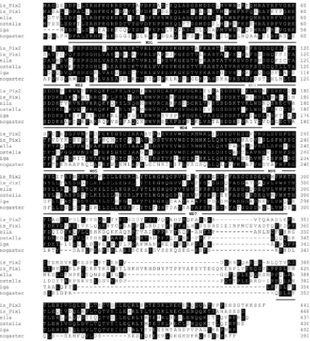
Figure 3. Sequence alignments of Pix proteins from representatives of major animal groups compared by ClustalW.Nematostella vectensis(Cnidaria, Anthozoa);Capitellasp. (Lophotrochozoa, Annelida);Drosophila melanobaster(Ecdysozoa, Arthropoda);Xenopus laevis, with two
Pixgenes (Deuterostomata, Vertebrata);Monosiga brevicollis(Choanoflagellida). Identical amino acids blocked in black and domains are identified according to theNematostellasequence, using the programs SMART and Coils at EMBL-EBI.
doi:10.1371/journal.pone.0003778.g003
reported to have flagellated cells in its life history. These organisms have not been intensively studied and it is possible that zoospores have been missed. Other Pelagophyceae have flagellated zoo-spores (Chrysonephos) or basal bodies (Pelagococcus), supporting the likelihood that a flagellated stage exists inAureococcus. This view is also supported by the presence in theAureococcusgenome of ab -tubulin with the C-terminal motif essential to form 9+2 cilia. Organisms without 9+2 cilia lack such tubulin. [19,20]. The single-celled algaChlorella, which is not known to have a flagellated stage, has a Pix homologue and ab-tubulin that is related to the flagellar type. This might support the idea of an unknown flagellated form ofChlorella, but the fact thatAureococcuscontains a variety of IFT genes but Chlorelladoes not, would make it more likely that, while the former has an undiscovered flagellated stage, Chlorelladoes not and uses Pix in some unusual way.
Other protists support the hypothesis that the presence of Pix homologues correlates with orthodox undulatory cilia/flagella. We have already mentioned the presence of a highly conserved Pix in a choanoflagellate. This is true of photosynthetic flagellates related to multicellular plants (Chlamydomonas, Volvox, Micromonas), indeed in Chlamydomonasthe Pix homologue (Poc1) has been identified in the flagellar proteome [21]. Pix is also present in other flagellates and ciliates, but is absent fromEntamoebaandDictyostelium, all of which lack cilia/flagella. InTetrahymenaa Pix (Poc1) homologue was identified in the basal body proteome. EM immunocytochemistry shows it to be localised to the basal end, or cartwheel of mature centrioles and to the amorphous assembly disc of newly forming centrioles [22]. This is different from Pix localisation in vertebrate centrioles, which is preferentially to the distal end [1]. This difference may be related to the absence of this cartwheel centriolar precursor in animals.
However, there are several protists which have flagella, but apparently lack Pix. These cases turn out to be provocative, because they make their flagella in an unusual way. Apicomplexans, such as the malarian parasite Plasmodium, have flagella that seem to be simpler than those of other eukaryotes, and their genomes lack IFT genes to transport components into the flagellum. In this case the axonemes are constructed within the main cell body [23], which is similar to the process by which the sperm axoneme is made in Drosophila, see below (review [24]). A second example is the centric diatomThalassiosira, which is deficient in Pix and IFT genes and has flagella with a 9(2)+0 axoneme [25].
Finally, there is direct evidence that the ciliary/centriolar function of Pix proteins is highly conserved in eukaryotes, since Pix proteins have been identified in the basal body proteome of Chlamydomonas[26] andTetrahymena[22]. Overall the presence of Pix, theb-tubulin motif, and undulatory cilia/flagella is correlated (Table S1), but there are several apparent exceptions which deserve further investigation.
Pix in Ecdysozoa
The absence of Pix in nematodes and its divergence in other Ecdysozoa has already been mentioned. One characteristic of Ecdysozoa is the absence of undulatory cilia, except in the sperm of some groups [27]. Ecdysozoa are characterised by an inert, moulting cuticle, which precludes the presence of ectodermal locomotory cilia [28]. Within the Ecdysozoa Drosophila has the most divergent Pix sequence identified in any Metazoan (1e-78) and several other insects are only a little less diverged (Figures 2, 4; Table S1). The crustacean Daphnia falls into the middle of this range. While, compared with other animals, there is some sequence conservation in the C-terminal region, in Diptera the Coils program predicts only a low probability that it will form a coiled-coil. On the other hand the probability is very high in Daphnia (Crustacea) and Apis (Hymenoptera), even though the
sequence is quite diverged. This suggests that this region of the protein may have lost its conserved function in dipteran flies, and that selection is relaxed in other Ecdysozoans.
What is special about Ecdysozoa? Neither nematodes nor most crustaceans, includingDaphnia, have flagellated sperm, although all have sensory primary cilia (9+0). Thus these organisms totally lack locomotory cilia. In insects the occurrence of primary cilia is restricted to Type I mechanoreceptors, so their dependence on sensory cilia is far less than in vertebrates [29]. In the insectDrosophila melanogaster sperm are flagellated, yet they are truly remarkable because the sperm tail is as long as the male. InDrosophila bifurcathey are forty times longer than the male, that is 58 mm [30]! It is hard to imagine that such sperm ever undulate in any organised way; rather the great length is likely to be an adaptation to sperm competition in a species in which females eject sperm before mating again with a new partner. Bees have more normal sperm length, but the axonemes are unusual in having 9+9+0 structure (Zama et al., 2005). Bee Pix is a little more like vertebrate Pix than that of Drosophila. This suggests that the axonemes of the flagella of insects may be different from those in other animals at a molecular level. Interestingly, the way in which sperm flagella are made inDrosophilais unusual and is more like that inPlasmodiumand diatoms described above, that is the axoneme is constructed in the main cytosol [31] without the function of IFT proteins [32,33]. On the other hand the sensory cilia ofDrosophilado require IFT proteins.
Thus, undulatory cilia/flagella have become simplified and less employed in the evolution of Ecdysozoa and they are absent in nematodes and most crustaceans. However, all members of the clade possess 9+0 primary, sensory cilia, although these are far less used than in animals like vertebrates. This suggests the generalisation that Pix proteins are essential to form typical motile cilia/flagella, but not necessarily the non-locomotory, primary kind. Since ecdysozoan Pix proteins are more diverged than in any members of the clade, including Choanoflagellates and multicel-lular animals, it suggests that the requirements of a Pix to make primary cilia are less demanding than in making normal undulatory cilia. In addition, the phylogenomics suggest that, at least in the long term, Pix proteins are only essential to make cilia/ flagella via an IFT-dependent mechanism. Since Pix proteins are localised within the lumen of the centriole, rather than in cilia/ flagella [1], it is reasonable to suggest that Pix proteins are needed to make the sort of centriole capable of making cilia/flagella via IFT transport processes. Of course other proteins are also needed to do this, includingb-tubulin with a specific tubulin motif (EGEF followed by 3 acidic residues; Table S1) [19,34]. These suggestions raise further questions about the core function of centrioles.
Centrioles and Pix in diverse organisms
with loss of the mitotic checkpoint protein, Mad2, even though Mad2 loss alone is not inviable (J. Raff, personal communication). Further, larval brains often develop malignant neoplasms in Dsas-4 deficient flies [39], suggesting that a role for centrioles in cell division remains important.
C. eleganslacks undulatory flagella, having amoeboid sperm, but has primary 9+0 sensory cilia. Its centrioles are of a single tubule, 9(1) kind [40]. Again this is consistent with the evolutionary loss of conventional cilia leading to a simplification of centrioles and a concomitant loss of Pix proteins. This simplification extends to the loss of other proteins from bothC. elegansandDrosophila, namelyd -ande-tubulin [41,42].
Together, these observations support a hypothesis that the core function of centrioles across eukaryotic phyla is to construct either the conventional motile 9(2)+2 secondary cilia or 9(2)+0 primary cilia. Pix is essential only for the former, and then only when they are made via an IFT-dependent mechanism.
Discussion
What are conventional centrioles for?
As explained, the presence of Pix, undulatory cilia/flagella and conventional centrioles correlate across the eukaryotic phyla.
Thus, when only primary cilia are present centrioles are simplified (C. elegans). InDrosophilathere are primary cilia and the sperm are flagellated, but their undulatory movement cannot be normal. Here Pix is divergent and in somatic tissues at least the centrioles are simplified. These organisms have primary, 9+0 cilia, for which a reduced centriole is sufficient. In advanced land plants and most fungi, without even primary cilia, the loss of all cilia has led to the loss of centrioles. Broadly speaking these correlations are supported across protists. Apparent exceptions like diatoms and apicomplexans have flagella, but no IFT genes and intra-cytosol manufacture of the axoneme.
[image:7.612.64.526.57.445.2]The main conclusion of these observations is that the core conserved function of centrioles is to construct flagella/cilia, but that if these are not of the undulatory 9(2)+2 kind a less sophisticated centriole will do (see also discussion by Marshall [5]). Without this function selection does not maintain centrioles at all, at least on evolutionary time scales. This makes sense because the structure of the centriole corresponds to that of the axoneme, indeed it blends into it from the basal body. On the other hand centriolar structure has no relationship to the microtubules nucleated by MTOCs. In this role centrioles merely act as a platform for aggregating MT nucleating proteins. Typically, in mammalian cells the centrioles organise a bipolar division spindle, prevent multipolar spindles
Figure 4. Comparison of Pix proteins in Ecdysozoa.ClustalW was used to compare the Pix proteins ofDrosophila melanogaster (Insecta, Diptera),Anopheles gambiae(Insecta, Diptera)Apis melifera(Insecta, Hymenoptera),Daphnia pulex(Crustacea, Cladocera). Details as in Figure 3. doi:10.1371/journal.pone.0003778.g004
forming, and control aspects of progression through the cell cycle. It is essential that these processes are precisely regulated or chromosomal missegregation may occur.
How did centrioles become associated with the division spindle?
Flagella clearly evolved in very early eukaryotic cells [43]. Ciliates are an early diverged offshoot, but typically the early protists would have had a single flagellum or a pair, in each case arising from a single basal body, or centriole. Although centrioles can arise de novo it would clearly be advantageous for each mitotic daughter cell to be able rapidly to assemble new flagella using a basal body, and hence to swim. Thus, there would have been selection for a robust mechanism to supply each daughter with a single centriole. On the one hand there would have to be robust control of centriolar replication, tightly linked to the cell cycle. On the other hand association of the centrioles with the spindle poles would have ensured that each centriole would arrive in a different daughter cell. One can envisage that there would have been progressive integration of the centrioles into other aspects of cell division. This might be compared to situations where parasitism evolves towards symbiosis. Initially, the centriole has ‘‘parasitized’’ the spindle, then the two have become mutually dependent. The tight linkage of centriolar replication to the cell cycle would have led to the centriole becoming a platform for molecules regulating the cell cycle and controlling the number of spindle poles, rather than simply using them for localisation. Such a role would be consistent with the observation thatChlamydomonaswithout centrioles can still divide, albeit with abnormal cell division and slow growth caused by disorganized mitotic spindles and cytoplasmic microtubules [44]. Of course plants and fungi tell us that while this role may be advantageous, without the role of centrioles in constructing cilia/ flagella their existence is unsupportable in the long term. It is noteworthy that these organisms have rigid cell walls, which may have enabled control of cell division by other means. Furthermore, while centrioles are largely dispensible in the later development of Drosophila, they are essential for the early divisions, when the embryos are syncytial [36]. This reduced dependence on centrioles may be aided by the fact that the requirement for centrioles is relaxed in ecdysozoans, but apparently centrioles are still absolutely necessary when there is not even a cell membrane for astral microtubule attachment.
To support these proposals further work is clearly needed. Exceptional situations, likeSelaginella, should be clarified.Drosophila provides an interesting test, since centrioles have different degrees of complexity in different tissues. If Pix is knocked out would it
affect only sperm, or sensory neurones, or other tissues as well? And are other centriolar proteins divergent or absent in a way that correlates with Pix? InChlamydomonascentrioles alternate between essential spindle roles and constructing flagella. So what would disruption of Pix do, indeed what is the precise function of Pix in any organism? While there are many experimental lines that need investigation, the argument for a core role of centrioles in making cilia/flagella, while largely non-experimental, is still a very strong one. Moreover the phylogenetic survey of centrioles certainly throws up interesting trends, particularly that of simplification of centrioles in the Ecdysozoa.
Supporting Information
Table S1 Survey across eukaryotes of centrioles, cilia, flagellum-specific b-tubulin and Pix homologues. Deuterostomia (white), Ecdysozoa (blue), Lophotrochozoa (grey), Cnidaria (yellow), Fungi (pink), plants and protistan sister groups (green), other protists (purple). Column 3, taxonomic groups are from the NCBI taxonomic database. The b-tubulin cilia/flagellum C-terminal domain was sought in genomes using BLAST with the Drosophila sequence (EGEFDED; the human sequence is EGEFDEE and consensus is EGEF+3 acidic residues[19]). The absence of this protein from the puffer fish genome is unlikely to be real. Column 8, Pix homologues were sought in genomes by BLAST with the Xenopus Pix2 sequence. The diagnostic feature of the Pix proteins was taken to be seven WD40 repeats plus homology in a coiled coil region in the C-terminus. The proteins were re-BLASTed again and their closest relatives were known Pix genes. The presence of the C-terminal coiled-coil region was confirmed using the program COILS (http://www.ch.embnet.org/software/COILS_form. html). The number of Pix homologues in genomes is shown in brackets. The similarity of Pix homologues is represented by a BLAST similarity score with the Xenopus Pix2 sequence. Found at: doi:10.1371/journal.pone.0003778.s001 (0.03 MB XLS)
Acknowledgments
We would like to thank R. Schmid (Leicester) for modelling of the Pix WD40 domain.
Author Contributions
Conceived and designed the experiments: HRW. Performed the experiments: HRW. Analyzed the data: HRW. Wrote the paper: HRW AMF.
References
1. Hames RS, Hames R, Prosser SL, Euteneuer U, Lopes CAM, et al. (2008) Pix1 and Pix2 are novel WD40 microtubule-associated proteins that colocalize with mitochondria in Xenopus germ plasm and centrosomes in human cells. Experimental Cell Research 314: 574–589.
2. Mikule K, Delaval B, Kaldis P, Jurcyzk A, Hergert P, et al. (2007) Loss of centrosome integrity induces p38-p53-p21-dependent G1-S arrest. Nat Cell Biol 9: 160–170.
3. Srsen V, Gnadt N, Dammermann A, Merdes A (2006) Inhibition of centrosome protein assembly leads to p53-dependent exit from the cell cycle. Journal of Cell Biology 174: 625–630.
4. Doxsey S, McCollum D, Theurkauf W (2005) Centrosomes in cellular regulation. Annu Rev Cell Dev Biol 21: 411–434.
5. Marshall WF (2007) What is the function of centrioles? J Cell Biochem 100: 916–922.
6. Dutcher SK (2003) Elucidation of basal body and centriole functions in Chlamydomonas reinhardtii. Traffic 4: 443–451.
7. Kuriyama R, Borisy GG (1981) Centriole Cycle in Chinese-Hamster ovary cells as determined by whole-mount electron microscopy. Journal of Cell Biology 91: 814–821.
8. Bettencourt-Dias M, Glover DM (2007) Centrosome biogenesis and function: centrosomics brings new understanding. Nature Reviews Molecular Cell Biology 8: 451–463.
9. Delattre M, Gonczy P (2004) The arithmetic of centrosome biogenesis. J Cell Sci 117: 1619–1630.
10. Hinchcliffe EH, Sluder G (2001) ‘‘It takes two to tango’’: understanding how centrosome duplication is regulated throughout the cell cycle. Genes Dev 15: 1167–1181.
11. Satir P, Christensen ST (2007) Overview of structure and function of mammalian cilia. Annu Rev Physiol 69: 377–400.
12. Afzelius BA (1999) Asymmetry of cilia and of mice and men. International Journal of Developmental Biology 43: 283–286.
13. Machado RJ, Moore W, Hames R, Houliston E, Chang P, et al. (2005)Xenopus Xpat protein is a major component of germ plasm and may function in its organisation and positioning. Developmental Biology 287: 289– 300.
15. Andersen JS, Wilkinson CJ, Mayor T, Mortensen P, Nigg EA, et al. (2003) Proteomic characterization of the human centrosome by protein correlation profiling. Nature 426: 570–574.
16. Renzaglia KS, Maden AR (2000) Microtubule organizing centers and the origin of centrioles during spermatogenesis in the pteridophyte Phylloglossum. Microscopy Research and Technique 49: 496–505.
17. Renzaglia KS, Bernhard DL, Garbary DJ (1999) Developmental ultrastructure of the male gamete ofSelaginella. International Journal of Plant Sciences 160: 14–28.
18. Garbary DJ, Renzaglia KS, Duckett JG (1993) The phylogeny of land plants - a cladistic-analysis based on male gametogenesis. Plant Systematics and Evolution 188: 237–269.
19. Nielsen MG, Turner FR, Hutchens JA, Raff EC (2001) Axoneme-specific beta-tubulin specialization: a conserved C-terminal motif specifies the central pair. Current Biology 11: 529–533.
20. Dutcher SK (2001) Motile organelles: The importance of specific tubulin isoforms. Current Biology 11: R419–R422.
21. Keller LC, Romijn EP, Zamora I, Yates Iii JR, Marshall WF (2005) Proteomic analysis of isolatedChlamydomonascentrioles reveals orthologs of ciliary disease genes. Current Biology 15: 1090–1098.
22. Kilburn CL, Pearson CG, Romijn EP, Meehl JB, Giddings TH Jr, et al. (2007) New Tetrahymena basal body protein components identify basal body domain structure. J Cell Biol 178: 905–912.
23. Sinden RE, Canning EU, Spain B (1976) Gametogenesis and fertilization in Plasmodium-yoelii-Nigeriensis - Transmission electron-microscope study. Proceed-ings of the Royal Society of London Series B-Biological Sciences 193: 55–&. 24. Avidor-Reiss T, Maer AM, Koundakjian E, Polyanovsky A, Keil T, et al. (2004)
Decoding cilia function: Defining specialized genes required for compartmen-talized cilia biogenesis. Cell 117: 527–539.
25. Montsant A, Allen AE, Coesel S, De Martino A, Falciatore A, et al. (2007) Identification and comparative genomic analysis of signaling and regulatory components in the diatom Thalassiosira pseudonana. Journal of Phycology 43: 585–604.
26. Merchant SS, Prochnik SE, Vallon O, Harris EH, Karpowicz SJ, et al. (2007) The Chlamydomonas genome reveals the evolution of key animal and plant functions. Science 318: 245–251.
27. Schmidt-Rhaesa A, Bartolomaeus T, Lemburg C, Ehlers U, Garey JR (1998) The position of the Arthropoda in the phylogenetic system. Journal of Morphology 238: 263–285.
28. Valentine JW, Collins AG (2000) The significance of moulting in Ecdysozoan evolution. Evolution & Development 2: 152–156.
29. Basto R, Lau J, Vinogradova T, Gardiol A, Woods CG, et al. (2006) Flies without centrioles. Cell 125: 1375–1386.
30. Pitnick S, Spicer GS, Markow TA (1995) How Long Is a Giant Sperm. Nature 375: 109–109.
31. Tokuyasu KT, Hardy RW, Peacock WJ (1972) Dynamics of spermiogenesis in Drosophila melanogaster.1. Individualization process. Zeitschrift Fur Zellforschung Und Mikroskopische Anatomie 124: 479–&.
32. Han YG, Kwok BH, Kernan MJ (2003) Intraflagellar transport is required in Drosophila to differentiate sensory cilia but not sperm. Current Biology 13: 1679–1686.
33. Sarpal R, Todi SV, Sivan-Loukianova E, Shirolikar S, Subramanian N, et al. (2003)DrosophilaKAP interacts with the Kinesin II motor subunit KLP64D to assemble chordotonal sensory cilia, but not sperm tails. Current Biology 13: 1687–1696.
34. Popodi EM, Hoyle HD, Turner FR, Xu K, Kruse S, et al. (2008) Axoneme specialization embedded in a ‘‘generalist’’ beta-tubulin. Cell Motility and the Cytoskeleton 65: 216–237.
35. Gonzalez C, Tavosanis G, Mollinari C (1998) Centrosomes and microtubule organisation during Drosophila development. Journal of Cell Science 111: 2697–2706.
36. Stevens NR, Raposo A, Basto R, St Johnston D, Raff JW (2007) From stem cell to embryo without centrioles. Current Biology 17: 1498–1503.
37. Rodrigues-Martins A, Riparbelli M, Callaini G, Glover DM, Bettencourt-Dias M (2008) From centriole biogenesis to cellular function - Centrioles are essential for cell division at critical developmental stages. Cell Cycle 7: 11–16.
38. Marshall WF, Rosenbaum JL (2000) How centrioles work: lessons from green yeast. Curr Opin Cell Biol 12: 119–125.
39. Castellanos E, Dominguez P, Gonzalez C (2008) Centrosome dysfunction in Drosophilaneural stem cells causes tumors that are not due to genome instability. Current Biology 18: 1209–1214.
40. Wolf N, Hirsh D, McIntosh JR (1978) Spermatogenesis in males of free-living nematode,Caenorhabditis elegans. Journal of Ultrastructure Research 63: 155–169. 41. Feldman JL, Marshall WF (2004) Centrioles: Bad to be bald? Current Biology
14: R659–R660.
42. Chang P, Stearns T (2000) delta-Tubulin and epsilon-tubulin: two new human centrosomal tubulins reveal new aspects of centrosome structure and function. Nature Cell Biology 2: 30–35.
43. Cavalier-Smith T (2002) The phagotrophic origin of eukaryotes and phyloge-netic classification of protozoa. International Journal of Systematic and Evolutionary Microbiology 52: 297–354.
44. Matsuura K, Lefebvre PA, Kamiya R, Hirono M (2004) Bld10p, a novel protein essential for basal body assembly in Chlamydomonas: localization to the cartwheel, the first ninefold symmetrical structure appearing during assembly. Journal of Cell Biology 165: 663–671.