THE ROLE OF ENDOTHELIAL CELLS IN PROMOTING
ADHESION, SPREADING AND MIGRATION OF B16F10
CELLS
Valerie Anne Ferro
A Thesis Submitted for the Degree of PhD
at the
University of St Andrews
1989
Full metadata for this item is available in
St Andrews Research Repository
at:
http://research-repository.st-andrews.ac.uk/
Please use this identifier to cite or link to this item:
http://hdl.handle.net/10023/14067
THE ROLE OF ENDQTHHÏ JA Ï. CELLS IN PROMOTING
OF B16F10 CELLS
A thesis submitted to the University of St. Andrews for the degree of
Doctor of Philosophy
by
VALERIE ANNE FERRO
Department of Biology and Preclinical Medicine, University of St. Andrews.
ProQuest Number: 10166584
All rights reserved
INFORMATION TO ALL USERS
The quality of this reproduction is dependent upon the quality of the copy submitted.
In the unlikely event that the author did not send a com plete manuscript and there are missing pages, these will be noted. Also, if material had to be removed,
a note will indicate the deletion.
uest
ProQuest 10166584
Published by ProQuest LLO (2017). Copyright of the Dissertation is held by the Author.
All rights reserved.
This work is protected against unauthorized copying under Title 17, United States C ode Microform Edition © ProQuest LLO.
ProQuest LLO.
789 East Eisenhower Parkway P.Q. Box 1346
PAGE NO.
Abstract Declaration Copyright
Academic record Acknowledgements Abbreviations Figures
Tables
Photographs
Scanning electron micrographs
I General Introduction 1-17
II Extravasation 18-66
III Adhesion 67-134
IV Spreading 135-160
V Migration 161-180
VI Conclusion 181-187
Appendices References
% %
4
.1
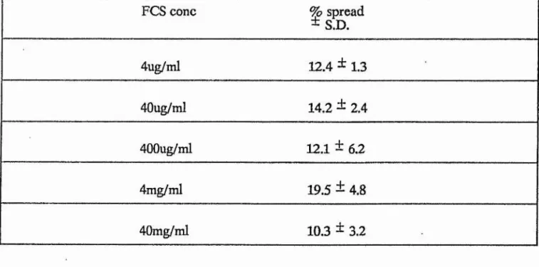
For the successful establishment of secondary tumours, blood-bome metastatic tumour cells must adhere and spread on the vascular endothelium before they can migrate through it to form secondary growths in the tissue beneath. In this study an in vitro assay was developed to study the behavourial interactions between B16F10 cells and Bovine aortic endothelial cells.
I, Valerie Anne Ferro, hereby certify that this thesis has been composed by myself, that it is a record of my own work, and that it has not been accepted in partial or complete fulfilment of any other degree or professional qualification.
Signed Date
I was admitted to the Faculty of Science of the University of St. Andrews under Ordinance General No. 12 in October 1985, and as a candidate for the degree of Ph.D. in October 1985.
Signed Date I. k
I hereby certify that the candidate has fulfilled the conditions of the resolution and Regulations appropriate to the Degree of Ph.D.
Signature of Supervisor Date
Unrestricted
In submitting this thesis to the University of St. Andrews I understand that I am giving permission for it to be made available for use in accordance with the regulations of the University Library for the time being in force, subject to any copyright vested in the work not being affected thereby. I also understand that the title and abstract will be published, and that a copy of the work may be made and supplied to any bona fide library or research worker.
j
I would firstly like to thank my supervisors, Dr. Jim Aiton (University of St Andrews) and Dr. Clive Evans (University of Auckland) for aU their help and advice. I would especially like to thank Dr. Gordon Cramb for his encouragement and helpful discussions over the past year. I would also like to thank Ken Thom, Dave Roche, Dave Ogden, Iain Laurie, Karen Johnstone, Irvine Davidson, Carol Voy and Mary Falls for their technical assistance, and the remaining members of the Biology Department for their assistance at various times.
BAE Bovine aortic endothelial cells BSA Bovine serum albumin
CE Crude BAE cell extract
CEM Conditioned BAE cell medium
CMF-PBS Dulbecco's calcium and magnesium free phosphate buffered saline EC Eagle’s complete medium
EDTA Ethylene diamine tetra acetic acid ELISA Enzyme linked immunosorbent assay FCA Freund’s complete adjuvant
FCS Fetal calf serum Fg Fibrinogen
FIA Freund’s incomplete adjuvant Fn Fibronectin
HAT RPM I1640 medium containing hypoxanthine, aminopterin and thymidine
HT RPMI 1640 medium containing hypoxanthine and thymidine IdU Iodine labelled deoxy uridine
Lm Laminin
O.D. Optical density
PAGE Polyacrylamide gel electrophoresis PEG Polyethylene glycol
RPMI RPMI 1640 medium SDS Sodium dodecyl sulphate SF-RPMI Serum free RPMI 1640 medium
TEMED N, N, N’,N’-tetramethyl ethylene diamine
Tn Tenascin
TSP Thrombospondin
Tween 20 Polyoxyethylene sorbitan monolaurate Vn Vitronectin
FI.G.UBE.8.
PAGE NO
Fig 1 spread of tumour cells 8
Fig 2 BAE-B16F10 cell interactions 46
Fig 3 Shows the zone of dead cells 55
Fig 4 Area covered by tumour cells 60
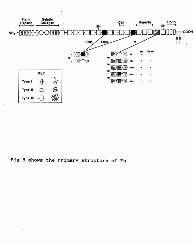
Fig 5 Primary structure of Fn 72
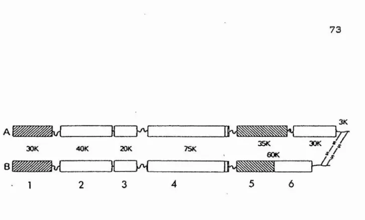
Fig 6 Functional domain structure of Fn 73
Fig 7 Effect of RGD peptide on attachment cell 74
Fig 8 Functional domain structure of Lm 77
Fig 9 Primary structure of Lm 77
Fig 10 Domain structure of vitronectin 81
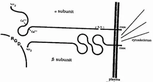
Fig 11 Structure of RGD-receptor 88
Fig 12 Extraction of cell extracts 99-100
Fig 13 Apparatus used in probing dot blots 105
Fig 14 Apparatus used in probing Western blots 106
Fig 15 Protocol used in cell attachment assay 107
Fig 16 Protocol used in production of hybridomas 112
Fig 17 Protocol used in inhibition of attachment 115
Fig 18 Shows the possible role of cell extracts 134
Fig 19 Protocol used in spreading and inhibition 142
Fig 20 Antibodies inhibiting spreading to CEM 157
Fig 21 Steps to extravasation 161
Fig 22 Boyden chamber apparatus 168
Fig 23 Migration response to Fn 176
TABLES
PAGE NO
TABLE 1 Interactions of ECM proteins 84
TABLE 2 Cross reactivity of anti -BAE EXT 117
TABLE 3 Test panel of antibody specificities 119
TABLE 4 X-reactivity and inhibition of adhesion 120
TABLE 5 Spreading dose response data - CE and CEM 145
TABLE 6 Spreading dose response data - Fn and Lm 148
TABLE 7 Spreading dose response data - FCS 149
TABLE 8 Surface areas of spread ce 1 Is 154
TABLE 9 Effect of antibodies on spread to CEM 156
TABLE 10 Chemotaxis, kinesis and random locomotion 169
TABLE 11 Haptotaxis locomotion 170
TABLE 12 Migration to Fn 175
TABLE 13 Migration to Lm 175
:l
EH.Q.XQGHABHS
PAGE NO
Photograph 1 B16F10 cells - day 3 35
Photograph 2 BAE cells - day 3 37
Photograph 3 B16F10 aggregate - day 10 41
Photograph 4 Compressed BAE cells - day 10 45
Photograph 5 BAE-B16F10 cells -,day 17 48
Photograph 6 BAE-B16F10 cells - day 17 50
Photograph 7 B16F10 aggregate - day 17 52
Photograph 8 B16F10 aggregate - day 17 52
Photograph 9 BAE-B16F10 cells - day 30 56
Photograph 10 BAE-B16F10 cells - day 30 58
Photograph 11 BAE-B16F10 cells - day 30 58
Photograph 12 SDS-PAGE analysis 124
Photograph 13 Spreading on CE 147
Photograph 14 Spreading on CEM 147
Photograph 15 Spreading on Fn 151
Photograph 16 Spreading on Fn 151
Photograph 17 Spreading on Lm 153
SCANNING ELECTRON MICROGRAPHS
PAGE NO
■4
SEM 1 B16F10 cells - day 3 35
SEM 2 BAE cells - day 3 39 |
SEM 3 BAE cells - day 3 39
SEM 4 B16F10 aggregate - day 10 41
SEM 5 BAE-B16F10 cells - day 10 43
SEM 6 BAE-B16F10 cells - day 17 48
m L i m m c . %
Tumour cells may tentatively be classified as either
benign or malignant. The classification may be based on
several factors, namely comparison with normal cells,
composition, morphology, growth and cell behaviour (Sherbert,
1982). Growth and cell behaviour properties are common
indices used to allow a reliable distinction to be made
between the two types of tumour cells. For a more detailed account of the differences between benign and malignant
tumours see the following reviews: Cole, 1973; Weiss, 1976;
Sherbert, 1982; Vincent, 1985 and Weiss, 1985.
Briefly, benign tumours usually grow by expansion
through cell division, remain localised and displace
surrounding tissue (Woodruff, 1980). Pressure atrophy to
cells surrounding the tumour, may result in the formation of a
fibrous coating, around the tumour. However, this is not a
universal feature of benign tumours (Sherbert, 1982). Notably, infiltration and destruction (invasion) of the tissue surrounding the tumour does not occur (Weiss, 1985).
i. Invasion of the primary tumour into surrounding tissues. ii. Spread of the tumour into body cavities and vessels and
release of tumour cells for metastasis.
iii. Arrest of tumour cells at particular sites and invasion into surrounding tissue.
iv. Manipulation of the new environment to promote tumour
cell survival, vascularisation and tumour growth.
metastasis (Vincent, 1985). A frequently cited exception to
this general rule, is basal cell carcinoma. This common form s
of skin cancer, rarely, if ever, metastasizes, although it is capable of invading widely and may be highly destructive (Levenne .«giLai.. 1982) . The reason that this tumour displays such a marked distinction between invasive and metastatic characteristics is not known.
Metastasis may be defined as the transfer and spread of tumour cells from a primary site to one or more remote
secondary sites (Roth al, 1976). A series of sequential
steps, leading to the formation of distant métastasés was proposed by Nicolson and Winklehake (1975):
%
i
The first steps leading to metastasis are extension and local invasion (reviewed by Fidler and Nicolson, 1981). Local invasion is usually a fundamental step leading to spread of
tumour. By breaching basement membranes (Trinkaus, 1976;
Nicolson, 1981) (cellular sheet-like structures) (Siegal .e..t a,l, 1981) and interstitial connective tissues (consisting of cells located in a matrix of collagen fibres, glycoproteins and proteoglycans) (Weiss, 1985), invasion of surrounding tissues can occur (Willis, 1973; Poste and Fidler, 1980).
The following observations, made .lo vltm, may help
account for the release mechanisms involved in invasion and ;|
are reviewed by Hart (1981):
(1) an increase in amoeboid activity
(2) a decrease in adhesiveness (3) loss of contact inhibition
(4) a possible release of lytic enzymes.
The relative importance of these mechanisms may vary
from tumour system to tumour system (Cole, 1973).
Susceptibility of host tissue to tumour cell invasion varies with the tissue type: cartilage, tendons, ligaments and arteries are relatively resistant, whereas veins, lymphatics, soft tissue and muscles are easily invaded (Willis, 1973). It would appear that tissues rich in ground substance or those containing dense elastic or collagen fibres, provide a more
successful barrier to malignant cells (Weiss, 1985). Normal
host interactions at the site of invasion may also be #
important (Woodruff, 1980).
Once primary malignant cell invasion has occurred, the next step appears to be cell detachment when single tumour
cells or tumour emboli, separate from the primary mass
(Sherbert, 1982). Release mechanisms (reviewed by Weiss,
1985), include adhesive weakness, local cell rupture and enzymatic destruction, among others.
SEBE.m._QE. XUMQ.URS.
There are three major pathways involved in tumour cell dissemination following local invasion, as outlined by Cole
(1973) and Willis (1973):
This route is taken particularly by carcinoma (tumours arising from epithelial cells) (Lindberg, 1972; Carter, 1978) and to a lesser extent by malignant melanoma (tumours arising from the melanin producing cells in the skin) (Carter, 1978;
1984). Primary tumours do not contain lymphatic vessels and so major points of entry are through small lymph vessels near the edge of the growing lesion (Carr e.t al, 1981) . Access is generally achieved through the structural features of these small vessels which lack a basement membrane and have a high proportion of gaps between the endothelial cells (Yoffey and
4
:i
Court ice, 1970) . As in the case of macrophages and |
lymphocytes, tumour cells may enter the vessels through these
gaps (Carr , 1976) and pass into the lymph nodes (Hewitt
and Blake, 1975) which are able to filter clumps of cells, but presumably not single cells (Cobb and Steer, 1987).
Tumour cells may also readily enter thin walled venules
(Papadimitriou and Woods, 1975) and these cells may
subsequently gain access to the lymphatic system via anastamoses between venules and small lymphatic vessels (Wood,
1958; Haagenson, 1972; del Regato, 1978). There is now
substantial evidence that malignant cells can pass freely
between the lymphatic and circulatory system (Fidler et al.
1978) and the belief that tumour spread occurs exclusively by one or other, is an oversimplification.
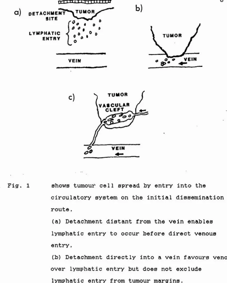
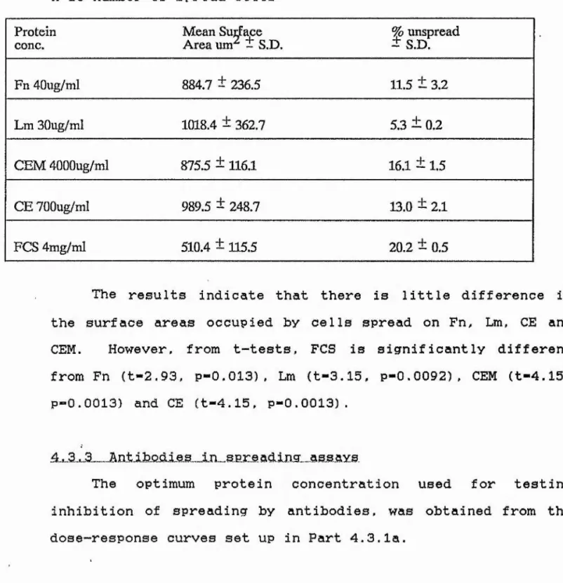
The most common route for metastasis is via the circulatory system. An example being melanoma (Carter, 1978). There are three routes into the circulatory system as shown in Fig, 1, p8 (taken from Weiss, 1985): lymphatic entry, direct venous invasion and invasion of tumour vessels.
This occurs if cell detachment from the primary tumour results prior to contact between tumour and blood vessel
I
Malignant cells may enter the bloodstream by invading blood vessels lying near the advancing edge of the tumour
(Haagensen, 1972). See Fig. 1(b). It is thought that
mechanical (Easty and Easty, 1978) and/or enzymatic factors,
i
occurring at the edge of growing tumours (Kleinerman and 4
Liotta, 1977), may contribute to penetration of the vascular system.
Vascularisation of the primary tumour, is thought to be
triggered off by tumour angiogenic factors (Folkman e.,fe Ml,
1971; Folkman, 1974). Invasion of these tumour vessels may
occur through defects in vessel walls (Willis, 1973; Warren,
1974). These vessels are often imperfectly formed with
irregular lamina, discontinuous or sometimes
abnormalendothelium and defective membranes which make them more penetrable (Carter, 1984). See Fig, 1(c).
.S.p.r„e,.a.4..„.by .s..ur.f. .ft,c.e..
Tumour cells implant readily on the endothelial surface of pleura and peritoneum (Cole, 1973). When this occurs on an extensive scale, it is invariably associated with an effusion
of fluid containing tumour cells (Woodruff, 1980).
Dissemination of cancer of the lung in the pleural cavity and of cancer of the ovary, stomach and colon in the peritoneal cavity, are examples (Willis, 1973).
Tumour cells may also implant on epithelial surfaces (Cole, 1973); this occurs in the urinary tract from papillary carcinoma of the renal pelvis and in the bladder from primary bladder tumours (Willis, 1973).
Spread also occurs in the gastointestinal tract (Cole,
1973), but appears to depend on a pre-existing breach in the j
continuity of the epithelium (Woodruff, 1980). Implantation M
of tumour cells in the subcutaneous tissue and in the wall of the colon may also occur during the course of surgical operations (Cole, 1973).
I
a ) D E T A C H M E N T S I TE
Trr iJ iT T T r i iT rTi i -ii
0
8
l y m p h a t i c
ENTRY
VEIN
b)
T UMOR VEIN
c) T U M O R
V A S C U L A R C L E F T
V E I N
Fig. 1 shows tumour cell spread by entry into the
circulatory system on the initial dissemination route.
(a) Detachment distant from the vein enables lymphatic entry to occur before direct venous entry.
(b) Detachment directly into a vein favours venous over lymphatic entry but does not exclude
lymphatic entry from tumour margins.
1.5 ARREST a n d DISTRIBUTION OF TUMOUR CELLS I.M BLOOD-BORNE
MIAS,TA.SI.S
In this study, we are primarily concerned with events which occur in the circulatory system. First let us consider the events which occur in blood-borne metastasis, following invasion and spread.
Some tumour cells are released from the primary mass, possibly by becoming transiently less adhesive (Vollmers and Birchmeier, 1983), or by hydrolase activity of necrotic tissue which is prevalent in large tumours (Weiss, 1977). or by shear
forces in the blood stream (Glucksmann and Cherry, 1964). In
order to form metastses, these released circulating tumour
cells must become arrested at particular sites, such as in
capillary beds (Wood .e,t &1. 1966) . As shown in clinical and experimental studies, the mere presence of circulating tumour
cells does not constitute metastsis (S'alsbury, 1975: Fidler ,e,.t
al, 1978).
The site of arrest on the endothelial lining of the blood vessels may be non-specific and determined by the size of the
clumps of tumour cells released from the primary mass (Liotta
1976) . Large clumps of cells can become mechanically
impacted in vessels and the size and deformabi1ity of the
tumour cells, the diameter and distensibility of the
capillaries and the interaction of the tumour cells with each
10
create multice1lular emboli are relevant in this context (Poste and Fidler. 1900).
However, the arrest of smaller aggregates and single cells cannot be accounted for by mechanical means alone. The experiments of Wood (1958; 1966), described the association of tumour cells with platelets, lymphocytes and thromboplastic
components during the arrest process. Various workers have
since shown that tumour cells do indeed undergo interactions with these host cells and components to form stable aggregates
(Pearlstien et .al. 1980; Fidler, 1978; Kohga and Tanaka,
1979). In addition, Stringfellow and Fitzpatrick (1979) and
Pearlstein ,e..t ,al (1980) suggest that prostaglandins may play a role in the arrest of many tumours. Roos and Dingemans (1979)
suugest that the inflammatory events involving fibrin
deposition and platelet aggregation around the tumour cells may be activated to protect the cancer cells from blood shear forces rather than in playing an active role in the arrest of
tumour cells. Some reviews on the role of inflammatory
reactions in metastsis include those by Gatspar (1977);
Hilgard ,et a,.l (1977); Sherbert (1982). In brief, arrest and emboli trigger events which couple an inflammatory response, blood coagulation and fibrinolysis, complement activation, and tissue damage, partially through products of the arachidonic
acid cascade. The effects of vessel wall damage enhances
further cancer arrest since exposure of the subendothelium results in platelets becoming adherent and activated.
11
processes are required for arrest, especially in view of the fact that clinical and experimental observations show that certain tumours consistently metastasize to particular organs (organ-specificity) (Willis, 1973; Cole. 1973: Fidler and
Nicolson, 1976). More recently, emphasis has been placed on
cell surface properties and an excellent demonstration of this
was provided by Hart and Fidler (1980). They showed that
B16F10 cells inoculated intravenously, form deposits on lung and ovary tissue even when these organs are transplanted to
new locations within the body. This suggests that perhaps
tumour cell surface determinants (Poste, 1980) or properties of the endothelial cells within different organs (Nicolson,
1982; Auerbach and Joseph, 1984) contribute to arrest.
Auerbach and Joseph (1984). postulated that capillary
endothelial cells differ in their surface antigens, the
differences reflecting their developmental history. These
distinct organ-associated antigens on the cell surfaces could
explain organ-specif icity. A report by Dietrich a..l (1983) .
suggested that heparan sulphates play a role in cell-cell recognition and adhesiveness since heparan sulphates are able
to alter their configuration. To investigate
organ-specif icity, Alby and Auerbach (1984) used endothelial cells from mouse brain or ovaries and tested them in an In v,l.tr.Q,
assay system for adhesion. They found that ovary-derived
teratoma cells and a testicular teratoma with ovary seeding properties adhered preferentially to ovary endothelial cells.
î
12
endothelial cells. At present, too few endothelial cell lines
are available to validate the studies. However, lymphocyte
homing studies demonstrate clearly the importance of
endothelial cell specificity in regulatin extravasation of
circulating cells (Stamper and Woodruff, 1977; Chen and
Singer, 1983; Streeter et a l, 1988). Lymphocyte adherence to, and penetration through specialised endothelial cells, depends
on the presence of specific adherence molecules on the
endothelial cells (Butcher et al. 1979). Moreover, there is
organ specificity in this lymphocyte-endothelial cell %
interactive system (Gallatin et a.1. 1983) .
Historically, two simple hypotheses have been used to account for organ specificity (reviewed by Weiss, 1985). The first, the "haemodynamic" or "mechanical" hypothesis, focuses
on the delivery of tumour emboli to target organs. The 4
second, the "seed-soi1" hypothesis, focuses on the
differential growth of arrested emboli in different target
organs subsequent to delivery. A contribution to the theory
of organ specificity was made by Fidler and Nicolson (1976), using B16F1 cells (tumour cells which show low arrest in the
lungs) and B16F10 cells (tumour cells which show high arrest in the lungs). Following either tail vein or left ventricular injections of '^^IdU-labelled melanoma cells, the initial
arrest of cells depended on the injection route; after
intravenous injection, more cells were arrested in the other 4
organs than after intracardiac injections. However, 24 hours after injection, in both cell lines, more viable cancer cells
13
were present in the lungs after intracardiac injections.
Fourteen days after the injection, similar numbers of
pulmonary tumours were seen following tail vein or
intracardiac administration. The results suggested that the
organ pattern of tumour colonisation was not determined by
non-specific arrest in the first organ encountered by the
injected cells.
At present, it appears that the "haemodynamic" hypothesis
I
accounts for the majority of metastatic patterns of |
development up to the stage of general arterial dissemination.
At the stage of general dissemination leading to end
metastasis, some features of pattern are clearly not
explicable solely in terms of the "haemodynamic" hypothesis. This implicates the "seed-soi1" hypothesis. It seems likely that the "seed-soi1" hypothesis accounts for the differential growth rate of métastasés in different target organs, as distinct from the incidence of métastasés in these organs.
.I..Â
E.aTABLI.SHMEH.T .QE MEXAETABEB
Ultrastructural studies indicate, that tumour cells gain
a secure foothold in the circulatory system by adhering to
endothelial cells and to the underlying basement membrane (Warren, 1974; Fidler eLai,. 1978: Auerbach and Joseph, 1984) . It is the initial arrest reactions which culminate in the
adhesion of tumour cells to the vascular endothelium: this
leads to exposure of the sub-endothelium which also acts as an
14
the vessel into surrounding tissue(s) in order to escape from
the hostile environment of the circulatory system.
Extravasation of malignant cells is believed to involve
mechanisms similar to those responsible for the initial
invasion of blood vessels fPoste and Fidler, 1980). Following
successful extravasation, small tumour cell lesions are
f ormed.
Throughout the metastatic process, malignant cells are
subject to attack by host defence systems which may be immune
or non-immune (Weiss, 1985). The chances of métastasés
developing from circulating tumour cells therefore are small (reviewed by Weiss, 1980) and may depend on:
(1) anatomical factors which influence the distribution of tumour embo1i;
(2) properties of the sites at which emboli are arrested and the extent to which they can satisfy the metabolic and other requirements of the particular tumour;
(3) properties of the tumour including its capacity to produce plasmin-like enzymes and
(4) various factors which influence local and general
resistance to the tumour and modify the complex process of metastasis in ways which, for the most part, remain to be elucidated.
Even following extravasation, only a small proportion of
tumour cells survive and are able to become established. Little is known about the factors involved in establishment and growth of métastasés in the target tissue. One factor may
""I
■! 15
cells are able to induce the^formation of blood vessels from the host circulatory system (Folkman and HaudenschiId, 1980) and are thought to be able to do this by releasing a tumour
angiogenic factor(s) (TAF). This factor(s), which was
isolated by Folkman ,e,.t m.i (1971) , appears to be produced by tumours both ln.,.,„ y i t m and ia.,.viyo. (Klagsburn et aJL, 1976; Phillips et .al, 1976; Brem ,et .a 1, 1977) .
The failure of the rest of the extravasated cancer cells to grow may be due to a number of reasons, including the inability of the tumour cells to overcome host defence mechanisms (reviewed by Poste and Fidler, 1980; Weiss, 1985) and a non-favourable host environment (Folkman, 1975). Horak e..t™ ,„al, (1986) showed that organs such as murine liver and thyroid gland are able to rapidly and effectively diminish the number of live tumour cells in vitro,, whereas murine lung and
ovary promote tumour cell attachment and survival. This
effect can also be exerted by cell-free organ conditioned medium and does not require previous contact with the tumour
cells (Nicolson and Dulski, 1986). This observation
implicates a soluble substance in determining which organs allows the survival of different tumour types.
It would appear that the ultimately successful cells- as
few as 0.1% of the circulating tumour cells (Fidler al.
1978) - would have to undergo rigorous selection in order to survive (Roos and Dingemans, 1979; Poste and Fidler, 1980;;
Poste ,e.,t al, 1981). It is possible that only a small
subpopulation of cells in the heterogenous primary tumour
possess the characteristics to establish a successful
16
metastasis (Alexander, 1984; Fidler, 1978). Fidler (1973),
showed that it is possible to obtain tumour lines with
increasing metastatic potential by subjecting the cell
population to selection procedure involving successive s
transplantation with intermittent in v itro culturing. Fidler injected viable B16 melanoma cells into the tail veins of C57BL/6 mice, isolated the tumour nodules which formed in the
lungs of these animals and grew these isolated cells in tissue 'I
culture. The cells of the first l.n vivo selection for lung
colonization were designated B16F1. The B16F1 cells were
injected back into syngeneic mice, and the pulmonary colonies that formed were isolated and adapted for culture (B16F2). The procedure was carried out ten times to obtain the B16F10
cell line. At each stage of the selection procedure, the
ability of the cells to survive and form colonies in the lungs increased (Fidler and Nicolson, 1976; Fidler and Kripke,
1977). This outcome is compatible with the view that the
original unselected primary tumour contained a number of variant lines differing widely in their potential for lung colonization.
The presence of heterogeneity within a primary tumour has profound implications for the way in which research should be carried out to identify the cellular properties responsible
for metastasis. Experiments to identify features unique to
metastatic subpopulations require comparisons to be made with
non-metastatic (but tumourigenic) subpopulations of cells from 4
17
its variant cell lines are a good, model to work with.
From clinical studies. it is clear that the formation of metastses, is an important step in the pathogenesis of cancer, since metastatic growths are the cause of most deaths in cancer patients (Nicolson et ai, 1976). A crucial step in the survival of tumour cells which have been disseminated through
the circulatory system is extravasation. A greater
understanding of this process may help in preventing the formation of métastasés.
Bearing this in mind, the aim of the work described in this thesis is to try and investigate in greater detail the interactions between tumour and endothelial cells, with a view to increasing knowledge on the process of extravasation.
18
.2..»,„1 1.N.TRQD.U.C.T.I.DK
Extravasation, as defined by Wood (1958), is the process by which malignant cells adherent to endothelial cells penetrate the endothelial cell layer, bind to and finally
invade the underlying basement membrane. In doing so the
tumour cells escape host defence mechanisms (Weiss, 1985) and
blood shear forces (Glucksmann and Cherry, 1964). This
definition was arrived at from micro-cinematography
observations made by Wood (1958) of cancer cells crawling out
from blood vessels in rabbit ear chamber experiments. In
these experiments, Wood injected V2 carcinoma cells into the central artery of a rabbit ear, proximal to a transparent
window implanted in the ear. The events following this
injection were photographed at high magnification.
"Successful" tumour emboli were noted to form aggregates readily and to adhere strongly to the endothelium in comparison with "unsuccessful" tumour emboli. Adhesion to the endothelium was followed by formation of thrombi around the cells. Within a variable period from a few minutes to a few hours, the endothelial cells underlying the tumour thrombus
appeared to become damaged. Leucocyte migration occurred
beneath the tumour thrombus and the cancer cells followed, passing through breaches in the endothelial lining caused by a variety of factors, including platelets, histamines and
leucocyte-mediated injury (Weiss, 1985).
19
1. Escape of tumour cells through defects in the
endothelial wall (Sherbert 1982; Carter, 1984).
2. Endothelial penetration by breakage of endothelial
intercellular junctions (Locker , 1970; Nicolson,
1981).
3. Proteolytic enzyme destruction of endothelial cells by
tumour cells (Liotta et al. 1980; Crissman, 1985). The enzymes are released from the pseudopodia of the tumour cells.
4. Penetration of endothelial cell cytoplasm by tumour cell
pseudopodia and by pinocytosis (Dingemans, 1974).
5. Endothelial wall rupture due to physical disruption as a
result of proliferation of tumour cells at the site of arrest by pressure of the growing mass (Basserga and
Saffioti, 1955; Chew 1976).
6. Changes in permeability of the endothelium e.g. during
inflammation, histamine release makes the endothelium
1 eaky (Majno , 1969; Ozaki ei-jSl, 1971).
Whatever the method of escape, the basement membrane is the major barrier to extravasation and it is likely that more than one mechanism is utilised in this process (Crissman, 1985).
20
investigation of the interaction between tumour and
endothelial cells. Workers using in. Klfcro. systems have added a further complication to the subject by confronting tumour cells of one species with endothelial cells of another species. The reasons for doing so are usually technical; but surprisingly, there is little evidence to show that the results obtained from studies of interactions between cells of different species and those obtained from systems utilising syngeneic tissues are markedly different (Easty and Easty,
1984) . There are obvious differences between and in
Ki.Èr.Q studies. In.... KiKQ. there is a complex interplay of hormonal, inflammatory and possibly immunological responses
(reviewed by Eccles and Alexander, 1974; Sherbert, 1982;
Weiss, 1985; Hanna, 1985; Robins, 1986) which may contribute to the magnitude and rates of interactions; ln...ici.tr.Q these are
usually absent. Thus, systems would appear to be the
more desirable. In practice, the complexity and
interdependence of reactions, and the difficulty of
examining the effects of a single component, renders analysis
of la, ,irJ.KO. systems very difficult, while the relative
simplicity of the la._._Kl,±r.o systems facilitates analysis of mechanisms, but may yield data which are not substantiated by
subsequent experiments in animals. Nevertheless, In yitro
experiments have proven invaluable in elucidating endothelial and tumour cell interactions.
21
leucocytes seeded onto monolayers of bovine aortic endothelial
cells (BAE) . BAE cells are commonly used In vXtXlQ
experiments, because these cells form intercellular junctions and an extracellular matrix in culture of similar composition to the basement membrane in K.iv..Q (Birdwell .e..t .ai ^ 1978). Using time-lapse photography, Kramer and Nicolson (1979) observed that, in general, the malignant cells, monocytes and
leucocytes, which can all invade the endothelium in..._.vi.K.o (Ward ..«1, 1979), firstly adhered to the endothelial cells. This was followed by the retraction of the endothelial cells at the site of adhesion. However, only cells adjacent to endothelial cell junctions were able to migrate between the undersurface
of the endiothelial and their extracellular matrices. The
endothelial juctions then re-sealed, walling off the invading cells. Similar results were obtained by Zamora .e.La.1. (1980), who inoculated monolayers of endothelial cells grown on collagen gels with multice1lular spheroids of mouse mammary carcinoma. Again, retraction of the endothelial cells in the vicinity of tumour cells was observed following adhesion, and the mammary tumour cells spread on the collagen, migrating under and over the endothelial cells and into the collagen. The steps seen in these experiments correspond well with the observations made in the i.o,,„..Yi.zo. situation (Wood, 1958; 1966; Crissman, 1985).
22
Due to some of the difficulties arising from studying this
type of interaction , an model system was
developed. B16F10 cells were used for a variety of reasons in addition to their availability;
1. The cells can be easily distinguished by their
morphology and colour (Kramer and Nicolson, 1979).
2. Comparison can be made with variants of the parent cell.
These include cells with differing metastatic potential and métastasés arising in a variety of organs, for
example brain and ovary (Brunson and Nicolson, 1978;
1979) and liver (Tao 1979) .
3. The cells are easily grown in culture as well as invivo
(Fidler and Nicolson, 1981).
Initially, B16F10 cells were seeded onto BAE monolayers as described by Kramer and Nicolson (1979) and observations
were made using light microscopy. This differed from the
Kramer and Nicolson experiments, in that time-lapse
photography was not used. The B16F10 cells adhered to the
monolayer within 30 mins and tumour cells near endothelial cell junctions destroyed the monolayer in the regions proximal to the tumour cells after 24h. The retraction and re-sealing of the endothelium, described by other workers, could not be identified, possibly since time-lapse photography and/or magnification higher than x600 were not used.
23
previously described (Kramer and Nicolson,. 1979; Zamora ,e.fe. a.l, 1980) is that the tumour cells are seeded on top of
endothelial monolayers. This limits the number of
observations which can be made, without making transverse
sections of the experimental material and thus introducing
artefacts and misinterpretations into the system. Therefore
the model system developed in this investigation allowed the study of interactions between the two cell types in a small volume, 10mm apart, onto different areas of a petri dish until the cells had attached (typically 30 min) . Unattached cells were aspirated off and the petri dish covered with medium. Observations of interactions between the two cell types as they grew towards each other were made under light and
scanning electron microscopy. One problem associated with
light microscopy is that identification of specific cells can
sometimes prove to be difficult. In order to overcome this
difficulty and to distinguish between the two cell types, cell-specific antibodies were used to stain the cells.
This model has some modifications from those previously
described. It offers the potential of studying a wide range
of tumour-endothelial cell interactions, including invasion,
extravasation and angiogenesis. In addition, more detailed
24
2,...2„.mTERI.ALS...AN.D....,METHODS.
B16F10 cells, supplied by Dr IJ Fidler (Frederick Cancer Research Centre, Frederick, MD, USA), were maintained in Eagle's minimum essential medium with Earle's salts and supplemented with benzyl penicillin (SOIU/lOOml), streptomycin sulphate (SOug/lOOml), glutamine (2mM), sodium pyruvate (ImM), essential vitamins, amino acids, and 10% heat inactivated fetal calf serum (FCS) (all from Gibco). This medium will be referred to as Eagle's complete (EC). Cells were subcultured every third day by the transfer of 2x10” cells per 9cm petri dish (Nunclon) after harvesting with 2mM EDTA in Dulbecco's calcium and magnesium-free phosphate buffered saline (CMF-
PBS) . (See Appendix 1) . The cells were counted using a
Coulter Counter Model ZB in conjunction with a Channelyzer CIOOO.
BAE cells, a gift from Dr JR Starkey, Montana State University, were also maintained in EC, but with the addition of insulin (0.013IU/ml) and endothelial growth factor (5ng/ml)
25
The amount of protein in solution was measured using a modification of the Lowry method (Larson ,e,.t..„...,al, 1986) ;
2m 1 stock reagent (1% CuS04, 10% 2M NaaCOs, 1% ION NaOH, 16.25% tartaric acid (D+) in distilled water) were mixed with 0.1ml protein sample and 0.2ml of a 50% dilution of Folin Ciocalteau reagent in plastic micro cuvettes and left to incubate for 3min. To complete the reaction in lOmin, 0.2ml 20mM ascorbic acid was added at room temperature and mixed
well. After lOmin, reading at 660nm were taken in a Unicam
SP1800 spectrophotometer and compared against a standard curve established using BSA ranging from 0.2mg/ml-2mg/ml.
Discs, 1cm in diameter, cut from surfactant-free nitrocellulose sheets (Millipore HATF 13750, pore size 0.45um), were fitted into the wells of a Nunc 24-well tissue culture grade plate. A confluent 9cm plate of BAE or B16F10 cells was harvested as described previously and resuspended in
1ml PBS (see Appendix 1). Samples of cell suspension (2ul)
were dotted around the circumference of each disc. To each
culture well was added 1ml of 10% Marvel (dried milk protein) (w/v) in PBS/0.05% Tween 20 (v/v) for 30min at 37®C and the plates incubated to block other available protein binding
sites on the nitrocellulose. The discs were then incubated
26
for 30min at 37-^C. The discs were washed twice with PBS/Tween and then once with PBS, prior to a 30min incubation with 1ml of the appropriate phosphatase conjugated second antibody
(Sigma) diluted 1:1000 with PBS at 37*C.
The discs were washed 3x with PBS and developed for 20min with Fast violet B salt (Img/ml) and beta-naphthyl phosphate (Img/ml) diluted in sodium borate buffer pH8.7.
(See Appendix 1). The discs were then rinsed several times in distilled water and left overnight in 1ml water to reduce background staining. The discs were dried at room temperature
on tissue paper. In order to score the intensity of the
colour of the discs, it was necessary to include positive
controls and negative controls. The positive controls
consisted of dotting the phosphatase conjugated second antibody onto a nitrocellulose disc, followed by blocking and
developing. The negative control consisted of dotting the
test protein solution onto a nitrocellulose disc, followed by blocking, incubation with phosphatase conjugated antibody and developing.
A confluent 9cm plate of BAE or B16F10 cells was
harvested as usual and resuspended in EC medium. The cells
were seeded at their usual density (see Part 2.2.1) in 96-well plates and grown to confluency. The first row was used as a blank and no cells were grown in these wells.
27
50ul of 3% glutaraldehyde in PBS/well. Excess fixative was
discarded and the plate washed twice with PBS/Tween (200ul/well) before incubating with lOOul/well of diluted
primary antibody for 60mins at 37®C. The plate was washed
twice with PBS/Tween before incubating with 50ul of phosphatase conjugated second antibody as outlined above (Part
2.2.3a) for 2h at 37®C. The plate was washed twice with
PBS/Tween and 50ul of p-nitrophenyl phosphate substrate (Sigma) (Img/ml in O.IM glycine buffer pHlO.4, O.OOIM MgCla
and O.OOIM ZnCla) was added. The colour was allowed to
develop for 30min. The developed solution was transferred to a new 96-well plate before reading on an ELISA plate reader. An O.D. three times higher than background was taken as positive.
2 .2.4 Polyclonal antibody production
A 1ml suspension of BAE or B16F10 cells (scraped from five 9cm plates) in PBS or 1ml protein (lOOug/ml) in PBS was
vortexed with 1ml FCA added dropwise. The mixture was
injected into a New Zealand white rabbit, at 10 subcutaneous injection sites on its back (0.2ml/site). Six weeks later a small sample of the serum obtained from an ear bleed was assayed in an ELISA system to check antibody specificity. The rabbit was reboosted with the same protein in FIA and left for
3 weeks before bleeding from the ear (30ml) . Serum was
28
centrifuged for lOmin at 680g. The supernatant obtained
(serum) was then purified as described below. If the activity of the antibody appeared to be decreasing the reboosting step was repeated.
Saturated ammonium sulphate solution (pH7.0) was added dropwise to antibody containing serum at 4®C and the mixture stirred continuously until 40% saturation was achieved. The mixture was stirred for a further 30min before being
centrifuged at 680g for lOmin. The pellet was dissolved in
10ml of PBS and dialysed overnight against PBS. In the
meantime, Ig of Protein A-sepharose CL-4B (Pharmacia)
resuspended in 4m1 PBS was poured into a plugged 10ml syringe. The column was equilibrated with 10ml O.IM glycine-HCl buffer
PH3.0 (eluting buffer) followed by 10ml PBS pH7.4. The
29
Confluent cells were fixed with 3% glutaraldehyde for
20min at 37®C. The fixative was washed off with PBS, before
adding the first antibody, diluted 1:100 in PBS, for 30min at 37® C.
The cells were washed gently with PBS before incubating with phosphatase-conjugated second antibody for 2h at 37®C. The cells were then washed once and the colour developed as described in Part 2.2.3a.
B16F10 and BAE cells were harvested as usual and
resuspended in EC medium with BAE supplements (see Part
2.2.1). The B16F10 cells were seeded in 2 areas (in 10-20ul of medium) on one side of 5cm petri dishes at 1x10* cells and BAE cells at 2.8x10* cells in 2 areas on the other side of the petri dishes keeping the four seeding areas about 10mm apart. The petri dishes were left at room temperature for 30min until
the cells had attached to the plastic. The dots were
aspirated and fresh medium gently placed into the dish. The medium was changed at three day intervals. Observations were
made using a light microscope. In order to obtain some
indication as to the viability of the cells, a trypan blue dye exclusion test was carried out. This involved placing 5ml of
0.1% trypan blue solution into the tested petri dish. After
30
cases the plates were stained with antibody as outlined in
Part 2.2.6. Scanning electron microscope observations were
made on some plates as outlined in Part 2.2.8.
The cells were grown as described in Part 2.2.7. The
cells were fixed with 3% glutaraldehyde in PBS, for 30min at room temperature. Discs were cut from the petri dish using a heated cork borer. The samples were sequentially dehydrated
in 50% ethanol (15min), 70% ethanol (15min), 96% ethanol (15min) and 100% ethanol (15min, 2 changes). The discs were then critical point dried and gold sprayed by Mr. I. Davidson, Department of Biology and Preclinical Medicine, St. Andrews. The scanner used was a JSM-35CF, and the photographic film 120
FPA. The photographs taken were examples of interactions
31
2...a...HES.ULIS
In this study an model was developed in order
to visualise interactions between B16P10 and BAE cells. Both cell types were seeded in the same petri dish and interactions were observed using light and scanning electron microscopy. Under the scanning electron microscope, specific cells were distinguished by their morphology and size, but under the light microscope this was not as easy to do. This problem was overcome by raising cell-specific polyclonal antibodies to be used to immunostain the two cell types.
One problem encountered was a result of the endogenous enzyme activity of the cells which resulted in false positives
in the antibody specificity tests (Goding, 1986). For
example, the BAE cells reacted with the alkaline phosphatase substrate (B-naphthyl phosphate and Fast violet B salt) used in developing the colour reaction of the antibody staining.
(See Part 2.2.3a). This was attributed to alkaline
phosphatase activity of the BAE cells. In order to try and
remedy this problem, peroxidase conjugated second antibodies and ■’ the appropriate substrate (o-phenylenediamine - see
Appendix 1) were tried. In this case the B16F10 always
stained positive. This was attributed to the endogenous
32
NAkane, 1981). However, due to the dénaturation of the
antigens on the cells by these chemicals, the primary
antibodies then did not interact with the cells. It was
eventually decided to use antibodies raised against B16F10 cells (a-FlO - 9.2mg/ml), with alkaline phosphatase conjugated second antibodies. This combination gave a slightly stronger staining for the B16F10 cells than the background staining for BAE cells and so it was possible to distinguish between the tumour and endothelial cells (See Photographs 1 and 2).
Observations were made daily. The first changes were
noted 3 days after seeding and then at intervals as indicated below. The following results are representative of ten dishes
for each day and a total of four repeat experiments. The
observations made were highly reproducible and typically 80- 90% of the dishes showed the interactions described.
On the third day of growth the B16F10 cells appeared as
two discrete compact clusters. (See Fig. 4). In the centre
of the clusters (which had doubled in size from the initial seeding) the cell density was very high and the cells were rounded and overgrown into thick aggregates (See Photograph
1) . On the outer edge of each cluster the cells grew
33
were elongated and lined sideways on (See Photograph 1 and
Scanning electron micrograph 1) . A few of the aggregated
cells from the centre had detached and become attached elsewhere in the petri dishes and a few single cells appeared to have "escaped" from the discrete dots; so colonied developed in the initially bare area of the petri dishes.
On the other hand, the BAE cells were not as compact as the B16F10 cells. The centre of each cluster of BAE cells had a typical cobblestone appearance, but at the outer edge, the cells were loosely arranged, with the cells having large widespread lamellipodia (See Photograph 2 and Scanning electron micrographs 2 and 3) . In the area of the petri dish between the tumour and endothelial cells there were large
numbers of single endothelial cells. However there were no
Photograph 1 shows the leading edge of the B16F10 cells in the confrontation assays after three days growth. Towards the top of the photograph dense aggregated cells can be seen. Magnification x240.
*
Ut
V
> ■ - * V
J.
Scanning electron micrograph 2 shows the BAE monolayer. Towards the top right hand corner the cells are confluent. But at the leading edge the cells are loosely arranged. Magnification xl72.
Scanning electron micrograph 3 shows the BAE cells at the leading edge in greater detail.
40
On the tenth day of growth, the B16F10 cells still appeared as discrete clusters (doubled in diameter, compared
with day 3) . (See Fig 4) . However, the centres contained
very dark aggregated cells (see Photograph 3 and Scanning electron micrograph 4). The outside edge was still compact. Only one half of the dishes was covered with tumour colonies and this area appeared to be in the area nearest to ’ the original tumour cell seeding areas. (See Fig 4).
By this time single BAE cells started to appear at the leading edge of the tumour cells. The morphology of the BAE cells appeared to change and it seemed as though the BAE cells were lying across the path of the B16F10 cells. (See Scanning electron micrograph 5).
Within the BAE monolayer, there were areas where the aggregated tumour cells from the centre of the tumour cell dots had landed after detachment. This could be equeated to
the ,in. J/TIk q situation where tumour cells detach from the
Photograph 3 shows a dense aggregate (A) of B16F10 cells which is able to detach and float to
other areas of the petri dish. (After ten days growth). Magnification x240.
Photograph 4 shows the compression of BAE cells (E) after ten days growth. Magnification x240.
1
I
%
Photograph 5 shows the BAE monolayer (H) being pushed back after seventeen days growth. Note that the B16F10 cells (T) have begun to aggregate (A) in the centre of the retraction area. Magnification xl60.
-,
\
7* V
n\ ’
À A!'
W
Photograph 7 shows an aggregate of B16F10 cells (A) on the BAE monolayer (E). The monolayer has started to develop vacuoles, but the cells are still viable
as revealed by a trypan blue dye exclusion test. Magnification xl60.
Photograph 8 shows the B16F10 aggregate in higher magnification. However, the nature of the
54
By day 30 of growth, the whole dish was covered with cells (see Fig. 4) and the whole endothelial cell layer appeared compressed. As the B16F10 cells began to move under the BAE monolayer they became elongated and began to palisade.
(See Photograph 9) . Once they had moved under the monolayer they regained a spread morphology which could be observed
through gaps in the monolayer. (See Photographs 10 and 11) .
Some of the BAE cells became so compressed, they appeared to
be fused into aggregates. However, by trypan blue dye
exclusion test, some of these cells were still viable but at the edge and 0,015mm into the compression, the cells were non- viable.
Photograph 9 shows the interaction between the two cell types after thirty days growth. Note that the B16F10 cells are exhibiting a palisading effect. Magnification xl70.
0.015mm
isading B16 cells
BAE cells
compressed BAE cells
I
Photograph 10 shows the interaction between the two cell types after thirty days of growth. It can be seen that the B16F10 cells under the endothelial cells are taking on a spread morphology.
Magnification x280.
$ *
T.
■
« *
-»o
T ;
S • (
r
'' .»v
#
■*] A .
' V
t,.* '.
. #■ f
' V
»
" \
\
® ’«<=o/o„,e3
dot
dot
^^^ndary of ceils
®ay Ï0
e^Edot
^Orriit,
‘^°^naaryof
® ’® co/o„,es
30
61
The model system developed in this study showed two types of confrontation between tumour and endothelial cells. The first type which resembled invasion, occurred between
substratum-attached tumour cells and the endothelial
monolayer. The second type, which resembled extravasation,
occurred between aggregated tumour cells and the endothelial mono1ayer.
This type of interaction occurred as the two cell types grew towards each other in a process similar to invasion of
the endothelium. It appeared as though initially, the two
cell types adhered to each other, followed by the B16F10 cells inserting pseudopodia under the BAE cells.
This interaction involved B16F10 cell aggregates and the endothelial cells. It would appear that at high densities the B16F10 cells become detached from the substrate and attached
to each other (as aggregates). Initially these aggregates
still adhered by one or two cells to the plastic and a trypan blue dye exclusion test revealed that these aggregated cells
were viable. Following aggregation, the cells were able to
detach from the substrate (although they were still
62
the plastic or onto the BAE monolayer. This is similar to the in. v.i.KQ. situation when clumps of cells become detached from the primary tumour and circulate in the vascular system
(Sherbert, 1982). The aggregated cells adhered strongly,
adjacent to endothelial intercellular junctions, and in these
areas the BAE cells were seen to peel back. This may be
compared with the events which occur in...y.i.KQ, prior to and
during extravasation (Wood, 1968, Crissman, 1985). At this
time changes observed in behaviour of substratum-attached and aggregated tumour cells were identical.
In both types of interaction it was observed that the
tumour cells adhered to the edges of the BAE cells. These
observations indicate the importance of the endothelium in the initial step of causing arrest of the tumour cells. It would
seem from these experiments, that the presence of
leucocytes and platelets are not necessary for interactions to occur between the tumour and endothelial cells. This is also
the view held by Warren (1976). Following adhesion, the
tumour cells penetrated the endothelium, not by piercing the cells (as described by Chew et al. 1976), not through defects in the surface of the endothelium (Carter, 1984), but by breaking intercellular junctions (as described by Nicolson, 1981). This was followed by the BAE cells being rolled away
from the tumour cells. The tumour cells then appeared to
spread and move on the extrace 1 lular matrix which had been
63
The results obtained in this study corresponded well with those of other workers (Kramer and Nicolson, 1979; Zamora
et ai, 1980). The difference between the observations made in this study and ' those of the other workers was that the retraction and re-sealing of the endothelial cells over the
tumour cells, could not be identified. It may be possible
that the re-sealing of the endothelium is a very transient
process, followed closely by damage of the endothelium. If
the processes follow one another closely the advantage of using time-lapse photography becomes apparent. In this study I observed that within 24 hours of tumour cell attachment, the BAE monolayer, proximal to tumour cell adhesion, was non- viable as determined by trypan blue staining. In the Kramer and Nicolson studies the experiments were stopped before this time, therefore damage was not mentioned. It is possible for B16P10 cells to damage the integrity of the endothelium by production of degradative enzymes. These include plasminogen activators and collagenase IV (Liotta .e±.„...ai., 1980; Kramer and
Nicolson, 1982; Reich 1988) . The conclusion I draw
from my observations is that once the endothelial cells become detached from the substrate by the action of tumour cells, re attachment by the endothelial cells to form a contiguous structure is not observed lo. Kltro (Young and Herman, 1985) . If it does, it cannot do so for long. On the other hand, it
is possible for re-sealing to occur , since repair
64
It is also interesting to compare some of the
observations made in this study with the description of in
vJ-KQ. extravasation of B16a (amelanotic) cells in C57 mice (Crissman, 1985), in which endothelial cells were gradually displaced by contact of tumour cells with the vascular
basement membrane. The mechanism of endothelial cell
displacment by tumour cells could not be identified, but appeared to occur at inter-endothelial cell junctions with gradual retraction of the endothelial cells as the B16a cells
increased their area of basal lamina contact. Ther was no
evidence of active migration by B16a cells. Extravasation
occurred through a combination of inravascular tumour cell proliferation and destruction of the vascular basement membrane by the B16a cells. Hence observations made in this thesis may well illustrate some of the mechanisms by which B16a cells extravasate
There are some limitations to the model which must be
considered. The tumour cells used were murine and the
endothelial cells bovine, but as stated earlier, experiments using different species show little difference from those using syngeneic tissues (Easty and Easty, 1984). Also, it is likely that the interactions between B16F10 and BAE cells will follow general principles and will not be affected by species
source of the cells. For future experiments, it will be
65
made follow a general trend or are specific to B16F10 and BAE
cells only. I have already carried out some preliminary
experiments using B16BL6 and B16F1 cells in confrontation with BAE cells. These experiments have yielded similar results as
those obtained for B16F10. This suggests that the
interactions between B16 tumour cell lines and BAE cells are representative of a general trend.
The model used in this study has allowed a unique observation of the interaction which occur between B16F10 and BAE cells and shows potential for further studies. Behavioural interactions between B16F10 and BAE cells have been illustrated which are not disimilar from those of initial vascular invasion and extravasation. It must be realised that a simplified model system such as this do not include the
complex haemodynamical, mechanical and electrostatic
interactions which occur in. KXV.Q. between tumour and
endothelial cells. However, models such as these may provide
some insight to the possible processes which occur in the in.
kriza metastatic cascade and perhaps eventually even to the way
these processes are controlled. This system also enables
addition of various factors such as platelets, histamine and prostaglandins to look at the effect that these components have on the interactions between tumour and endothelial cells.
66
occurring, in greater detail. The first question to be
addressed was whether the composition of the cancer cell or
endothelial cell surface affects tumour arrest. From the