BIOCHEMICAL AND MUTANT ANALYSIS OF NITRITE
REDUCTION IN BARLEY
Euan Duncanson
A Thesis Submitted for the Degree of PhD
at the
University of St Andrews
1991
Full metadata for this item is available in
St Andrews Research Repository
at:
http://research-repository.st-andrews.ac.uk/
Please use this identifier to cite or link to this item:
http://hdl.handle.net/10023/14465
IN BARLEY
by
EUAN DUNCANSON
A thesis submitted to the University of St. Andrews in application for the degree of Doctor of
Philosophy
Molecular Genetics Unit
August 1990
University of St. Andrews
Harold Mitchell Building
St. Andrews
ProQuest Number: 10166331
All rights reserved
INFORMATION TO ALL USERS
The quality of this reproduction is dependent upon the quality of the copy submitted.
In the unlikely event that the author did not send a com plete manuscript and there are missing pages, these will be noted. Also, if material had to be removed,
a note will indicate the deletion.
uest
ProQuest 10166331
Published by ProQuest LLO (2017). Copyright of the Dissertation is held by the Author.
All rights reserved.
This work is protected against unauthorized copying under Title 17, United States Code Microform Edition © ProQuest LLO.
ProQuest LLO.
789 East Eisenhower Parkway P.Q. Box 1346
ABSTRACT
The object of this study was to isolate and characterise barley mutants that lacked a functional nitrite reductase activity. This work should complement previous studies on nitrate reductase to develop a fuller understanding of nitrate assimilation in barley.
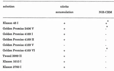
30 nitrite reduction-deficient barley plants (azide-treated in the M^) were identified as nitrite accumulators after treatment with nitrate. Biochemical analysis of selections revealed that leaf tissue from 9 selected plants lacked detectable nitrite reductase protein.
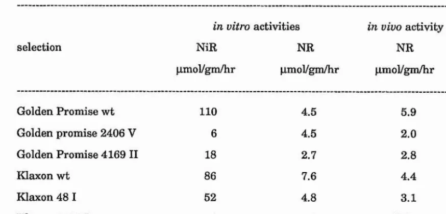
Progeny from 4 of the selected plants (Golden Promise 2406, Tweed 3999, Klaxon 1010 and Klaxon 2760) inherited the phenotype of a lack of leaf nitrite reductase protein. These plants also lacked significant in vitro nitrite reductase activity. However, in vitro nitrate reductase and nitrate accumulation were comparable with wild type controls.
Root tissue from nitrate-treated progeny of Golden Promise 2406, Tweed 3999, Klaxon 1010 and Klaxon 2760 also lacked nitrite reductase protein.
Loss of nitrite reductase protein in selection Tweed 3999 was caused by a single recessive nuclear gene mutation.
Thus, these plants are defective in nitrite reduction due to the inherited loss of nitrite reductase protein molecules in both leaf and root after treatment with nitrate in the light. This defect is caused by a mutation within a single nuclear gene in selection Tweed 3999.
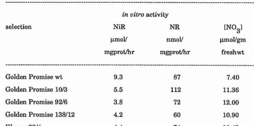
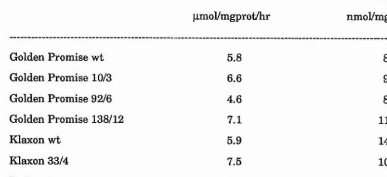
Analysis of wild type barley cv. Golden Promise revealed that increases in nitrite reductase activity in response to treatment with nitrate and light in leaf tissue and with nitrate in root tissue are due to de novo synthesis of enzyme molecules.
page Contents Acknowledgements Declaration Academic record Certificate Copyright Abbreviations 1 vii viii ix X xi xii
Chapter 1 Introduction
1.1. Introduction
1.1.1. Nitrate assimilation 2
1.1.2. Nitrate uptake 3
1.1.3. Nitrate reductase 3
I.I.3.I. Prosthetic groups 4
I.I.3.2. Catalytic activities associated with the nitrate reductase holoenzyme 5
1.1.3.3. Location 5
1.1.3.4. Environmental control 6
1.1.4. Nitrite reductase 6
I.I.4.I. Prosthetic groups 8
I.I.4.2. Location 9
1.1.4.3. Environmental control 12
1.1.5. Nitrite reductase in non-chlorophyllous tissue 13
1.2. Molecular analysis of nitrate assimilation
1.2.1. Nitrate reductase
1.2.2. Nitrite reductase
14 14 14
1.3. Nitrate assimilation mutants
1.3.1. Mutant analysis
1.3.2. Regulation
1.3.3. Nitrate uptake
1.3.4. Nitrate reduction
1.3.5. Nitrite reduction
16 16 16 17 17 17
11
Chapter 2 Materials and methods 20
Materials 21
2.1.1. Chemicals 21
2.1.2. Seed and plant growth materials 23
Methods 24
2.2. Mutant production, isolation and maintenance 24
2.2.1. Seed mutagenesis 24
2.2.2. Growth of M^ plants and collection of Mg seed 24
2.2.3. Screening for in vivo nitrite accumulators 24
2.2.3.1. Bulk-harvested populations 24
2.2.3.2. Spike-harvested populations 26
2.2.4. Screening for in vitro nitrite accumulators 26
2.2.5. Growth of nitrite accumulating plants 26
2.3. Genetic analysis 28
2.3.1. Cross pollination 28
2.3.2. M^ spike analysis 28
2.4. Biochemical analysis 29
2.4.1. Plant growth 29 j
2.4.1.1. Wild type plants 29 |
2.4.1.2. Mg progeny derived from Mg selections 29 !
2.4.2. Analysis of tissue from hydroponically-maintained Mg plants 29 J
2.4.3. Tissue extraction 30 j
2.4.3.1. Tissue micro-extraction 30 i
2.4.4. Enzyme assays 31 ;
2.4.4.1. /n mYro NADH-nitrate reductase 31
2.4.4.2. methyl viologen nitrate reductase 31 ;
2.4.4.5. Total-extract NAD H-nitrate reductase 31 i
2.4.4.4. In vivo nitrate reductase 32
2.4.4.5. In vitro methyl viologen nitrite reductase 32
2.4.4.6. Total-extract methyl viologen nitrite reductase 33 I
2.4.5. Protein determination 33
2.4.6. Nitrate determination 33 '
2.4.7. Non-denaturing polyacrylamide gel electrophoresis 34
2.4.8. Gel staining for nitrite reductase activity 34
2.4.9. SDS polyacrylamide gel electrophoresis 34
2.4.10. Protein electrotransfer (western blotting) 35 '
2.4.11. Development of electroblots (western blots) for nitrite reductase 35
2.5. Immunolocalisation of nitrite reductase 36
2.5.1. Fixation of tissue 36
2.5.2. Embedding of tissue 36
2.5.3. Grid coating 36
2.5.4. Microtome sectioning 37
2.5.5. Immunolabelling of ultrathin sections 37
2.5.6. Post-embedding staining and electron microscopy 37
Chapter 3 Selection of nitrite accunm lating barley plants 38
3.1. Introduction 39
3.1.1. . Mutant analysis 39
3.1.2. Chemical mutagenesis 39
3.1.3. Nitrite reductase-deficient mutants 41
3.2. Results 42
f" 3.2.1. Selection of nitrite accumulators 42
3.2.2. Selection growth 43
3.2.2.I. Bulk-harvested selections 43
3.2.2.2. Spike-harvested selections ■ : % 43
3.22.3. Mg progeny growth 44
3.2.3. Chlorophyll-deficient mutants 44
3.2.3.1. Frequency of chlorophyll-deficient seedlings 44
3.2.3.2. Nitrite accumulating chlorophyll-deficient seedlings 44
3.3. Discussion . ' 46
3.3.1. Nitrite accumulating selections
3.3.2. Maintenance of selected nitrite accumulators 45
L. * 3.3.3. Chlorophyll-deficient mutants
Chapter 4 Biochem ical characterisation of selected 49
nitrite accumulators
4.1. Introduction 50
4.1.1. Biochemical analysis of selected nitrite accumulators ' 50
4.2. Results 51
4.2.1. Analysis bf Mg selections 51
4.2.2. Mg bulk-harvested selections ^ 51
4.2.3. Mg spike-harvested selections 51
4.2.3.1. Nitrite rèâuctase activity ^ 51
4.2.3.2. Nitrate reductase activity / _ 51
4.2.3.3. Nitrite reductase cross reacting material 52
4.2.4. v^âîysis of Mg progeny derived from bulk-harvested selections ' 53
IV
4.2.4.2. In vitro nitrite accumulation 53
4.2.4.S. Nitrite reductase activity 53
4.2.4.4. Nitrate reductase activity 53
4.2.4.5. Nitrate accumulation 54
4.2.4.6. Nitrite reductase activity staining in non-denaturing 54
polyacrylamide gels
4.2.4.7. Nitrite reductase cross reacting material 54
4.2.5. Analysis of Mg progeny derived from spike-harvested selections 55
4.2.5.1. In vivo nitrite accumulation 55
4.2.5.2. In vitro nitrite accumulation 55
4.2.5.3. Nitrite reductase activity 55
4.2.5.4. Accuracy of in vitro nitrite reductase assay 55
4.2.5.5. Mg progeny nitrite reductase activity 56
4.2.5.6. Nitrate reductase activity 57
4.2.5.7. NADH-nitrate reductase 57
4.2.5.8. Methyl viologen nitrate reductase 57
4.2.5.9. In vivo nitrate reductase 57
4.2.5.10. Nitrate accumulation 58
4.2.5.11. ^^N-nitrate incorporation into leaf protein 58
4.2.5.12. Nitrite reductase activity in non-denaturing polyacrylamide gels 58
4.2.5.13. Nitrite reductase cross reacting material 58
4.3. Discussion 59
4.3.1. Bulk-harvested selections
4.3.2. Spike-harvested selections
4.3.3. Sensitivity of in vitro nitrite reductase assay 4.3.4. Nitrite accumulation screen selection pressure
59 60 61 63
Chapter 5 Genetic analysis of selected lines 66
5.1. Introduction
5.1.1. Genetic analysis of nitrite reduction
5.1.2. Isozyme analysis
5.1.3. Mutant analysis
67 67 67 5.2. Results 5.2.1. 5.2.1.1. 5.2.1.2. 5.2.1.3. 5.2.2. 5.2.3. 5.2.3.1. 5.2.3.2. 5.2.3.3.
Segregation within Fg generations In vivo nitrite accumulation
Nitrite reductase cross reacting material In vitro nitrite accumulation
In vivo nitrite accumulation by Fg selections
Identification of Mg heterozygotes carried on selected M^ spikes Mg family in vivo nitrite accumulation
5.3.3. Segregation within Mg families 76
5.3.4. Segregation of chlorophyll-deficient Mg seedlings 76
5.3.5. Allelic analysis 77
Chapter 6 Environmental control of nitrite reductase 78
6.1. Introduction 79
6.1.1. Environmental influences on nitrite reductase 79
6.1.2, Root nitrite reductase 81
6.2. Results 82
6.2.1. Nitrite reductase polyacrylamide gel electrophoresis 82
6.2.2. Effect of nitrate 82
6.2.3. Effect of light 83
6.2.4. Effect of end products 84
6.3. Discussion 86
6.3.1. Leaf and root nitrite reductase 86
6.3.2. Influence of nitrate and light 86
6.3.4. Influence of ammonium ions and glutamine 88
6.3.5. Accurate measurement of low levels of nitrite reductase activity 88
6.3.6. Environmental control of nitrate assimilation 89
Chapter 7 Immunolocalisation of nitrite reductase 90
7.1. Introduction 91
7.1.1. In vivo location of nitrite reductase 91
7.1.2. Immunolocalisation and electron microscopy 91
7.1.3. Mutant analysis 93
7.2. Results 94
7.2.1. Leaf tissue fixation 94
7.2.2. Immunogold localisation 94
7.2.3. Intra-chloroplastic immunolocalisation 95
VI
Chapter 8 Discussion 100
1.1. Isolation of nitrite accumulating barley plants 101
1.2. Maintenance of selections 102
1.3. Biochemical characterisation 103
1.4. Genetics of defect 109
1.5. Spike screening 109
1.6. Root nitrite reduction 110
1.7. Environmental control 110
1.8. Further work 111
ACKNOWLEDGEMENTS
I would like to thank Dr. John Wray for his advice and criticism throughout this work. I would also like to express my thanks to Mrs. Amanda Gilkes for her work in screening M^ spikes for nitrite accumulators and her help in maintaining selected plants in hydroponic culture. My thanks go also to Mrs. Anne Holyoake and Miss Amanda Wilshin for their work in screening bulk-harvested Mg seed for nitrite accumulators and Mr. Denis Kirk for his help in harvesting M^ barley spikes.
I would like to thank Dr. Roger Wallsgrove, Rothamsted Experimental Station, for providing bulk-harvested M^ seed and the production and growth of M^ barley plants from which M^ spikes were harvested. I would also like to thank Dr. William Thomas, Scottish Crop Research Institute, for providing growing facilities for M^ populations of barley from which M^ spikes were harvested.
V lll
DECLARATION
I, Euan Duncanson, hereby certify that this thesis has been composed by myself, that it is a record of my own work, and that it has not been accepted in partial or complete fulfilment of any other degree or professional qualification.
ACADEMIC RECORD
I was admitted to the Faculty of Science of the University of St. Andrews under Ordinance General No 12 on 1®^ October 1986 and as a candidate for the degree of Ph.D. on 1^^ October 1986.
CERTIFICATE
I hereby certify that the candidate has fulfilled the conditions of the Resolution and Regulations appropriate to the Degree of Ph.D.
COPYRIGHT
XII Abbreviations cv. GP IX IT IN IV DO CO DI M l Mg Mg M4 NiR NR CRM PAGE BCIP BSA EDTA NaFe EDTA FAD GARG15 MES MV NADH NADPH NBT cultivar Golden Promise Klaxon Tweed Natasha Vista Doublet Corniche Digger
Population derived from cross pollinations between selected Mg nitrite accumulators and wild type plants
Generation derived from self pollination of F i population Population treated with chemical mutagen
Generation derived from self pollination of M i population Generation derived from self pollination of Mg population Generation derived from self pollination of Mg population Nitrite reductase
Nitrate reductase Cross reacting material
Polyacrylamide gel electrophoresis 5-bromo-4-chloro-3-indolyl phosphate Bovine serum albumin
ethylenediaminetetra-acetic acid
ethylenediaminetetra-acetic acid, ferric monosodium salt Flavin adenine dinucleotide
Goat anti-rabbit IgG-gold conjugate (15nm) 2[N-morpholino]ethanesulphonic acid
Methyl viologen (l,l'-dimethyl-4-4'-bipyridinium chloride) P-nicotinamide adenine dinucleotide, reduced form
NED SDS
Szechrome NAS*' TEMED
: N-l-naphthylethylenediamine dihydrochloride : Sodium dodecyl sulphate
: Diphenylamine sulphonic acid chromogene : N,N,N',N'-tetramethyl-ethylene diamine
Chapter 1
Nitrate is the major nitrogen source for most cultivated crop plants grown under normal field conditions. Although in natural ecosystems ammonium as well as nitrate is formed by mineralisation of organic soil nitrogen, and there is considerable input of ammonium fertiliser nitrogen in agricultural ecosystems, the oxidation of ammonium by autotrophic nitrifying bacteria such as Nitrosomonas and Nitrobacter species ensures that in most well-aerated soils inorganic nitrogen is mainly available to the plant as nitrate (reviewed in Haynes, 1986).
Nitrogen and carbon constitute approximately 2 per cent and 40 per cent respectively 9
of the dry weight of plant material. On a worldwide scale 200x10 tons of carbon are fixed annually (Galston, 1961) which on a basis of approximate analysis would require the
9
incorporation of 10x10 tons of nitrogen per year. Biological dinitrogen fixation accounts for only a small proportion of nitrogen incorporation with the total, annual amount of dinitrogen
9
fixed in the biosphere estimated at 0.175x10 tons (Burns and Hardy, 1975). Thus, the majority of nitrogen incorporated into plant material occurs through the production and subsequent assimilation of nitrate.
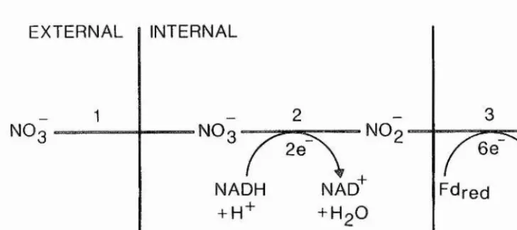
Nitrate assimilation, defined here as the uptake of nitrate into the plant and its subsequent two-step reduction to ammonium ions by the enzymes nitrate reductase and nitrite reductase, requires eight electrons (Fig 1.1). In non-chlorophyllous tissue these eight electrons are derived from carbohydrate oxidation (Beevers and Hageman, 1980). In chlorophyllous tissue two electrons are derived from carbohydrate oxidation while six are derived from the trapping of light energy (via ferredoxin) (Beevers and Hageman, 1980). The reduction of carbon dioxide to the level of carbohydrate requires four electrons. Higher plants utilise, on average, about 25 per cent of the energy required for the fixation of carbon dioxide on the assimilation of nitrate (Guerrero et aZ., 1981). The ammonium produced must then be incorporated into organic compounds converting ammonium-nitrogen into a-amino-nitrogen. Thus, the overall assimilation of nitrate into a-amino-nitrogen requires a massive amount of
Fig 1.1. Nitrate assimilation pathway
EXTERNAL INTERNAL
NH
NO NO
6 e 2e
NAD + HoO
cytoplasm chloroplast
[image:21.613.125.489.175.337.2]remobilisation within the plant are the most poorly characterised steps of the nitrate assimilation pathway. Uptake into the root is likely to be a carrier-mediated process utilising metabolic energy since it occurs against an electrochemical potential gradient, with anaerobic conditions (Trought and Drew, 1981), uncouplers of oxidative phosphorylation (Rao and Rains, 1976) and low temperature (Clarkson and Warner, 1979) being inhibitory.
Nitrate uptake rates follow a dual-phase relationship with the concentration of nitrate available to the plant. Both phases appear to be hyperbolic in accordance with Michaelis-Menten kinetics and suggest the existence of at least two uptake mechanisms operating at high and low nitrate concentrations respectively (Rao and Rains, 1976; Doddema and Telkamp, 1979). One system is likely to be "constitutive" and operates in plants which have not been exposed to nitrate, while the other is an "inducible" system and is characterised kinetically by experiments which show that maximal rates of net uptake occur after a lag of from one to a few hours after exposure of roots to nitrate (Jackson et al., 1986).
The development of maximal rates of net nitrate uptake from ambient solutions by roots is prevented by inhibitors of RNA and protein synthesis (Tompkins et uZ., 1978; Rao and Rains, 1976) suggesting that perhaps some component of the induced nitrate uptake system is synthesised de novo in response to the presence of nitrate.
1.1.3. Nitrate reductase
The nitrate reduction step is without doubt the best characterised step in nitrate assimilation at the physiological, biochemical and molecular level and has been the subject of numerous reviews (Hewitt, 1975; Beevers and Hageman, 1980; Guerrero et aL, 1981; Campbell and Smarelli, 1986; Wray, 1988).
Higher plant nitrate reductases are flavohaemomolybdoproteins which catalyse the two electron reduction of nitrate to nitrite (Hewitt and Notton, 1980). Almost all higher plants so far examined possess NADH-nitrate reductase (EG 1.6.6.1.) which has a pH optimum around 7.4 and a Michaelis constant for nitrate and NADH of about 200pM and 2 |liM respectively (Beevers and Hageman, 1980; Guerrero et aL, 1981). Some plants also
Biochemical and physiological evidence suggests that NAD(P)H-nitrate reductases are distinct species from NADH-nitrate reductases, possessing a higher Km for nitrate (4mM) and a lower pH optimum (6.5) (Campbell, 1976). The two forms can be separated by chromatography on blue dextran Sepharose (Redinbaugh and Campbell, 1981) and show different developmental (Orihuel-Iranzo and Campbell, 1980) and induction patterns (Shen et al., 1976). Most work has been carried out on the NADH-nitrate reductases and this is outlined below.
Attempts to purify the enzyme in its native state have been made difficult by its extreme sensitivity to proteolytic modification (Brown et aL, 1981; Wray and Kirk, 1981; Campbell and Wray, 1983). Estimated molecular weights of the holoenzyme vary from about 200.000 for spinach (Notton and Hewitt, 1979), tobacco (Mendel and Muller, 1980), barley (Small and Wray, 1980) through 220,000 or 230,000 for barley (Kuo et aL, 1980) or squash (Redinbaugh and Campbell, 1985) to 270,000 for spinach (Nakagawa et aL, 1985).
Nitrate reductase is usually assumed to be a homodimer with subunit molecular weights of 100,000 (Campbell and Wray, 1983) or 110,000 (Kuo et aL, 1982) in barley, 105.000 and 114,000 (Nakagawa cZ uZ., 1985) or 110,000 and 120,000 (Fido and Notton, 1984) in spinach, and 115,000 in squash (Redinbaugh and Campbell, 1985).
I.I.3.I. Prosthetic groups
Nitrate reductase contains flavin adenine dinucleotide (FAD) (Hewitt and Notton, 1980; Redinbaugh and Campbell, 1985). The activity of nitrate reductase is stimulated by exogenous FAD (Maretski et aL, 1967; Schrader et aL, 1968) suggesting that the flavin is readily dissociable. Purified nitrate reductase from spinach (Notton et aL, 1977), tobacco (Mendel and Muller, 1980), barley (Somers et aL, 1982) and squash (Redinbaugh and Campbell, 1985) have spectra indicative of the presence of a b-type cytochrome (cytochrome bggy). The first definitive evidence for the presence of molybdenum was obtained with the spinach enzyme (Notton and Hewitt, 1971) although it had previously been shown that in barley a non-functional form of the enzyme was synthesised in the presence of the molybdenum analogue, tungsten (Wray and Filner, 1970).
Molybdenum is carried as a molybdenum cofactor (MoCo) which is a complex between molybdenum and a phosphorylated pterin, molybdopterin (Johnson and Rajagopalan, 1982). The pterin acts as a chelator of molybdenum, interfacing it to the protein and conferring on it biological activity and is also responsible for dimérisation of the protein subunits (Ketchum et a l, 1970).
1.1.3.2. Catalytic activities associated with the nitrate
reductase holoenzym e
In addition to the overall physiological reaction (NADH-dependent nitrate reduction) the higher plant enzyme carries a dehydrogenase (diaphorase) function usually assayed as cytochrome c reductase as well as flavin mononucleotide and reduced viologen dye nitrate reductase activities. These partial activities are considered to be catalysed by specific regions of the nitrate reductase molecule (Wray and Fido, 1990).
Studies show that the dehydrogenase activity is catalysed by the proximal part of the electron transport chain, perhaps encompassing the flavin domain (Brown et al., 1981) and is an activity of the flavohaemoprotein subunit, independent of the nitrate binding site (Wray and Fido, 1990).
Electrons from flavin mononucleotide and probably from reduced viologen dyes are donated to a later, but undefined, site in the electron transport chain. Since nitrate reductase partial activities are molybdenum-dependent, flavin mononucleotide and reduced viologen dye nitrate reductase activities are functions of the distal part of the electron transport chain.
1.1.3.3. Location
1.1.3.4. Environmental control
In general, nitrate reductase activity is high in cells or plants grown on nitrate in the light (Beevers and Hageman, 1969; Gupta and Beevers, 1983). While nitrate application leads to increases in nitrate reductase levels nitrate is not an obligatory requirement since several species such as soybean (Lahav et aL, 1976) and tobacco (Muller, 1983; Buchanan and Wray, 1982) possess considerable nitrate reductase activity even if never exposed to nitrate.
The level of nitrate reductase activity in leaf tissue is influenced by the intensity of illumination (Sanderson and Cocking, 1964a; Beevers et al., 1965), with nitrate reductase activity remaining at basal levels in pea leaf tissue treated with nitrate in the dark (Gupta and Beevers, 1983). Immunological studies (Somers et aL, 1983) show clearly that the increase in nitrate reductase activity seen after nitrate treatment of plants in the light is due to de novo synthesis of nitrate reductase molecules. Studies using cDNA species, encoding the nitrate reductase apoprotein gene, as a hybridisation probe show that the steady state level of the nitrate reductase apoprotein mRNA increases markedly (over 100-fold) after nitrate application (Cheng et aL, 1986; Crawford et aL, 1986), demonstrating that nitrate brings about its effect at the mRNA level. However it is, as yet, unclear whether nitrate regulation occurs at the level of transcription, or acts to modify the stability of the mRNA transcript.
1.1.4. Nitrite reductase
The ferredoxin .-nitrite oxidoreductase (EC 1.7.7.1.) (nitrite reductase) enzyme catalyses the six-electron reduction of nitrite to ammonium ions in the third step of the nitrate assimilation pathway (Fig 1.1). Nitrite reductase has been purified to apparent homogeneity from leaf tissue in a number of species including spinach (Ho and Tamura, 1973; Ida and Morita, 1973; Ida et aL, 1976; Vega and Kamin, 1977; Ida, 1977; Hirasawa and Tamura, 1980; Ida and Mikami, 1986), Cucurbita pepo (Hucklesby et aL, 1976), barley (Serra et aL, 1982; Ip et aL, 1990), wheat (Small and Gray, 1984) and the bean Phaseolus angularis (Ishiyama and Tamura, 1985; Ishiyama et aL, 1985).
These results are at variance with the observation that purified spinach nitrite reductase of molecular weight 63 KDa can utilise ferredoxin and methyl viologen equally effectively as an electron donor (Joy and Hageman, 1966; Ida and Mikami, 1986).
The amino acid composition for the enzyme has been determined in spinach (Vega and Kamin, 1977; Ida and Mikami, 1986), Cucurbita pepo (Hucklesby et ah, 1976) and barley (Ip et aL, 1990) producing nearest integer values of 564, 558, 565 and 575 amino acid residues respectively. The calculated molecular weight of leaf nitrite reductase from the amino acid composition lies within the range 60-63 KDa while the spinach structural gene encodes a mature protein of molecular weight 63 KDa (Back et aL, 1988).
The enzyme can use purified ferredoxin (reduced with dithionite) as electron donor for in vitro nitrite reduction (Joy and Hageman, 1966), with ferredoxin widely accepted as the physiological electron donor in leaf tissue, reduced in vivo by electron transport associated with photosystem I (Neyra and Hageman, 1974). The enzyme can also utilise reduced viologen dyes (methyl and benzyl viologen) as electron donors (Hageman et aL, 1962; Joy and Hageman, 1966) for in vitro nitrite reduction, but is unable to use reduced pyridine nucleotides without the addition of the separate activity NADPH-ferredoxin reductase and ferredoxin (Hageman et aL, 1962).
Purified spinach leaf nitrite reductase possesses a greater affinity for nitrite with ferredoxin as electron donor, with a Michaelis constant of lO.SpM (Ida and Mikami, 1986) compared to 360pM (Ida and Morita, 1973) and llOpM (Ida and Mikami, 1986) possessed by the methyl viologen-linked activity. Serra et aL, (1982) reported a Km value for nitrite of 250pM obtained using either ferredoxin or methyl viologen as electron donor.
The pH optimum of the enzyme from spinach leaves is 7.5 (Ida and Morita, 1973) but lower in barley leaf where the optimum lies within the range pH 6.0-6.5 (Serra et al., 1982). The enzyme has a low thermostability (Ho and Tamura, 1973; Serra et al., 1982) with enzymic activity almost completely destroyed after treatment at 60°C for 5 minutes (Ho and Tamura, 1973).
1,1.4.1. Prosthetic groups
Nitrite reductase contains sirohaem (Murphy et al., 1974; Vega and Kamin, 1977) which has also been found in several sulphite reductases and shown to be an iron tetrahydroporphyrin of the isobacteriochlorin type (Murphy et al., 1974). Sirohaem expresses a visible spectrum with absorption maxima in spinach nitrite reductase at 276, 386 (soret), 573 (a) and 690nm (Vega and Kamin, 1977). This characteristic spectrum has been found in purified nitrite reductase from other species including spinach (Ho and Tamura, 1973; Ida, 1977; Ida et al., 1976; Hirasawa and Tamura, 1980; Ida and Mikami, 1986; Hirasawa-Soga and Tamura, 1981), Cucurbita pepo (Hucklesby et al., 1976), bean (Ishiyama et al., 1985) and barley (Serra et al., 1982; Ip et al., 1990).
Spectral shifts occur when the enzyme is treated with dithionite, nitrite, cyanide and carbon monoxide (Hucklesby et al., 1976; Vega and Kamin, 1977) indicating some form of interaction with the haem moiety. Electron paramagnetic resonance (EPR) studies show that
3+
the untreated oxidised enzyme contains a high-spin Fe haem (sirohaem) and that the addition of nitrite or cyanide results in a spin-state change of the haem (Aparicio et al., 1975; S toller et al., 1977; Cammack et al., 1978). Cyanide, a competitive inhibitor of nitrite, is assumed to bind to nitrite reductase and interact with the sirohaem (Aparicio et al., 1975),
Nitrite reductase also contains an iron-sulphur centre, originally thought to be a Pe^Sg centre (Vega and Kamin, 1977), but shown later to be a tetranuclear Fe^S^ centre (Lancaster et al., 1979). Haem ligands such as carbon monoxide and cyanide were found to modify both the reducibility and the EPR signal line shape of the Fe^S^ centre with the addition of carbon monoxide resulting in a 10-fold increase in intensity of the reduced iron-sulphur centre EPR signal (Lancaster et al., 1979). This indicates an interaction between the haem and the Fe^S^ centre of the active site.
spectrum of the complex with the appearance of a g=1.94 type EPR, spectrum typical of a reduced centre (Wilkerson et al., 1983). This suggests that the two prosthetic groups of nitrite reductase may interact in some way at the catalytic centre of the enzyme, with the sirohaem moiety binding nitrite and accepting electrons from the Fe^S^ centre which is reduced, in vivo, by ferredoxin or in vitro by methyl viologen/dithionite.
Nitrite reductase-linked sirohaem from spinach leaves possesses a midpoint potential of -50mV (Stoller et al., 1977) compared to -120mV for purified nitrite reductase from
Cucurbita pepo leaves (Cammack et al., 1978) and +80mV for barley leaf nitrite reductase (Ip et al., 1990). However, the midpoint potential of the iron-sulphur centre of purified nitrite reductase from spinach leaves is extremely electronegative, approximately -550mV (Stoller et al., 1977) and estimated at -570mV at pH 8.1 (Cammack et al., 1978) who found the midpoint potential appeared to be pH-dependent in the purified enzyme from cucurbita pepo leaves. Ip et aZ. ,(1990) also experienced difficulty in determining the midpoint potential of such an electronegative centre in nitrite reductase from barley leaves but used a computer generated best fit procedure to determine a value of -517mV. This raised the difficulty of how ferredoxin, with a midpoint potential of -420mV could reduce the iron-sulphur centre in vivo. It was suggested by Stoller et al., (1977) that the midpoint potential of the iron-sulphur centre, under physiological conditions, shifts to a more electropositive value, thus allowing its reduction by ferredoxin.
1.1,4.2. Location
Nitrite reductase is widely accepted to be located within the chloroplasts in higher plant leaf tissue with fractionation studies generally demonstrating the sedimentation of nitrite reductase coincidentally with chloroplast marker enzymes and chlorophyll.
10
However, Grant et al., (1970) using high rates of CO^ fixation and evolution as a measure of chloroplast integrity found that nitrite reductase activity was present mainly in the supernatant fractions after differential centrifugation studies on spinach and sunflower. Lips and Avissar, (1972) used density gradient centrifugation to suggest that nitrite reductase was located in the peroxisomes in tobacco leaves.
The main difficulty in these studies was the fragility of the organelles which caused leakage or rupture during the isolation procedure and so allowed cross contamination between the various fractions. Adsorption and occlusion of enzymes by organelles may also produce cross contamination resulting in the conflicting reports outlined above.
Indirect evidence for the chloroplastic location of nitrite reductase has come from functional studies on isolated chloroplasts. Paneque et al., (1963) demonstrated that spinach grana with added chloroplast extract could reduce nitrite in the light, while Magalhaes et al., (1974) reported that intact spinach chloroplasts, capable of fixing COg at high rates, reduced nitrite and synthesised amino-nitrogen de novo when supplied with only nitrite and light.
Nitrite reduction is functionally associated with electron transport arising from the light reactions of the chloroplast in spinach and maize (Neyra and Hageman, 1974). These workers used methyl viologen and phenazine methosulphate (photosystem I electron acceptors), 3-(3,4-dichlorophenyl)-l,l-dimethylurea (DCMU) an electron-transport inhibitor and methylamine a photophosphorylation uncoupler to show that nitrite reduction in isolated chloroplasts is associated with photo system I, with ferredoxin the most likely physiological electron donor in leaf tissue. Purified nitrite reductase from spinach and maize would accept electrons from ferredoxin reduced either by illuminated chloroplasts or with dithionite (Joy and Hageman, 1966).
(0.9-1.35), suggesting nitrite was assimilated to glutamate via the chloroplastic enzymes nitrite reductase, glutamine synthetase and glutamate synthase (Anderson and Done, 1978). Thus, the biochemical and physiological evidence suggests that nitrite reductase is located within the chloroplast in higher plants.
Since nitrite reduction is considered to occur within the chloroplasts several workers have attempted to determine whether the nitrite reductase structural gene is coded by DNA within the nucleus or wdthin the plant organelles. Attempts have been made to infer the origin of nitrite reductase mRNA using the protein synthesis inhibitors cycloheximide and chloramphenicol which inhibit the peptidyl transferase activity of the nuclear and organellar small ribosomal subunits respectively. Cycloheximide inhibits the induction of nitrite reductase activity in Lemna minor when presented to the tissue before nitrate (Stewart, 1967). When cycloheximide was added after exposure to nitrate all further induction of nitrite reductase activity was prevented, suggesting that nitrite reductase was synthesised on cytoplasmic ribosomes and was coded by nuclear DNA. This view was supported by Sluiters-Scholten, (1973) who showed that the induction of nitrite reductase activity by nitrate in green leaves of Phaseolus vulgaris in the light was inhibited by cycloheximide. Both cycloheximide and chloramphenicol inhibited the induction of nitrite reductase activity in etiolated leaves (Sluiters-Scholten, 1973). However, chloramphenicol did not inhibit the induction of nitrite reductase activity when presented to seedlings after 24 hours illumination. Sluiters-Scholten (1973) concluded that nitrite reductase was synthesised on cytoplasmic ribosomes, and therefore presumably coded by nuclear DNA, but some form of chloroplast development is required before production of nitrite reductase molecules can occur.
Molecular studies (Small and Gray, 1984; Gupta and Beevers, 1987; Back et al., 1988) suggests that nitrite reductase shares common properties with nuclear-encoded chloroplast proteins. Such proteins are usually synthesised as larger molecular weight precursors containing an amino-terminal sequence, the transit peptide (Chua and Schmidt, 1979) which is proteolytically cleaved to generate the mature functional protein during or after transport into the chloroplast (Grossman et al., 1982; Robinson and Ellis, 1984; Smeekin et al., 1985).
12
cleaved, in a two step process, to a peptide of the same size as that of the native enzyme by a proteinaceous extract from chloroplasts (Gupta and Beevers, 1987).
This evidence for a precursor protein has been confirmed by the molecular cloning of spinach nitrite reductase cDNA species by the use of oligonucleotide probes based on partial amino acid sequence data and through immunoscreening of a cDNA library in the expression vector lamda g tll, (Back et al., 1988). These studies show that the precursor protein for nitrite reductase is 594 amino acids long (molecular weight 66,394) and has a 32 amino acid extension at the N-terminal end of the mature protein (molecular weight 62,883) which probably serves as the transit peptide.
I.I.4.3. Environm ental control
Environmental control of nitrite reductase levels has not been investigated as extensively as for nitrate reductase. However, nitrite reductase activity is high in leaf tissue from plants grown on nitrate in the light (Gupta and Beevers, 1983; Small and gray, 1984; Ogawa and Ida, 1987). Nitrate is not obligatory for expression of nitrite reductase activity. Plants grown in the light in the absence of nitrate possess low, but measurable levels of leaf nitrite reductase activity (about 10-30 per cent of nitrate-grown plants) (Gupta and Beevers,
1983; Ogawa and Ida, 1987).
The level of nitrite reductase activity in leaf tissue is also influenced by light. Plants exposed to nitrate in the dark possess low levels of leaf nitrite reductase activity (about 30-50 per cent of light-grown, nitrate-treated plants) (Gupta and Beevers, 1983; Ogawa and Ida, 1987).
1.1.5. Nitrite reductase in non-chlorophyllous tissue
Nitrite reductase activity has been found in non-chlorophyllous tissues such as maize scutellum, where two isozymes were identified (Hucklesby et al., 1972; DallingeZ al., 1973) with similar properties to each other and leaf nitrite reductase.
Nitrite reductase activity also occurs in root tissue and is believed to be located within root plastids (Bailing et al., 1972; Miflin, 1974; Ernes and Fowler, 1979). The enzyme is similar to leaf nitrite reductase, utilising dithionite-reduced viologen dye as reductant in in vitro assays (Sanderson and Cocking, 1964b). Nitrite reductase activity is increased in plastids isolated from root tissue from nitrate-grown pea which also possess an increased flow of carbon through the pentose phosphate pathway compared to root plastids from peas grown in the absence of nitrate (Ernes and Fowler, 1983). Plastids from root tissue from nitrate-grown barley plants also contain a glucose-6-phosphate and NADP^-linked nitrite reductase system (Oji et al., 1985) with both benzyl and methyl viologen enzymatically reduced by plastid extract in the presence of glucose-6-phosphate and NADP^. The identification of a pyridine nucleotide reductase immunologically similar to spinach leaf ferredoxin-NADP^ reductase (Suzuki et al., 1985), and of a ferredoxin-like electron carrier (Ninomiya and Sato, 1984; Suzuki et al., 1985), support the suggestion that the supply of reducing power for in vivo root nitrite reduction originates from the pentose phosphate pathway.
Purified nitrite reductase from pea roots has a molecular weight of about 60 KDa and exhibits absorption maxima at 278, 384, 573 and 695nm (Bowsher et al., 1988) similar to spinach leaf nitrite reductase (Vega and Kamin, 1977). The electron paramagnetic resonance spectrum is indistinguishable from that of the Cucurbita pepo leaf enzyme (Bowsher et al., 1988), while reduction of the purified enzyme with dithionite in the presence of cyanide allows the appearance of a signal around g=1.94, characteristic of an iron-sulphur centre (Bowsher et al., 1988).
14
1.2. Molecular analysis of nitrate assim ilation
1.2.1. Nitrate reductase
Complementary DNA encoding nitrate reductase has been isolated in barley (Cheng et ah, 1986) squash (Crawford et ah, 1986) and tobaco (Calza et ah, 1987). These cDNA clones have been used as probes to isolate the nitrate reductase structural gene and for Northern blot analysis to study the regulation of nitrate reductase (Caboche et ah, 1989; Crawford and Davis, 1989) and in the characterisation of nitrate reductase-deficient mutants (Kleinhofs et ah, 1989).
1.2.2. Nitrite reductase
Complementary DNA encoding nitrite reductase has been isolated from spinach (Back et ah, 1988) and maize (Lahners et ah, 1988). The cloned nitrite reductase cDNA from spinach codes for a precursor protein that contains 594 amino acids with a deduced molecular weight of 66,394 (Back et ah, 1988). The nitrite reductase precursor protein is 32 amino acids longer than the mature protein which contains 562 amino acids and has a deduced molecular weight of 62,883 (Back et ah, 1988). This value corresponds well with the apparent molecular weight of 61kDa (Ho and Tamura, 1973; Vega and Kamin, 1977). The additional amino acids precede the mature protein and are likely to act as a transit peptide sequence in directing the nuclear-encoded nitrite reductase protein into the chloroplast (Back et ah, 1988).
The amino acid sequence deduced from the spinach cDNA sequence contains 4 cysteine residues in positions 473, 479, 514 and 518 (Back et ah, 1988) that are probably involved in the binding of the tetranuclear iron cluster (Aparicio et ah, 1975; Lancaster et ah, 1979). These cysteines are conserved in the maize cDNA sequence in exactly the same location in the protein (Lahners et ah, 1988).
Nitrate induction of nitrite reductase activity due to de novo synthesis of enzyme molecules has been well documented for several plant species (Beevers and Hageman, 1969; Gupta and Beevers, 1984, 1987; Small and Gray, 1984, Ogawa and Ida, 1987). Northern blot analysis has shown that nitrite reductase mRNA increases considerably in spinach leaves (Back et ah, 1988) and maize leaf and root tissue (Lahners et al., 1988) from plants treated with nitrate.
There is a rapid induction of nitrite reductase mRNA after the addition of nitrate in maize with significant increases in the level of nitrite reductase mRNA in root tissue after 30 minutes and in leaf tissue after 90 minutes (Kramer et al., 1989). This timing difference may reflect the time at which inducing concentrations of nitrate become available to the different organs.
The stability of nitrite reductase mRNA is very low in maize, having a half-life of less than 30 minutes in root and 40 minutes in leaf tissue (Kramer et al., 1989). Thus the level of nitrite reductase mRNA decreased rapidly after a maximum RNA level was reached (Kramer et al., 1989) even though the plants were still exposed to nitrate.
16
1.3. Nitrate assim ilation mutants
1.3.1. Mutant analysis
The isolation and characterisation of mutant alleles which define genetic loci is a powerful tool in the study of metabolism as evidenced by its classical and continued application to microbial systems and, more recently, its application to higher plants (reviewed in Blonstein and King, 1986). Mutant analysis allows not only the identification of genetic loci whose existence can be deduced a priori from a knowledge of the biochemical characteristics of the pathway but also it should allow the identification of genes which encode previously unknown components of the pathway. Of special interest among these being genes whose products contribute to the regulatory networks controlling metabolism. Thus, mutant analysis may lead to the identification (and subsequent manipulation) of single genes of agronomic importance allowing more efficient crop production.
1.3.2. Regulation
There have been no reports of regulatory mutations affecting nitrate assimilation in higher plants so far. In Nicotiana tabacum two cnx A alleles (Cnx 68 and Cnx 101) produce constitutive levels of nitrate reductase apoprotein and also show an increased constitutive nitrite reductase, as do the nia allele Nia 102 and cnx B alleles (Mendel and Muller, 1979; Mendel et al., 1984; Schiemann and Muller, 1985). Whether the constitutive levels of nitrate reductase and nitrite reductase activity are a consequence of the MoCo or apoprotein gene mutation is not clear. However in Aspergillus nidulans some mutations causing loss of a functional nitrate reductase molecule lead to constitutive synthesis of both nitrate and nitrite reductase and suggest an autoregulatory role for functional nitrate reductase molecules in the regulation of synthesis of both nitrate reductase and nitrite reductase (Pateman et al.,
1967; Cove and Pateman, 1969).
1.3.3. Nitrate uptake
Nitrate uptake mutants are poorly represented amongst the reports of nitrate assimilation mutants so far identified (Doddema and Telkamp, 1977; Wallsgrove, 1987). The lack of a positive selection scheme for the identification of nitrate uptake mutants forces the adoption of total isolation procedures (Wallsgrove, 1987) which are both labour intensive and time consuming,
1.3.4. Nitrate reduction
A great number of mutants altered in nitrate reduction have been isolated in higher plants. These mutants carry mutations either in the nitrate reductase apoprotein structural genes or in genes involved in molybdenum cofactor synthesis and insertion (reviewed in Dunn-Coleman et al., 1984; Kleinhofs et al., 1985; Wray, 1986; Wray 1988). That such a large number of mutants altered in nitrate reduction have been isolated is undoubtedly due to the availability of chlorate as a positive selection agent (Oostindier-Braaksma and Feenstra, 1972; Braaksma and Feenstra, 1982a,b) allowing the identification of plants lacking a functional nitrate reductase activity by a simple visual screening procedure.
1.3.5. Nitrite reduction
The only plant variants defective in nitrite reduction were described in Haplopappus gracilis (Gilissen et al., 1985). They selected a callus (Ao) which was able to grow in the presence of normally toxic levels of asparagine. After X-irradiation this line was plated on medium containing alanine as nitrogen source and calli growing at wild type rates were selected. Two lines failed to grow on medium containing nitrate as a nitrogen source and excreted nitrite. While in vitro nitrate reductase activity was similar to wild type, both in vivo nitrate reductase and in vitro nitrite reductase activities were only 20-40 per cent of the wild type level. Attempts at plant regeneration were unsuccessful and the molecular basis of the defects are unknown.
18
yet, there have been no reports in the literature of whole plant mutants defective in nitrite reduction.
1.4. Aims
The aims of the work described here were:
i. To isolate mutant barley plants defective in nitrite reduction by identifying individuals which accumulate nitrite after treatment with nitrate.
ii. To biochemically characterise selected plants to determine the basis for nitrite accumulation.
iii. To genetically characterise selected plants by allelism tests through cross pollinations between selected nitrite accumulators and the identification, by subsequent progeny segregation, of heterozygous lines carried on selected spikes, or produced by cross pollination between selected nitrite accumulators and wild type plants.
20
Chapter 2
Materials
2,1.1. Chemicals
All biochemicals and common chemicals used were of standard laboratory grade unless otherwise stated. Where appropriate, chemical formulae and common names used in the text are given.
5-bromo-4-chloro-3-indolyl phosphate, disodium salt (BCIP), Goomassie brilliant blue G-250, cysteine HCl, 1,1-dimethyl-4-4'-bipyridinium chloride (methyl viologen), diphenyl-3,3'-(3,3'-dimethoxy-4,4'-diphenylene) di tétrazolium chloride grade III, crystalline (nitroblue tétrazolium, NBT), flavin adenine dinucleotide disodium salt, (FAD), gentamicin sulphate, glutamine, glycine, goat antirabbit alkaline phosphatase conjugate, molecular weight markers (prestained kit MW-SDS-BLUE 27,000-180,000) for SDS gel electrophoresis (triosephosphate isomerase (rabbit muscle), 26,600 Da, lactic dehydrogenase (rabbit muscle), 36,500 Da, fumarase (porcine heart), 48,500 Da, pyruvate kinase (chicken muscle), 58,000 Da, fructose-6-phosphate kinase (rabbit muscle), 84,000 Da, p-galactosidase (E.Coli), 116,000 Da, ag-macroglobulin (human plasma), 180,000 Da), P-nicotinamide adenine dinucleotide, reduced form, disodium salt from yeast, grade III, (p-NADH), 2[N-morpholino] ethanesulphonic acid (MES), triphenyl tétrazolium chloride were obtained from Sigma Chemical Go. Ltd. Poole, Dorset, England.
22
Formvar powder (polyvinyl formal), glutaraldehyde (EM grade), lead nitrate, L.R, White resin (medium grade), nickel specimen grids (100 mesh hexagon, 340pm opening), osmium tetroxide, paraformaldehyde (EM grade), Spurr resin kit, uranyl acetate were obtained from Agar Scientific, Stansted, Essex, England.
Goat anti-rabbit IgG-gold conjugate (15nm, GAR G15) was obtained from Janssen Life Sciences Products, Wantage, Oxon, England.
2.1.2. Seed and plant growth materials
Barley seed was obtained from William Watt, Seed Merchants, Cupar, Scotland and the Scottish Crop Research Institute, Invergowrie, courtesy of Dr W. Thomas.
Bulk-harvested M^ barley seed, cv. Golden Promise, Klaxon, Patty, Apex, Maris Mink (azide-treated in the M^) was obtained from Rothamsted Experimental Station, Harpenden, Herts, courtesy of Dr R. Wallsgrove during the period 1980-1985.
Spike-harvested M^ barley seed (cv. Corniche and Digger, 1987) was produced by mutagenesis with sodium azide in the M^ generation and grown in field plots at Rothamsted Experimental Station, Harpenden, Herts., courtesy of Dr R. Wallsgrove.
Spike-harvested M^ seed cv. Golden Promise, Klaxon, Tweed, Natasha, Vista, Doublet, 1987 (azide-treated in the M^) was grown at the Scottish Crop Research Institute, Invergowrie, courtesy of Dr. W. Thomas.
24
Methods
2.2. Mutant production, isolation and m aintenance
2.2.1. Seed m utagenesis
Wild type seeds of barley (M^ seed) were mutagenised by chemical mutagenesis using the procedure described by Kleinhofs et al., (1978). Seed of various cultivars were hydrated in trays of tap water at 0-4®C for 16 hours and then transferred to 10 litre flasks containing 3-4 litres of tap water which was aerated vigorously for a further 8 hours at room temperature. The flasks were placed in a fume cabinet and the water was replaced with 3 litres O.IM potassium phosphate buffer pH 3 and sodium azide solution added to the buffer to give a final concentration of ImM. After aeration of this solution for a further 2 hours, the seeds were washed in several changes of tap water for 30 minutes, blotted to remove excess liquid and redried.
2.2.2. Growth of plants and collection of seed
Barley seed mutagenised courtesy of Dr.R.Wallsgrove as described above was grown in field plots at Rothamsted Experimental Station, Harpenden, Herts. Seed mutagenised as described above was also grown in field plots at the Scottish Crop Research Institute, Invergowrie, courtesy of Dr.W.Thomas.
Bulk-harvested single seed was collected with a combine harvester, or individual spikes carrying seed were collected manually. 60,000 spikes were dried and then individually bagged and numbered.
2.2.3. Screening for in vivo nitrite accumulators
2.2 3.1. Bulk-harvested populations
Bulk-harvested seed was sown in trays of vermiculite to give a germination rate of about 200 seedlings per tray, and watered with nitrate-less half-strength Hoaglands nutrient solution (0.2mM NaFe EDTA, 0.5mM KH^PO^, ImM M g80^.7Hg0, 0.4pM Zn80^.7Hg0, 0.2pM MnSO^.SH^O, 0.02mM H^BOg, O.OSpM NagMo^0^.2HgO; Hoagland and Amon, 1938) in the dark. After 4 days the germinated seedlings were transferred into
-2 -1
f
light overnight. The following day the 7 day old seedlings were screened for defects in nitrite reduction by testing leaf tissue for accumulation of nitrite.
Seedlings were screened for nitrite accumulation by firstly dividing each tray into sections of about 50 plants with wooden markers. The leaf tips (about 10mm in length) from each plant within the section were pooled and then homogenised by grinding the tissue in a mortar and pestle in 1ml HgO, 1ml 1% sulphanilamide in 3N HCl and 1ml 0.02% NED. The homogenate was centrifuged for 5 minutes at l,200g in a bench centrifuge. Nitrite present in the supernatant, due to in vivo nitrite accumulation, reacts with the acidified sulphanilamide and NED in a diazo-coupling reaction (Snell and Snell, 1949), to form an azo dye with a characteristic pink colour.
When pools of leaf tissue producing a positive nitrite reaction were identified all plants within the pool were individually rescreened for nitrite accumulation. In the process of initial section screening it was possible to determine which plants had already been sampled since the top 10mm of the primary leaf had been cut off, while those yet to be sampled remained intact. However, during individual rescreening, all plants lacked the top 10mm of the primary leaf. Therefore, a numbered marker was placed beside each plant as it was separately rescreened to ensure every plant within the section was rescreened and to facilitate the identification of the plant or plants responsible for the positive nitrite reaction (Fig 2.1).
2.2.S.2. Spike-harvested populations
Spike-harvested Mg seed was sown in grids placed in trays of vermiculite such that up to 5 seed from each spike were sown together in a unique position in the numbered grid (Fig 2.2). Once all the grid positions were full the seed was lightly covered with vermiculite so that the grid was still visible. The trays were watered with nitrate-less half-strength Hoaglands nutrient solution and left in the dark. After 4 days the germinated seedlings were transferred into the light (115pEm s ) for 2 days. The green seedlings were then watered with half-strength Hoaglands nutrient solution containing 25mM KNOg and maintained in the light overnight. On the following day the 7 day old seedlings were screened for in vivo nitrite accumulation.
26
sulphanilamide in 3N HCl and 1ml 0.02% NED. The homogenate was centrifuged for 5 minutes at l,200g in a bench centrifuge. Nitrite present in the supernatant, due to in vivo nitrite accumulation, reacts with the acidified sulphanilamide and NED in a diazo-coupling reaction (Snell and Snell, 1949), to form an azo dye with a characteristic pink colour.
When pools of leaf tissue producing a positive nitrite reaction were identified leaf tissue from the plants within each separate grid position from the section were pooled and rescreened for nitrite accumulation. Thus, it was possible to identify which grid position the nitrite accumulating plant, or plants, occupied and so from which spike the nitrite accumulating plant, or plants, were derived. Each plant within the grid position responsible for the positive nitrite reaction was then individually rescreened to identify the individual plant, or plants, responsible for the nitrite accumulation.
2.2.4. Screening for in vitro nitrite accumulators
Individual seed was sown in unique positions in a numbered grid placed in a tray of vermiculite then given a light covering of vermiculite so that the grid was still visible. The seedlings were germinated in the dark for 4 days then transferred to the light (llSpEm s" ) for 2 days growth and watered with nitrate-less half-strength Hoaglands nutrient solution.
The leaf tips (about 10mm in length) from each plant were placed in separate test tubes to which 1ml O.IM KNOg was added. The tubes were then placed in the light
-2 -1
(115)xEm s ) for 16-20 hours. The following day the solution is decanted into clean test tubes to which 1ml 1% sulphanilamide in 3N HCl and 1ml 0.02% NED is added. Nitrite accumulation is identified by the production of an azo dye with a characteristic pink colour (Snell and Snell, 1949). It was then possible to identify the plant or plants responsible for the nitrite accumulation.
2.2.5. Growth of nitrite accum ulating plants
28
2.3. Genetic analysis
2.3.1. Cross pollination
Plants identified as nitrite accumulators, and which had been maintained to flowering in the hydroponic system, were reciprocally cross pollinated with wild type plants by hand. The plant destined to be the female parent (pollen recipient plant) was emasculated when the tiller had swollen and the awns on the spike appeared up to one inch from the top of the flag leaf. Spikes were emasculated by cutting open every seed sac carried on the spike and plucking out the anthers (3 anthers per seed sac), while still green, with fine forceps. The spike was left for 2-3 days covered by a glassine bag to prevent uncontrolled cross pollination from other plants. If self pollination had inadvertantly occurred, fertilisation and initiation of seed formation was visible after 2-3 days. In these cases the spike was discarded. Once the seed sacs were gaping and the ovaries ready to accept pollen, as indicated by their white fluffy appearance, anthers were individually removed, with fine forceps, from the plant destined to be the male parent (pollen donor plant) and gently tapped over the ovaries and then left in the seed sac. A spike is ready for pollen donation when the anthers (yellow and swollen in appearance) protrude from the seed sac within 10-15 minutes of the sac being cut open. Once pollination of the whole spike was complete, it was again covered with the glassine bag and the spike left to produce seed.
2.3.2. spike analysis
2.4. Biochem ical analysis
2.4.1. Plant growth
2.4.1.1. Wild type plants
Wild type barley seeds used in the study of the regulation of nitrite reductase under varying conditions of light and nitrogen nutrition were sown in trays of vermiculite that had been thoroughly washed in several changes of distilled water until the excess rinsing solution gave a negative nitrite reaction. The seeds were germinated in the dark at 25^0 for 4 days,
-2 -1
after which they were placed in the light (115 jxEm s ) for 2 days and watered with nitrate-less half-strength Hoaglands nutrient solution by which time the leaves were fully expanded and green. The plants were then exposed to various growth conditions with light, dark and nitrogen-containing compounds.
2.4.1.2. Mg progeny derived from Mg selections
Mg progeny seeds from Mg nitrite accumulating selections were sown individually in small ipots or in unique positions within a grid placed in trays of washed vermiculite. The seeds were germinated in the dark at 25° C for 4 days, and watered with half-strength Hoaglands. nutrient solution without nitrate. The seedlings were then transferred into the
-2 - 1
light (115pEm s ) for 2 days by which time the leaves were fully expanded and green. The plants were then watered with half-strength Hoaglands nutrient solution containing 25mM KNOo and after a further 24 hours in the light the plants were harvested for analyses.o
.
2.4.2. Analysis of tissue from hydroponically-maintained plants
30
Tissue (about lOmg) was floated on 30ml O.IM KNOg in separate 100ml conical flasks which were and placed in a dessicator. A vacuum was applied until air bubbles were forced from the tissue and the solution began to bubble. This was repeated twice more. The flasks containing the vacuum infiltrated tissue and O.IM KNOg solution were placed in the light (115)iEm ^s overnight.
Some 16-18 hours later tissue was analysed by western blotting, nitrate reductase and nitrite reductase enzyme assays.
2.4.3. Tissue extraction
Plant tissue (leaf or root) was extracted in 50mM Tris buffer pH 7.6, 10 mM EDTA, lOmM P-mercaptoethanol and 10% glycerol (NiR extraction buffer (Ip et al,, 1990» by grinding in a mortar and pestle. Tissue to buffer ratios varied between 1:3 to 1:10 (wt:vol) depending on the amount of tissue available for analysis. The homogenate was centrifuged at 30,000g in a MSE High Speed Super 18 centrifuge for 15 minutes and the supernatant collected as the enzyme source. All procedures were carried out at 4°G.
2.4.3.I. Tissue micro-extraction