Burkhard Ostertun, Helmut K. Wolf, Manuel G. Campos, Cristian Matus, Laszlo Solymosi, Christian E. Elger, J. Schramm, and Hans H. Schild
PURPOSE: To evaluate dysembryoplastic neuroepithelial tumors (DNTs) on MR and CT studies and to compare DNT with other frequently encountered epileptogenic glioneuronal lesions. METH-ODS: We analyzed the MR images and CT scans of 16 patients who had complex partial epilepsy and DNT with respect to tumor location, size, CT density, MR signal intensity, mass effect, contrast enhancement, and heterogeneity, and compared these features with CT and MR findings in 51 cases of ganglioglioma and 33 cases of glioneuronal malformation. RESULTS: DNTs were located in the temporal lobe in 14 patients and in the frontal lobe in 2 patients. The cortex was involved in all cases and the subcortical white matter in 10 cases. Fifty percent of the tumors had poorly defined contours. On MR images, 14 DNTs had multiple cysts and 2 had single cysts. Contrast enhancement was observed in 6 DNTs, and mass effect was present in 9. CT scans disclosed moderately hypodense lesions in 7 patients and markedly hypodense cystic lesions in 6 patients. Two DNTs were calcified. Tumor hemorrhage with perifocal edema was observed in 1 case. Contrary to previous reports, slow but definite tumor growth was present during a 13-year period in 2 of 6 patients in whom serial CT or MR studies were obtained. CONCLUSION: A multicystic appearance on MR images is a characteristic feature of DNT and corresponds to its myxoid matrix and multinodular architecture. This feature is rare in gangliogliomas and glioneuronal malforma-tions, and, as such, may help differentiate DNTs from these disorders.
Index terms: Brain neoplasms, magnetic resonance; Seizures
AJNR Am J Neuroradiol17:419 –430, March 1996
The term dysembryoplastic neuroepithelial
tumor (DNT) was introduced by Daumas-Du-port et al in 1988 (1) and has recently been included as a mixed neuroglial tumor in the World Health Organization classification of brain tumors (2, 3). Clinically, DNT is usually associated with chronic epilepsy in adolescents and young adults. DNTs are most commonly located in the temporal lobe (1) and usually cause no neurologic deficit. Histopathologi-cally, DNT is a benign multinodular lesion com-posed of glial and neuronal elements (1, 4 –11). DNTs share several important features with
gangliogliomas and glioneuronal malforma-tions, such as glioneuronal hamartomas and hamartias (small glioneuronal malformations) (10). These consist of glial cells and highly dif-ferentiated ganglion cells and are frequently as-sociated with chronic focal epilepsy.
In a series of 216 temporal lobe resections for chronic focal epilepsy, 34 gangliogliomas, 6 DNTs, and 32 glioneuronal malformations were found histopathologically (9). Therefore, pre-surgical criteria for the differentiation of these glioneuronal lesions are of interest. A few stud-ies have mentioned the appearance of DNTs on magnetic resonance (MR) and computed tomo-graphic (CT) studies (6 – 8, 12–19). These pub-lications describe lesions with nonspecific low signal intensity on T1-weighted images and high signal intensity on T2-weighted images, similar to other gliomas. We present the MR (n
5 16) and CT (n 510) findings in 16 patients with DNT and discuss criteria to distinguish DNT from gangliogliomas and glioneuronal malformations.
Received July 11, 1995; accepted after revision September 27. From the Departments of Neuroradiology/Radiology (B.O., C.M., L.S., H.H.S.), Neuropathology (H.K.W.), Neurosurgery (M.G.C., J.S.), and Epi-leptology (C.E.E.), University of Bonn, Germany.
Address reprint requests to Burkhard Ostertun, MD, Department of Neuroradiology/Radiology, University of Bonn Sigmund-Freud-Str 25, D-53127 Bonn, Germany.
AJNR 17:419–430, Mar 1996 0195-6108/96/1703–0419
qAmerican Society of Neuroradiology
Material and Methods
Clinical records, 10 CT scans, and 16 MR studies of 16 patients with a histopathologic diagnosis of DNT were analyzed retrospectively to determine the CT and MR char-acteristics of DNTs as well as their clinical presentation and postoperative course. All patients had surgery during the period from December 1988 to March 1995. Ten pa-tients were female, 6 were male. All papa-tients suffered from epilepsy, with a mean duration of 10.5 years (range, 0.2 to 31 years). Complex partial seizures were the only symp-tom except in 1 patient with insular DNT who also had a minimal motor deficit. The mean age at onset of epilepsy was 18.4 years (range, 1.5 to 60.5 years) (Table 1), and the average seizure frequency was 25 per month (range, 3 to 120 seizures per month). At surgery, the patients’ mean age was 28.9 years (range, 4.5 to 62.5 years).
Preoperative MR imaging was performed at various stitutions from 1988 to 1994. The imaging studies in-cluded at least T1-weighted images obtained before and after intravenous administration of contrast material, and proton density–weighted and T2-weighted images in at least one plane. In 8 cases, preoperative CT studies were obtained before and after intravenous administration of contrast medium. Two patients had only an unenhanced CT examination. In 6 patients, two or more CT or MR studies had been obtained over a period of 1.5 to 13 years preoperatively. The following criteria were evaluated: ex-act location (gyrus), mass effect, edema, size, shape, sharpness of margin, homogeneity, and signal intensity on T1-weighted, proton density–weighted, and T2-weighted spin-echo images, and T1-weighted inversion recovery images. Contrast enhancement and calcifications were
also noted. If solid and cystic components of the lesion could be identified, they were measured and characterized separately and their location within the whole lesion was characterized as central or excentric. Cystic components were described as monocystic or multicystic and they were categorized as small (,2 mm in diameter) or large (.2 mm in diameter). The MR and CT findings were compared with those of 51 gangliogliomas and 33 glioneuronal mal-formations that had been evaluated according to the same criteria (20, 21).
Clinical records were reviewed with respect to the his-tory, preoperative clinical presentation, and postoperative outcome. The latter was scored according to Engel’s clas-sification (22) as class I (seizure free or only auras), class II (rare seizures; ie, less than two per year), class III (75% or more reduction in frequency of seizures), and class IV (unchanged; ie, less than 75% reduction).
Results
Fourteen DNTs were located in the temporal lobe, 13 medially within the lobe. In 4 patients, the hippocampus was the only affected struc-ture. In addition to the hippocampus, 5 tumors involved the parahippocampal gyrus, 5 the un-cus, 3 the amygdaloid nucleus, 2 the tempo-rooccipital gyrus, and 1 the temporal pole re-gion (Table 1). Two tumors affected only the parahippocampal gyrus and amygdaloid nu-cleus with involvement of temporopolar tissue in 1 case. The only DNT in our series that
ex-TABLE 1: Epilepsy history, tumor location and extension, and postoperative outcome of 16 patients with dysembryoplastic neuroepithelial tumor
Patient/Sex Age at
Onset, y Duration, y
Age at
Surgery, y Tumor Location Size, mm
Mass Effect Sharp Border Edema Outcome Class (Engel)
1/F 17 1 18 R H,PG,TOG 60 c1s 1 1 2 . . .
2/F 4 23.5 27.5 R H 20 c 2 2 2 4
3/M 18.4 0.2 18.6 L TL 30 c1s 2 1 2 3
4/M 60.5 2 62.5 R PG,AN 12 c 2 1 2 . . .
5/F 17.5 10 27.5 L H,PG 60 c1s 2 2 2 1
6/F 8 21 29 R FP 40 c1s 1 1 1 1
7/M 15 8 23 L H,PG,U,AN,TOG 40 c1s 1 2 2 2
8/F 13.5 31 44.5 R H,PG,U,AN 25 c1s 1 2 2 2
9/F 10 21 31 L H 60 c 1 1 2 1
10/F 1.5 3 4.5 R I 55 c1s 1 1 2 1
11/F 37 4 41 R H 18 c 2 2 2 1
12/M 25 4 29 L H,U 25 c 2 2 2 1
13/M 23 5 28 R H 20 c 1 2 2 1
14/F 16 11 27 R H,U,AN,TP 45 c1s 1 2 2 1
15/M 16 8 24 R H,PG,U 50 c1s 1 1 2 1
16/F 12 16 28 L PG,AN,TP 25 c1s 2 1 2 . . .
Mean 18.4 10.5 28.9 37
Range 1.5–60.5 0.2–31 4.5–62.5 12–60
[image:2.612.58.556.111.332.2]tended to the brain surface was located in the middle temporal gyrus. No abnormality of the adjacent skull was noted in this or any other case. The frontal lobe was affected by 1 fronto-polar and 1 insular DNT. Cortical involvement was present in all cases, additional involvement of the subcortical white matter was found in 10 cases. The mean tumor size was 37 mm (range, 12 to 60 mm).
MR images showed signal abnormalities in all 16 patients. DNTs appeared as space-occupy-ing lesions with displacement of the adjacent
parenchyma or cerebrospinal fluid (CSF)
spaces in 9 cases. Five of these lesions had only a slight mass effect. The mass effect of a frontal tumor was caused mainly by surrounding edema. This was the only DNT with massive calcification and perifocal edema as determined by CT and MR imaging (Fig 1). The other 7 DNTs did not show any edema or tumor-related mass effect on MR images. The margins be-tween DNT and normal tissue were sharp in 8 cases, whereas 8 lesions had poorly defined borders.
All DNTs appeared as lesions with inhomo-geneous signal intensities on MR images, indi-cating a heterogeneous structure. The most consistent MR feature in all cases was the pres-ence of one or multiple areas of moderately (n
5 14) or markedly (n 5 2) reduced T1-signal
and markedly (n 5 15) or moderately (n5 1)
increased T2-signal, similar to CSF. This signal pattern is typical of cystic or semiliquid struc-tures. On proton density–weighted images these cysts appeared as areas of isointensity (n
5 2), moderate hyperintensity (n 5 10), or
marked hyperintensity (n54). In 9 DNTs, mul-tiple large cysts were seen, most often with a diameter of 10 to 30 mm (Fig 2). A single cyst was found in 2 cases. One tumor appeared to contain multiple small cysts. Multiple small and large cysts were found in 4 DNTs (Figs 3 and 4). In 1 temporomesial DNT (Fig 3), cysts were also found in the white matter lateral to the temporal horn of the lateral ventricle and corti-cally in the temporopolar region. These cysts appeared to be separate from the main tumor mass. Histopathologically, multifocal tumor in-volvement was found in this case.
Solid components were identified in 11 tu-mors. They were distinguishable from cystic ar-eas either because of contrast enhancement (n
5 6) or because of their isointense signal on
T1-weighted images (n 5 7). Proton density–
weighted images usually (n58) showed
mod-erate signal increase, whereas T2-weighted
im-ages showed moderate (n56) or marked (n5
5) signal increase. Their location was excentric in relation to the tumor as a whole in 7 cases. In 3 DNTs, the solid tumor elements formed a spongiform structure in between multiple cystic areas. Multifocal nodular enhancement was identified in 2 DNTs. The enhancing areas were completely separate in 1 patient (Fig 5) and partly confluent in the other patient. One DNT showed ring enhancement of a solid compo-nent. No disruption of the blood-brain barrier could be detected in 5 solid-appearing nodules. The CT scans of 10 patients with DNT re-vealed 9 lesions with sharp margins and a poorly defined margin in 1 case. The tumors appeared homogeneous in 4 cases. Inhomoge-neous tumor density was present in the other 6 DNTs. In 1 patient with head trauma, an area of central hyperdensity was temporarily apparent on CT scans, whereas several MR studies over some years did not show such changes. This CT finding was consistent with hemorrhage related to the head trauma, or it may have represented
a spontaneous asymptomatic intratumoral
hemorrhage. Mass effect was noted by CT in 7 patients. Markedly hypodense areas resembling CSF appeared in 6 cases. Three of these cyst-like lesions were associated with less markedly hypodense solid structures. In the other 3 cases, the cystlike lesions were the only visible tumor component on CT scans.
Moderately hypodense tumor compounds,
resembling solid low-grade gliomas, were
present in seven cases of DNT. Moderate con-trast enhancement was noted adjacent to a cys-tic lesion in one solid tumor part, that was
in-visible because of its isodensity on the
unenhanced CT scan. The three solid tumor components that were associated with cystic parts were located centrally in relation to the whole lesion. Irregular central calcification and perifocal edema was detected in one frontal DNT (Fig 1), which also showed a mass effect. A small oval area of peripheral calcification was detected in one temporomesial tumor (Fig 5).
the comparison of CT scans obtained from 1981, 1986, and 1994 showed significant growth of a calcified tumor area with progres-sive mass effect. The preoperative CT and MR studies in 1994 also showed significant peril-esional edema as compared with the previous CT scans (Fig 1). The mass effect was associ-ated with a new cystic component showing dis-crete marginal hypointensity on T2-weighted images, which may have represented tumor hemorrhage. Histopathologically, this tumor showed intralesional hemorrhage. A malignant transformation of this DNT was considered, but a review of the neuropathologic appearance
[image:4.612.57.557.86.515.2]confirmed the diagnosis of a typical DNT with-out signs of malignancy. In a 5-year-old girl (patient 10) with insular tumor, MR examina-tions were carried out over a period of 3 years. The comparison of the MR studies from 1991 and 1993 (Fig 6) showed growth of the cystic tumor parts with extension into the lower insular cortex and further into the deep white matter. Moreover, circumscribed tumor enhancement was noted for the first time in 1994. Again, histopathologic evaluation showed no signs of malignant transformation. No changes of imag-ing findimag-ings over time were noted in the other four patients.
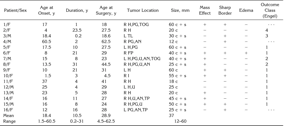
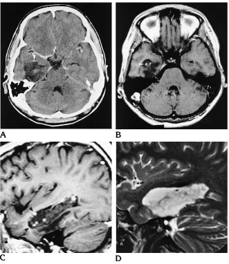
Fig 1. Right frontal DNT (patient 6).
Fig 1, continued.
G–L, From 1994, precontrast (G, J) and postcontrast (H, K) T1-weighted MR images and T2-weighted (I,L) MR images show a central calcification (white arrow) surrounded by multiple cysts (black arrows), enhancing tumor nodules, and perifocal edema (arrowheads). M, Low-power photomicrograph shows the nodular growth pattern and the loose matrix of this tumor. Note the focal calcification (arrow). Many small capillaries and hemosiderin-laden macrophages are at the surface of this nodule, suggesting an organizing intratumoral hemorrhage (arrowheads) (hematoxylin-eosin; magnification3165).
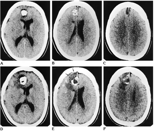
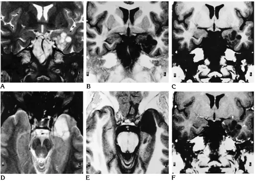
[image:5.612.57.556.83.629.2]Fig 2. Right temporomesial DNT (patient 13).
A–C, Coronal T2-weighted fast spin-echo (A), heavily T1-weighted inversion-recovery (B), and axial postcontrast T1-weighted spin-echo (C) MR images show multiple large cysts (arrowheads) that appear to replace the head and body of the hippocampus without subcortical extension. There is only slight mass effect.
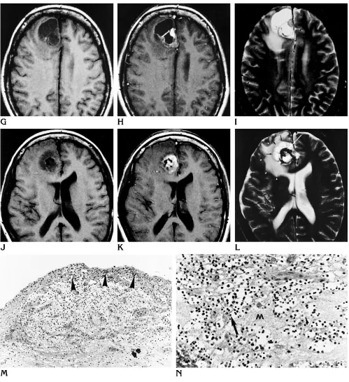
Fig 3. Right temporal DNT (patient 14).
[image:6.612.222.552.352.703.2]The postoperative outcome was evaluated with clinical follow-up examinations in 13 pa-tients. One patient died perioperatively of an unexplained diffuse cerebral hypoxia, while in 2 patients the postoperative phase is less than 6 months so far. Nine patients are in class I (sei-zure free), 2 patients are in class II, and 1 patient each is in class III and IV (Table 1).
Discussion
High-resolution MR imaging has significantly contributed to the recognition of low-grade tu-mors and tumorlike lesions as a major cause of chronic focal epilepsy. Modern MR techniques with high resolution and high contrast permit detection of lesions measuring no more than a few millimeters in size, and because of this, MR should be routinely used to examine patients
with chronic focal epilepsy. The neuroradiologic characterization of such lesions is essential for the treatment of these patients because it aids in identifying those who are likely to benefit from epilepsy surgery. Following hippocampal scle-rosis, benign tumors are the second most frequent brain lesions that are associated with epilepsy (10). The high prevalence of gang-liogliomas in patients with chronic focal epi-lepsy contrasts with the fact that they constitute only 0.4% to 1.3% of all brain tumors in general neurosurgical series (23, 24). In our epilepsy surgery series, which presently includes 469 resection specimens, there were 138 tumors, including 17 DNTs (13%), 62 gangliogliomas (45%), and 59 astrocytomas (mixed gliomas and oligodendrogliomas) (42%).
Histopathologically, DNT is a benign,
[image:7.612.59.558.87.445.2]pre-dominantly intracortical lesion composed
Fig 4. Left temporomesial DNT (patient 16).
mainly of a population of oligodendrocyte-like cells with admixtures of mature ganglion cells and astrocytes that are located in a myxoid or dense neurofibrillary matrix (Fig 1C and D) (1, 4 –11). Involvement of the cortical gray matter and a multinodular growth pattern are typical findings. The cortex adjacent to a DNT shows a disordered architecture. On the basis of its his-topathologic appearance and benign clinical course, it has been suggested that DNT may be
a malformation rather than a neoplastic lesion; however, significant proliferative activity as de-termined by Ki-67 labeling has been reported in some DNTs (8, 11). Histopathologically, DNTs can be distinguished from gangliogliomas by their multinodular architecture, myxoid matrix, and predominant population of oligodendro-cyte-like cells. In addition, DNTs lack the bi-zarre neurons, the abundant connective tissue stroma, and the perivascular lymphocytic
infil-TABLE 2: Preoperative MR and CT studies in six patients with dysembryoplastic neuroepithelial tumor over a period of up to 13 years
Patient Date of First Examination
Date of Subsequent Imaging Studies
Last Preoperative Control
Date of Surgery
Observation Period, y
5 CT 2/86 MR 1/91 MR 3/93 8/93 7
6 CT 2/81 CT 8/86, 2/94 MR 2/94 3/94 13
7 CT 4/83 MR 4/89 MR 4/90 6/90 7
10 MR 1/91 MR 6/93, CT 12/93 MR 3/94 3/94 3
13 MR 11/91 . . . MR 10/93 10/93 2
[image:8.612.224.555.86.468.2]16 MR 10/93 CT 12/94 MR 3/95 3/95 1.5
[image:8.612.59.554.502.595.2]trates that are frequently encountered in gan-gliogliomas (1). Other epileptogenic lesions that are composed of randomly scattered glial and neuronal elements are glioneuronal malfor-mations. Some glioneuronal malformations are large and may resemble a neoplasm grossly. These are classified as glioneuronal toma. Histopathologically, glioneuronal hamar-tomas may show great similarity with cortical tubers of tuberous sclerosis. Other glioneuronal malformations measure only a few millimeters in size and are termed glioneuronal hamartias. Glioneuronal malformations lack the markedly increased cellularity and tumor matrix that are so characteristic of true glial and glioneuronal neoplasms. For a review of this subject, see the article by Wolf and Wiestler (10). Because of
the different biological natures of these different epilepsy-associated glioneuronal lesions (tu-mor versus malformation), it is important to establish criteria that may help to distinguish these entities radiologically.
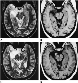
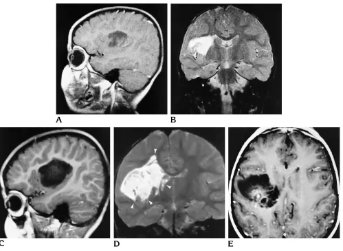
[image:9.612.62.561.85.450.2]A large series of 39 DNTs was published by Daumas-Duport et al in 1988 (1). Other articles in peer-reviewed publications report fewer than 8 cases each (5–7, 12–16, 18). Daumas-Duport et al performed a retrospective analysis of the histopathologic features of tumors resected be-tween 1963 and 1988 and coined the term DNT for a subset of supratentorial superficial cortical tumors that typically present with focal epilepsy before 20 years of age. The authors briefly men-tion MR (n 518) and CT findings (n532) and report cortical hypodense lesions on CT scans Fig 6. Right insular DNT (patient 10).
A–D, MR images from 1991 (A, B) show a cystic tumor that seems to replace the suprasylvian insular cortex and the frontal subcortical white matter, reaching the striatum but respecting its border. MR images from 1993 (C, D) show the tumor extending significantly farther below, into the insular cortex, and farther above and medially, into the white matter (arrowheads).
and prolonged T1 and T2 times on MR images, findings that are seen in most brain tumors. In a recent review, Daumas-Duport (8) describes a megagyri-like appearance of DNT on MR im-ages and mentions a possible pseudocystic ap-pearance of DNT on CT scans. Injection of ga-dopentetate dimeglumine was done in only a few of these cases, so that enhancement was detected only rarely.
In our series of 16 DNTs, the lobar distribu-tion, with a striking temporal lobe predomi-nance (86%) similar to that found in ganglio-gliomas (84%) and glioneuronal malformations (85%), and the clinical presentation, with sei-zures in all patients, were consistent with de-scriptions found in the literature. The absence of neurologic deficit as found in all but one of our patients has been described previously as typi-cal of DNT (2). Only 4 patients had their first seizure after 20 years of age, which is consistent with rare reports of cases with late seizure onset in the literature (2, 17, 25). The average dura-tion of epilepsy before surgery was 10.5 years (range, 0.2 to 31 years). This indicates a benign clinical course. Fourteen of the 16 patients had pharmacoresistant epilepsies for more than 2 years before surgery.
Imaging findings reported so far (1, 8, 6) con-sistently describe well-demarcated hypodense lesions on CT scans that rarely calcify or en-hance. In our analysis, CT revealed moderate hypodensity of presumably solid tumor compo-nents and marked hypodensity of cystic com-ponents of DNT. Calcification was noted in only 2 (20%) of 10 DNTs, whereas CT showed cal-cification in 41% of gangliogliomas (21) and in 25% of glioneuronal malformations (20). All DNTs but one were sharply demarcated; the majority had inhomogeneous density and were space-occupying, but showed no edema. Thus, CT did not provide specific features of DNT but showed the nonspecific features of a benign tumor.
MR studies allow a much more detailed eval-uation of the different tumor components. Al-though previous studies (2, 3) have reported cystic changes only in a minority of the cases, all lesions in our study contained cystic compo-nents, which often exhibited shorter T1 than CSF and appeared hypointense, isointense, or hyperintense on proton density–weighted im-ages (Figs 3–5). This signal pattern reflects the abundant myxoid interstitial matrix that is typi-cal of DNT. The high protein content of the
myxoid matrix explains signal differences on T1-weighted and proton density–weighted im-ages as compared with CSF. The cystic, in most cases even multicystic, appearance on MR im-ages is a typical feature of DNT despite the absence of true cysts intraoperatively or on mi-croscopic examination.
A cystic signal pattern is also found in other lesions that are frequently associated with chronic epilepsy, such as gangliogliomas, gli-oneuronal malformations, and other gliomas. In recently published series of 51 gangliogliomas (21) and 33 glioneuronal malformations (20), 2 gangliogliomas (4%) appeared completely cys-tic, 21 (41%) had both solid and cystic compo-nents, and 28 gangliogliomas (55%) were purely solid on MR images. A multicystic ap-pearance was present in only 5 (10%) of 51
gangliogliomas. Glioneuronal malformations
were associated with other lesions, mainly gan-gliogliomas in 16 cases. Isolated glioneuronal malformations were purely solid in 62% and had solid and cystic components in 38% of cases. Only 1 glioneuronal malformation was multi-cystic on MR images. Thus, although the mul-ticystic appearance on MR images is not en-tirely specific for DNT, it is a typical and frequent finding, which helps to distinguish DNT from gangliogliomas and glioneuronal malfor-mations.
The signal pattern of solid DNT components on MR images is similar to that of solid ganglio-gliomas, which appeared isointense on T1-weighted images in 32 (73%) of 44 cases in one study (21). Approximately half of the solid DNT elements show contrast enhancement, a finding that is observed in 44% of gangliogliomas (21), whereas enhancement is never observed in solid parts of glioneuronal malformations (20). Multifocal nodular enhancement was found in only 2 DNTs, but in none of the gangliogliomas of World Health Organization grade I. In contrast to the previously reported CT findings, MR im-ages showed poorly defined tumor contours in 8 DNTs. Calcification, which is best detected by CT, is less frequent in DNTs (2 [14%] of 14) than in gangliogliomas (7 [41%] of 17) and in glio-neuronal malformations (2 [25%] of 8). In con-trast to DNTs, gangliogliomas may occur
infrat-entorially and may be malignant both
histopathologically and clinically (26).
addi-tional cystic formation with perifocal edema could be explained by tumor hemorrhage and may not necessarily be the result of tumor growth. CT also showed significant growth of a large solid calcified tumor component. His-topathologically, the lesion contained abundant
capillaries and hemosiderin-laden
macro-phages, indicative of previous hemorrhage. To date, there is no evidence of recurrence 1 year after surgery in this patient.
The observation of a growing cystic tumor with mild mass effect and enhancement over time in another case (Fig 6) is unusual. It raises the possibility of a malignant transformation; however, review of the histopathologic findings showed no evidence of malignancy. The patient is seizure free 1 year after surgery, although complete tumor removal could not be achieved. In all other cases, the imaging studies yielded no evidence of growth or edema. However, our study includes some cases with approximately 20-year-old CT scans and first-generation MR images, which make the evaluation of possible changes in tumor size and demarcation difficult. Deformities of the calvaria adjacent to super-ficially located DNTs were described in 11 of 12 cases by Daumas-Duport (8). In our series, only 1 DNT extended to the cortical surface. This tumor was not associated with a deformity of the calvaria. The other 15 tumors were located temporomesially or deeply in the insula, so that remodeling of the skull would be unlikely.
Ten of 16 DNTs extended into the subcortical white matter. In some cases, such tumor exten-sion was observed without mass effect, suggest-ing replacement of brain tissue by DNT. This evolution was graphically documented in a case of insular DNT with 3 years of imaging follow-up (Fig 6). The observation of a patient who had two cysts that were clearly separate from the main tumor mass in a temporomesial DNT (Fig 3) may also raise the question of a multifocal tumor origin in such cases.
In conclusion, our analysis of the MR findings with special attention to signs of multinodularity and multicystic appearance closely reflects the well-documented histopathologic growth pat-tern of DNT. Together with the typical clinical presentation and the history of chronic focal epilepsy, MR findings may strongly suggest the preoperative diagnosis of DNT and may help to avoid inadequate therapy, such as radiation in-stead of surgical removal, which is the therapy of choice.
Acknowledgments
We gratefully acknowledge the editorial assistance of Monika Nolden and Hanne Storma and the photographic assistance of Wolfgang Nettekoven.
References
1. Daumas-Duport C, Scheithauer BW, Chodkiewiez JP, Laws ER Jr, Vedrenne C. Dysembryoplastic neuroepithelial tumor: a surgically curable tumor of young patients with intractable partial seizures. Neurosurgery1988;23:545–556
2. Kleihues P, Burger PC, Scheithauer BW. The new WHO classifi-cation of brain tumors.Brain Pathol1993;3:255–268
3. Kleihues P, Burger PC, Scheithauer BW.Histological Typing of Tumors of the Central Nervous System.2nd ed. Berlin: Springer, 1993:23
4. Honovar M, Ansari S, Janota I, Polkey CE. Dysembryoplastic neuroepithelial tumor.Neuropathol Appl Neurobiol1991;17:242– 243
5. Prayson RA, Estes ML, Morris HH. Coexistence of neoplasia and cortical dysplasia in patients presenting with seizures.Epilepsia 1993;34:609 – 615
6. Koeller KK, Dillon WP. Dysembryoplastic neuroepithelial tumors: MR appearance.AJNR Am J Neuroradiol1992;13:1319 –1325 7. Kirkpatrick PJ, Honovar M, Janota I, Polkey CE. Control of
tem-poral lobe epilepsy following en bloc resection of low grade tu-mors.J Neurosurg1993;78:19 –25
8. Daumas-Duport C. Dysembryoplastic neuroepithelial tumors. Brain Pathol1993;3:283–295
9. Wolf HK, Campos MG, Zentner J, et al. Surgical pathology of temporal lobe epilepsy: experience with 216 cases.J Neuropathol Exp Neurol1993;52:499 –506
10. Wolf HK, Wiestler OD. Surgical pathology of chronic epileptic seizure disorders.Brain Pathol1993;3:371–380
11. Wolf HK, Wellmer J, Mu¨ller WB, Wiestler OD, Hufnagel A, Pietsch T. Glioneuronal malformative lesions and dysembryoplastic neu-roepithelial tumors in patients with chronic pharmacoresistant epilepsies.J Neuropathol Exp Neurol1995;54:245–254 12. Hirose T, Scheithauer BW, Lopes MB, Vanden Berg SR.
Dysem-bryoplastic neuroepithelial tumor (DNT): an immunohistochemi-cal and ultrastructural study. J Neuropathol Exp Neurol1994;53: 184 –195
13. Jay V, Becker LE, Otsubo H, Hwang PA, Hoffman HJ, Harwood-Nash D. Pathology of temporal lobectomy for refractory seizures in children.J Neurosurg1993;79:53– 61
14. Kuroiwa T, Kishikawa T, Kato A, Ueno M, Kudo S, Tabuchi K. Dysembryoplastic neuroepithelial tumors: MR findings.J Comput Assist Tomogr1994;18:352–356
15. Leung SY, Gwi E, Ng HK, Fung CF, Yam KY. Dysembryoplastic neuroepithelial tumor: a tumor with small neuronal cells resem-bling oligodendroglioma.Am J Surg Pathol1994;18:604 – 614 16. Morris HH, Estes ML, Gilmore R, Van Ness PC, Barnett GH,
Turn-bull J. Chronic intractable epilepsy as the only symptom of pri-mary brain tumor.Epilepsia1993;34:1038 –1043
17. Prayson RA, Estes ML. Dysembryoplastic neuroepithelial tumor. Am J Clin Pathol1992;97:398 – 401
18. Sato T, Takeichi M, Abe M, Tabuchi K, Hara T. Frontal lobe tumor associated with late onset seizure and psychosis: a case report. Jpn J Psychiatry Neurol1993;47:541–544
20. Ostertun B, Solymosi L, Zentner J, et al. MR- und CT-Charakter-istika glioneuronaler Hamartien. Klin Neuroradiologie 1994;4:34 – 40
21. Zentner J, Wolf HK, Ostertun B, et al. Gangliogliomas: clinical, radiological and histopathological findings in 51 patients.J Neurol Neurosurg Psychiatry1994;57:1497–1502
22. Engel J, Van Nees PC, Rasmussen TB, Ojemann LM. In: Engel J, ed.Surgical Treatment of the Epilepsies.2nd ed. New York: Raven Press, 1993:609 – 621
23. Zu¨lch KJ. Atlas of Gross Neurosurgical Pathology. Berlin: Springer, 1975:49 –50
24. Drake J, Hoffmann HJ, Kobayashi HJ, et al. Surgical manage-ment of children with temporal lobe epilepsy and mass lesions. Neurosurgery1987;21:792–797
25. Gottschalk J, Korves M, Skotzek-Konrad B, Goebel S, Cervos-Navarro J. Dysembryoplastic neuroepithelial micro-tumor in a 75-year-old patient with long-standing epilepsy.Clin Neuropathol 1993;12:175–178