Phase-Contrast MR Angiography with Three-dimensional Acquisition
and Phase Display
Mario Mascalchi, Nello Quilici, Giampiero Ferrito, Salvatore Mangiafico, Fabio Scazzeri, Paolo Torselli, Pasquale Petruzzi, Mirco Cosottini, Carlo Tessa, and Carlo Bartolozzi
PURPOSE: To determine whether identification of the feeding arteries of spinal vascular lesions with phase-contrast MR angiography benefits from the higher spatial resolution of three-dimen-sional (volume) acquisitions and flow-direction information provided by the phase reconstruction of two-dimensional acquisitions. METHODS: Fifteen patients with high- or low-flow spinal vascular lesions proved by spinal arteriography underwent MR angiography with phase-contrast techniques. Arteriographic and MR angiographic studies were reviewed to identify the arterial feeders of spinal vascular lesions. RESULTS: On modulus reconstructions of coronal 2-D or 3-D acquisitions, three of four arteries feeding high-flow lesions and three of 14 arteries feeding low-flow lesions were identified as hypertrophic vessels joining the parent intercostal or cervical arteries. Of 11 intradural veins draining dural arteriovenous fistulas, three were identified on coronal 2-D acquisitions and six on coronal 3-D acquisitions as vessels that coursed from a neural foramen to a midline tangle of vessels. Phase reconstruction showed ascending and descending flow patterns in two patients with intramedullary arteriovenous malformations, and diverging flow in perimedullary veins draining a hemangioblastoma. In nine patients with dural arteriovenous fistulas, phase reconstruction pro-vided information as to the level of the arterial feeders. Phase reconstruction in coronal plane acquisitions also provided evidence of centripetal flow. CONCLUSION: Three-dimensional acqui-sitions and phase display of 2-D acquiacqui-sitions improved the visibility of arterial pedicles of spinal vascular lesions at phase-contrast MR angiography.
Index terms: Arteriovenous malformations, spinal; Magnetic resonance angiography; Spine, mag-netic resonance
AJNR Am J Neuroradiol18:351–358, February 1997
Selective spinal arteriography is the ultimate technique for use in diagnosing, classifying, and planning therapy for spinal vascular lesions (1). However, it is invasive, time consuming, expen-sive, and dependent on the skills of the operator. Modulus reconstruction of two-dimen-sional phase-contrast magnetic resonance
(MR) angiographic acquisitions in the sagittal plane is a useful and rapid complement to MR imaging for the identification of abnormal ves-sels within the spinal canal (2, 3). Modulus re-construction of three-dimensional (volume) phase-contrast MR angiographic acquisitions in the coronal plane was reported to show abnor-mally dilated midline serpiginous vessels in a single case of dural arteriovenous fistula (4). One goal in the use of MR angiography is the identification of arterial feeders of vascular le-sions of the spine. Such an identification may direct the selective spinal arteriographic exam-ination to specific spinal levels, thus shortening catheterization time. In a previous study (3), modulus reconstruction of 2-D phase-contrast MR angiographic acquisitions in the coronal Received April 23, 1996; accepted after revision August 14.
From the Cattedra di Radiologia, Universita’ di Pisa (M.M., P.T., P.P., M.C., C.T., C.B.); the Department of Neuroradiology, Ospedali Riuniti, Livorno (N.Q., G.F., F.S.); and the Department of Neuroradiology, Osped-ale Careggi, Firenze (S.M.); Italy.
Address reprint requests to Mario Mascalchi MD, PhD, Cattedra di Radiologia, Universita’ di Pisa, Via Roma 67 56100, Pisa, Italy.
AJNR 18:351–358, Feb 1997 0195-6108/97/1802–0351 ©American Society of Neuroradiology
plane provided evidence of arterial feeders of high-flow malformations, including intramedul-lary arteriovenous malformations (AVMs) and perimedullary arteriovenous fistulas (AVFs), whereas it often failed to show the source of low-flow dural AVFs, which are the most com-mon vascular lesions in adults or elderly per-sons (5).
We investigated whether the identification of feeding arteries of spinal vascular lesions via phase-contrast MR angiography benefits from the higher spatial resolution of 3-D (volume) acquisition (6) and flow-direction information provided by phase reconstruction of 2-D acqui-sitions.
Materials and Methods
From September 1994 to March 1996, 15 patients (seven women and eight men; 21 to 84 years old) with native (n 5 12), residual (n 5 1), or recurrent (n5 2) spinal vascular lesions proved by arteriography were ex-amined with the use of either a 1.5-T (n512) or a 0.5-T (n5 3) MR imaging unit. Selective spinal arteriographic diagnoses included intramedullary AVMs (n 5 2), high-flow perimedullary AVF (n51), dural AVFs (n511), and hemangioblastoma (n 5 1). All patients previously had enhanced MR imaging at different centers on 0.5-T or 1.5-T systems.
MR angiography included sagittal 5-mm-thick T1-weighted spin-echo sequences (360 –500/20 –25/2– 4 [repetition time/echo time/excitations]) with a 25-cm field of view and a 160 3 224 matrix. Since paramagnetic contrast agents reduce the effects of spin saturation in phase-contrast MR angiography, thereby enabling visual-ization of small low-flow vessels and improving the intra-vascular signal/background noise ratio (7), most (n513) of our examinations were performed after intravenous bo-lus injection of (0.3 mmol/kg) gadopentetate dimeglu-mine. Single-section 2-D phase-contrast MR angiography was then performed in the midsagittal plane with a gradi-ent-recalled echo sequence (38/11/8 –12 with a flip angle of 30°) incorporating bipolar flow-encoding gradients. Amplitude and duration of the bipolar gradients were set to optimally detect velocity of 20 to 30 cm/s in high-flow malformations and 6 to 10 cm/s in low-flow malformations (3). Three acquisitions with bipolar gradients encoding flow along superoinferior, anteroposterior, and left-right body axes were acquired. Section thickness was 15 mm with a 25-cm field of view and a 1603224 matrix. Ante-rior presaturation slabs, 80- to 120-mm thick, were used for thoracolumbar spinal MR angiographic examinations.
Coronal 2-D (38/11/8 –12 with a flip angle of 20° to 30°) and 3-D (29/11/1–2 with a flip angle of 20° to 30°) phase-contrast MR angiograms were then obtained with single 15-mm-thick sections and 28 to 60 sections 1 to 1.5 mm thick. Coronal acquisitions were centered on the
ab-normal intraspinal vessels detected on sagittal 2-D phase-contrast images. No spatial presaturation was used for coronal acquisitions. Flow was encoded along the supero-inferior, anteroposterior, and left-right axes with amplitude and duration of flow-encoding gradients identical to those used for sagittal 2-D MR angiography.
In all phase-contrast acquisitions, first-order flow com-pensation was used; projection dephasing gradients were not used. Total examination time was less than 1 hour for all patients. Phase-contrast MR angiograms were recon-structed using complex-difference or phase-difference techniques (7). The resulting superoinferior, anteroposte-rior, and left-right flow modulus images were then com-bined in a total flow modulus image. This phase-difference technique also provides phase images in which flow along each encoded axis is displayed as black or white according to its direction, whereas background stationary tissues are gray. In our MR systems, superoinferior flow is displayed as white and inferosuperior flow as black, anteroposterior flow is white and posteroanterior flow is black; right-left and left-right flow appears white and black. All 3-D coronal phase-contrast acquisitions were reconstructed using the complex-difference technique and the maximum-intensity projection (MIP) algorithm. Sagittal (n512) and coronal (n 5 9) 2-D acquisitions were reconstructed with the phase-difference technique in 12 patients and with the complex-difference technique in three patients. Hard cop-ies of selective spinal arteriograms and MR angiograms were reviewed jointly by two observers to try to identify the arterial feeders of spinal vascular malformations.
Results
Abnormal vessels within the spinal canal were demonstrated by sagittal 2-D MR angiog-raphy in all patients. On corresponding unen-hanced or enunen-hanced MR images, the abnormal vessels were identified in 11 patients (Figs 1–5), whereas they were barely detectable in four pa-tients with dural AVFs.
The Table provides a summary of the results of attempts to identify the arterial feeders of the spinal vascular lesions on phase-contrast MR angiograms. On the total flow modulus images of coronal 3-D acquisitions, three of four feed-ing arteries in three patients with high-flow vas-cular malformations were identified as hairpin, S-shaped, or short hypertrophic vessels joining the intercostal (n52) or cervical (n51) parent arteries (Figs 1 and 2). Total flow modulus re-construction images of coronal 2-D acquisitions in three patients showed only two arterial feed-ers, with lesser conspicuity and sharpness (Fig 2). One feeder of a high-flow perimedullary AVF was missed on both 2-D and 3-D acquisitions (Fig 1).
On total flow modulus reconstruction images of coronal 2-D or 3-D acquisitions, only three of 14 feeding arteries from low-flow vascular le-sions were identified (Figs 3 and 4).
Of 11 single intradural veins draining dural AVFs, three were identified on coronal 2-D and 3-D acquisitions as large vessels that coursed from the region of a neural foramen to the mid-line tangle of serpiginous vessels. In three pa-tients, tiny vessels approaching the midline tan-gles were barely seen on the 3-D acquisitions (Fig 5) and were missed on the 2-D acquisi-tions.
On coronal modulus 2-D or 3-D images, pre-vertebral or retropre-vertebral vessels that were in-advertently included in the section or volume of interest could have been misinterpreted as feed-ing arteries (Fig 3) when they approached or crossed the midline tangle of abnormal vessels; however, axial reconstructions of the 3-D data set using the MIP algorithm helped to avoid such pitfalls. Targeted MIPs in the coronal plane of 3-D acquisitions more clearly showed arterial feeders or intradural draining veins than did MIP reconstructions of the whole data set, but in no case did they reveal vessels undetected in the latter.
Phase reconstruction of superoinferior flow in sagittal or coronal 2-D acquisitions showed as-cending and desas-cending flow patterns in two patients with intramedullary AVMs and diverg-ing flow in the perimedullary veins draindiverg-ing the hemangioblastoma (Figs 2 and 3). In nine pa-tients with dural AVFs, phase reconstruction of the superoinferior flow provided an indirect clue as to the level of the arterial feeders by showing diverging (n 5 4), ascending (n 5 3), or de-scending (n52) flow direction in the perimed-ullary plexus (Fig 5). The point of divergence corresponded to the anastomosis between the medullary vein draining the fistula and the peri-medullary venous plexus. This point was always located within two levels of the arterial feeder. Phase reconstruction of the superoinferior flow direction in the dilated perimedullary vessels constituted the only clue as to the level of the arterial feeder in four patients with dural fistula whose intradural draining vein was missed on coronal 2-D or 3-D modulus reconstructions.
[image:3.587.51.548.84.273.2]Phase images of left-right flow in the coronal plane provided evidence of centripetal flow, that is, from outside to inside the spinal canal, in the case of the feeding arteries of high-flow lesions (n 5 2) (Fig 2) and intradural veins draining Fig 1. Perimedullary AVF.
A, Unenhanced sagittal T1-weighted (400/20/2) MR image shows large round area of signal void (arrow) displacing and deforming the conus medullaris.
B, Modulus reconstruction of contrast-enhanced 3-D phase-contrast MR angiogram (29/11/1, 20° flip angle) with velocity encoding of 30 cm/s in the coronal plane shows an S-shaped vessel (open arrow) joining a varix (solid arrow) from above.
C, The feeding artery is not seen on this modulus reconstruction of enhanced 2-D phase-contrast MR angiogram (38/11/10, 20° flip angle) with velocity encoding of 30 cm/s in the coronal plane.
D, Selective spinal arteriogram (frontal projection) with catheterization of right L-1 artery shows the arterial feeder (open arrow) joining a large venous pouch (solid arrow) without an intervening nidus, and is consistent with high-flow perimedullary fistula.
Fig 2. Intramedullary AVM partially occluded by glue embolization 1 year before present MR examination.
A, Unenhanced sagittal T1-weighted (360/20/2) MR image shows tiny areas of signal void within (solid arrow) and in front of (open arrows) the spinal cord.
BModulus reconstruction of contrast-enhanced 3-D phase-contrast MR angiogram (29/11/1, 20° flip angle) with velocity encoding of 20 cm/s in the coronal plane shows the feeder (open arrow), which arises from the left T-9 intercostal artery (arrowheads) and joins the partially occluded nidus and the descending draining vein (solid arrow).
C, In modulus reconstruction of enhanced 2-D phase-contrast MR angiogram (38/11/10, 20° flip angle) with velocity encoding of 20 cm/s in the coronal plane, the arterial feeder is visible (arrow) but the abnormal intraspinal vessels are less sharply defined.
D, Phase reconstruction ofCshows ascending (solid black arrow) and descending (solid white arrow) superoinferior flow in the proximal and distal portions of the feeding artery, and descending flow (open white arrow) in the draining vein.
E, Reconstruction ofCshows left-to-right flow in the proximal portion of the arterial feeder (arrow). F, Phase reconstruction ofCshows anteroposterior flow is noncontributory.
G, Selective spinal arteriogram (anteroposterior projection) with catheterization of left T-9 shows the arterial feeder (open arrow) nourishing the partially occluded nidus and the descending vein (solid arrow). Note artery of Adamkievicz (arrowheads), which was missed on all phase-contrast MR angiographic acquisitions.
dural AVFs (n51) and of centrifugal flow in the case of radicular veins draining an intramedul-lary AVM (n51). Phase images of the left-right flow in the sagittal plane and anteroposterior flow in the sagittal and coronal planes were noncontributory in helping to define the feeding arteries to the vascular lesions.
Discussion
[image:5.587.50.548.84.275.2]Identification of the arterial pedicles of in-tramedullary AVMs is not usually possible with MR imaging; however, the intramedullary nidus is well shown (8) and can be regarded as a rough indicator of the level of the arterial feeder. Identification of the arterial feeders of spinal perimedullary and dural AVFs is not always possible with MR imaging. Since perimedullary AVFs lack a nidus, and the nidus of dural AVFs is often microscopic, enlarged perimedullary vessels extending over a considerable length of the spinal canal are the only finding at MR im-aging (5, 8, 9). This precludes any a priori iden-tification of the arterial pedicles and leads to catheterization of many spinal vascular pedicles before the fistula is seen.
Fig 4. Dural AVF.
A, Sagittal T2-weighted (2000/100/1) spin-echo MR image shows high signal intensity in the thoracic spinal cord and conus medullaris with retromedullary serpiginous areas of signal void.
[image:5.587.306.548.416.612.2]B, Modulus reconstruction of unenhanced 3-D phase-contrast MR angiogram (29/11/2, 30° flip angle) with velocity encoding of 6 cm/s in the coronal plane shows a small vessel (open arrow) that from the right T-7 intercostal artery courses toward faint serpiginous midline vessels (solid arrow). Selective spinal arte-riogram with catheterization of right T-7 intercostal artery con-firmed the arterial feeder of the dural fistula and the intradural draining vein (not shown).
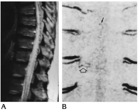
Fig 3. Intramedullary hemangioblastoma.
A, Contrast-enhanced sagittal T1-weighted (360/20/2) MR image shows intramedullary nodule within an extensive septated syrinx. Tiny retromedullary areas of signal voids (arrow) are seen below the lesion.
B, Modulus reconstruction of contrast-enhanced 2-D phase-contrast MR angiogram (38/11/12, 20° flip angle) with velocity encoding of 6 cm/s in the coronal plane shows extensive tangle of serpiginous vessels joining the left T-7 vascular pedicle through a small vessel (arrow).
C, The abnormal vessels are defined better on this modulus reconstruction of enhanced 3-D phase-contrast MR angiogram (29/11/1, 20° flip angle) with velocity encoding of 6 cm/s in the coronal plane. No connection of the left T-7 vascular pedicle is seen, whereas vessels apparently joining the right T-7 pedicle (open arrow) and the left T-9 intercostal vein (solid arrow) are present.
The distinction between high- and low-flow spinal vascular lesions is fundamental in plan-ning phase-contrast MR angiography because the value of flow-velocity encoding needs to be chosen before the examination begins. Our study confirms that identification of arterial feeders with the use of phase-contrast MR an-giography is easier in high-flow than in low-flow lesions (3). Our results indicate that modulus reconstruction of 3-D acquisitions improves de-tection of the arterial feeder of spinal vascular lesions as compared with modulus reconstruc-tion of 2-D phase-contrast acquisireconstruc-tions, pre-sumably because of the intrinsically poor spa-tial resolution of the latter. In only one of our patients was the arterial feeder identified on coronal modulus 2-D acquisitions, and it was missed on the 3-D acquisitions. Because this was a low-flow lesion (hemangioblastoma), we speculate that saturation phenomena, which are more prominent in 3-D than in 2-D acquisi-tions, may have accounted for this exception. The 3-D modulus acquisitions enabled us to directly or indirectly identify the arterial feeders in seven of 11 patients with dural AVFs. Our data are similar to those reported by Bowen et al (10), who identified the intradural draining vein and indirectly the source of dural AVFs in six of eight patients by using enhanced 3-D
[image:6.587.50.552.84.274.2]time-of-flight MR angiography. It is noteworthy that whereas those authors were able to see the en-tire course of intradural draining veins, we were able to show only portions of the intradural draining vein in some of our patients (see Fig 5). We speculate that this difference presum-ably reflects the fact that in enhanced time-of-flight imaging, T1 shortening induced by the paramagnetic contrast agent is to some extent independent of flow. While this may create some problems in differentiating enhanced sta-tionary tissue (10), it will considerably help to delineate very low flow vessels (11). Unen-hanced and enUnen-hanced phase-contrast MR an-giographic techniques rely entirely on flow to demonstrate the vessels (6, 7), and, as such, it is possible that they may not show very low flow vessels or portions of them (at least with the velocity-encoding value used in our study). The fact that, ultimately, enhanced time-of-flight imaging might seem to be more sensitive to very slow flow than is phase-contrast MR an-giography supports the observations made in recent reports that normal veins anterior and posterior to the spinal cord can be seen on contrast-enhanced time-of-flight MR angio-grams and MR images of the thoracolumbar spine in subjects without spinal vascular lesions at selective spinal arteriography (11, 12). These Fig 5. Dural AVF (imaged at 0.5 T).
A, Sagittal contrast-enhanced T1-weighted (400/20/4) MR image shows diffuse enhancement of the lower thoracic spinal cord and retromedullary vessels of low signal intensity.
B, Modulus reconstruction of contrast-enhanced 2-D phase-contrast MR angiogram (38/11/12, 30° flip angle) with velocity encoding of 6 cm/s in the sagittal plane shows retromedullary serpiginous vessels.
C, Phase reconstruction ofBshows diverging superoinferior flow in the retromedullary vessels (arrow) at T-12.
D, On modulus reconstruction of enhanced 3-D phase-contrast MR angiogram (29/11/2, 30° flip angle) with velocity encoding of 6 cm/s in the coronal plane, a tiny vessel (arrow) in the right paramedian site at T-12 joins the midline tangle of serpiginous vessels (confirmed by selective spinal arteriography).
veins correspond to the median veins and med-ullary or radicular veins, and may actually cre-ate some problems in determining a differen-tial diagnosis for true vascular lesions, in particu-lar for dural AVFs (11). By using the enhanced phase-contrast techniques described in this study, we observed normal posterior median veins in only two of 10 patients with normal arte-riographic findings (personal unpublished obser-vations). Interestingly, these two patients had
spi-nal cord astrocytoma and lupus myelitis, two conditions that can increase spinal cord blood flow. In the present study, considerable informa-tion as to detecinforma-tion of arterial feeders was pro-vided by phase reconstruction of sagittal and coronal 2-D acquisitions, which showed su-peroinferior and left-right flow direction in the abnormal intraspinal vessels. This information was always indirect in the patients with dural AVFs, focusing on flow direction in the
perimed-Arterial feeders of 15 spinal vascular lesions and their identification with phase-contrast MR angiography
Case Age, y/Sex
Selective Spinal Arteriography Phase-Contrast MR Angiography
Diagnosis Arterial Feeder
3-D Modulus Reconstruction
2-D Modulus Reconstruction
2-D Phase Reconstruction
1 49/F Perimedullary AVF
R L-1 L-4 L-1 arterial feeder
. . . .
2 24/F Intramedullary AVM (after incomplete treatment)
L T-9 Arterial feeder Arterial feeder Arterial feeder
3* 21/F Intramedullary AVM
L SIA Arterial feeder Arterial feeder Arterial feeder
4 21/M Hemangioblastoma L T-7 . . . Arterial feeder Diverging flow in perimedullary veins 5* 34/M Dural AVF R T-5 T-7 Arterial feeder . . . . 6 84/F Dural AVF
(recurrence)
R T-12 Intradural draining vein
. . . Ascending flow in perimedullary veins 7 73/F Dural AVF R L-1 Intradural
draining vein
Intradural draining vein
. . .
8 82/F Dural AVF L T-9 T-11 . . . Diverging flow in perimedullary veins 9 73/M Dural AVF R T-12 Intradural
draining vein
Intradural draining vein
Ascending flow in perimedullary veins 10 72/M Dural AVF R T-12 Intradural
draining vein
Intradural draining vein
Ascending flow in perimedullary veins 11 58/F Dural AVF R T-7 . . . Diverging flow in
perimedullary veins 12 72/M Dural AVF R T-12 Intradural
draining vein
. . . Diverging flow in perimedullary veins 13 74/M Dural AVF L T-9 . . . Descending flow
in perimedullary veins
14 71/M Dural AVF R T-5 . . . Descending flow in perimedullary veins
15 55/M Dural AVF R T-8 Arterial feeder; intradural draining vein
. . . Diverging flow in perimedullary veins
* Patients who did not receive contrast administration before MRA.
ullary plexus, but it proved particularly valuable in the four patients in whom only dilated peri-medullary slow-flow vessels were seen on 3-D modulus reconstructions.
In cases of dural AVFs, selective spinal arte-riograms usually show a single ascending drain-ing vein, which joins the perimedullary plexus on the dorsal surface of the spinal cord (1). The flow in the perimedullary plexus commonly di-verges from the point of anastomosis. Since the flow in, or the caliber of, vessels craniad or caudad to the anastomosis can be asymmetric, visibility of these vessels with phase-contrast MR angiography may differ with appreciation of only ascending or descending flow, as it did in five of nine of our patients. Admittedly, some problems in the use of phase display for the identification of arterial feeders may be fore-seen. In particular, the panoramic simultaneous visibility of arterial and venous structures on phacontrast MR angiograms with lack of se-lectivity and temporal resolution of selective spinal arteriograms may create a problem in evaluating the entangled vascular anatomy typ-ical of some spinal vascular lesions (see Figs 1 and 4). This may be especially true when thick sagittal or coronal 2-D sections, such as those used in the present study, are obtained. Phase reconstruction of multiple thin sections ac-quired with 2-D or 3-D techniques may help to improve spatial resolution and overcome these problems; but this was not done in our investi-gation.
Although, other approaches can be used to identify arterial feeders, including dynamic con-trast-enhanced T2*-weighted imaging (13) or MR myelography, we believe that the flow-di-rection information provided by phase display, possibly combined with 3-D acquisition schemes, is most specific and promising.
In conclusion, the deficient spatial resolution of currently available MR angiographic tech-niques renders this an inadequate substitute for careful, multiple-level, selective spinal arteriog-raphy in diagnosing and planning treatment for
spinal vascular lesions (1). However, our results indicate that 3-D acquisitions and phase display of 2-D acquisitions independently improve the visibility of arterial feeders of spinal vascular lesions at phase-contrast MR angiography. This information may have significant impact on the planning and execution of selective spinal arte-riography, and possibly on its duration.
References
1. Berenstein A, Lasjaunias P. Endovascular treatment of spine and spinal cord lesions. In:Surgical Neuro-Angiography.Berlin, Ger-many: Springer-Verlag; 1992:5:1– 85
2. Gelbert F, Guichard JP, Mourier KL, et al. Phase-contrast MR angiography of vascular malformations of the spinal cord at 0.5T.
J Magn Reson Imaging1992;2:631– 636
3. Mascalchi M, Bianchi MC, Quilici N, et al. MR angiography of spinal vascular malformations.AJNR Am J Neuroradiol1995;16: 289 –297
4. Provenzale JM, Tien RD, Felsberg GJ, Hacien-Bey L. Spinal dural arteriovenous fistula: demonstration using phase contrast MR an-giography.J Comput Assist Tomogr1994;18:811– 814
5. Terwey B, Becker H, Thron AK, Vahldiek G. Gadolinium-DTPA enhanced MR imaging of spinal dural arteriovenous fistulas.
J Comput Assist Tomogr1989;13:30 –37
6. Dumoulin CL, Hart HR Jr. Magnetic resonance angiography. Ra-diology1986;161:717–720
7. Tu R, Kennel T, Turski P, Polzin J, Korosec F, Mistretta C. Prelim-inary assessment of gadodiamide-enhanced, complex difference phase-contrast magnetic resonance angiography. Acad Radiol
1994;S47–S55
8. Turski P, Korosec F. Technical features and emerging clinical applications of phase-contrast magnetic resonance angiography.
Neuroimaging Clin North Am1992;2:785– 800
9. Dormont D, Gelbert F, Assouline E, et al. MR imaging of spinal cord arteriovenous malformations at 0.5T: study of 34 cases.
AJNR Am J Neuroradiol1988;9:833– 838
10. Bowen BC, Fraser K, Kochan JP, Pattany PM, Green BA, Quencer RM. Spinal dural arteriovenous fistulas: evaluation with MR an-giography.AJNR Am J Neuroradiol1995;16:2029 –2043 11. Bowen BC, DePrima S, Pattany PM, Marcillo A, Madsen P,
Quencer RM. MR angiography of normal intradural vessels of the thoracolumbar spine.AJNR Am J Neuroradiol1996;17:483– 494 12. Lane JI, Koeller KK, Atkinson JDL. MR imaging of the lumbar spine: enhancement of the radicular veins.AJR Am J Roentgenol
1996;166:181–185
13. Thorpe JW, Kendall BE, Mac Manus DG, McDonald W, Miller D. Dynamic gadolinium enhanced MRI in the detection of spinal arteriovenous malformations.Neuroradiology1994;36:522–529