Sharon Seltzer 1 Alexander S. Mark2 Scott W. Atlas3
Received April 6, 1990; revision requested July 11, 1990; revision received April1 0, 1991; accepted May 6, 1991.
Presented at the annual meeting of the American Society of Neuroradiology, Los Angeles, March 1990.
'Department of Radiology, George Washington University Medical Center, Washington, DC 20037. 2 Department of Radiology, Washington Hospital
Center, 110 Irving St., N.W., Washington, DC 20010. Address reprint requests to A. S. Mark.
3 Department of Radiology, Hospital of the
Uni-versity of Pennsylvania, Philadelphia, PA 19104.
0195-6108/91/1206-1227
10 American Society of Neuroradiology
CNS Sarcoidosis:
Evaluation
with
Contrast-Enhanced
MR
Imaging
1227
Reports of findings on unenhanced MR images and contrast-enhanced CT scans in patients with intracranial sarcoidosis have suggested that MR imaging without contrast enhancement may miss meningeal involvement, which is a frequent and prominent finding in neurosarcoidosis. We studied 14 patients with CNS sarcoidosis with T1- and T2-weighted pre- and postcontrast sequences and T1-weighted postcontrast se-quences. Eight of 12 patients with intracranial sarcoidosis and one of two with spinal sarcoidosis had meningeal involvement that was not apparent on the unenhanced scans. Eight of 12 patients had intraaxial areas of high signal intensity on T2-weighted images, although only two of these lesions enhanced. Three patients had enhancing extraaxial masses mimicking meningiomas on postcontrast T1-weighted images. In two patients, the lesions decreased markedly in size after steroid treatment. In one patient with sarcoidosis of the optic nerve, the lesion decreased in size and the patient's vision returned to normal after Cytoxan therapy. In five of 14 patients, CNS findings were the initial clinical manifestation of the disease. In nine of 14 patients, the diagnosis of neurosarcoidosis was suggested only after administration of contrast agent.
Use-of gadopentetate dimeglumine greatly enhances the sensitivity of MR imaging in the detection of CNS sarcoidosis.
AJNR 12:1227-1233, November/December 1991; AJR 158: February 1992
Symptomatic involvement of the CNS has been reported in 5% of patients with sarcoidosis, although autopsy results indicate that neurologic involvement is more
common [1-3). Neurosarcoidosis may be the presenting manifestation of this idiopathic granulomatous disease [1, 3], and CNS involvement may include men-ingeal disease, cranial neuropathy, hypothalamic and pituitary dysfunction, and both intra-and extraaxial masses [1, 3-6]. Although response to therapy is variable [1, 3, 5, 6] and few patients die from sarcoidosis, CNS involvement is one of the more common causes of sarcoid-related mortality [7]. Medical or surgical therapy can be palliative [1, 3, 5, 6, 8).
Reports of unenhanced MR imaging and contrast-enhanced CT [5, 8-11] have suggested that MR without contrast enhancement may miss meningeal involve-ment, which is a frequent and prominent finding in neurosarcoidosis. We report here the appearance of CNS sarcoidosis on contrast-enhanced MR images in 12
patients with intracranial involvement and two patients with spinal cord involvement. In our series, the addition of contrast material greatly enhanced the sensitivity of MR in the detection of CNS sarcoidosis and helped to evaluate patients' response to therapy.
Materials and Methods
... 1\) 1\) 0) TABLE 1 : MR Findings in 14 Patients with CNS Sarcoidosis• Precontrast MR Imaging Postcontrast MR Imag i ng Case Ag e S e x S ymp t oms Proof of Hydro-E x traa x ial Lesions lntraa x ial Lesions No . ( yr ) Diagnosis cephal us Meningeal Extraa x ial lntraaxial T1-Weighted T2-We i ghted T1-Weighted T2-Weighted Enhancement Enhancement Enhancement 1 . 18 F Amenorrhea C X R -Slightly thickened Increased signal -Hypothalamus , chiasm infundibulum in chiasm , hy-pothalamus 2 . 34 F Headaches CXR -Thickened hypo-Increased signal Fal x , frontal -Hypothalamus, infun-thalamus a n d in chiasm, hy-dura , dibulum, pituitary infundibulum potha l am u s
plenum dura
3 . 42 F Los s of balance ; Brain biopsy -Superior sagittal Increased signal Posterior -Rt. posterior parietal altered mental sin u s th r ombo-in rt. poste-fossa / lesions status sis rior parietal tento-and lt. cere-rium , in-bellar masses terhemis- pheric tis-sure 4 . 29 M Headache ; sus-Lacrimal -Increased signal Diffuse pected herpes gland bi-i n tensity at men
in-(/) m
encephalitis, opsy , gray-white geal en -r not improved uveitis junction
hance--l N
on acyclovir ment
m
5. 25 F Loss of vision Brain biopsy Rt. tem-Low signal -Rt. temporal L eft optic nerve :D OS poral intensity mass m mass -l 6 . 32 M Ata x ia ; intermit-CXR + -Increased signal Diffuse bas -Hypothalamus )> tent dementia in posterior ilar men-! internal cap-ingeal e n -sule hance- me n t 7 . 41 M Head and Brain biopsy -Lt . parase l-Low signa l Mass effect Vasogenic Lt . m i ddle Left parase l -speech prob-lar mass intensity edema lt. fossa lar to mid-lems temporal l obe die cranialfossa mass
8 . 32 F Headache ; in-Lu n g biopsy -Cerebellar -Hypothalamus creased intra-meninges cranial pres-sure 9. 57 M Hearing loss Lymph node -Increased signal -Lt . temporal lobe
)> c._
and ti n nitus biopsy in rt. middle mass; rt . middle z A D ; de-cerebellar pe-cerebe ll ar pedunc l e ~ creased visual d u ncle mass; epe n dyma 1\) ac u ity
z 0
10 . 38 F Headache CXR -Increased signal l nterhem
-<
"'
bilateral oc-ispheric 3 cipital regions fiss u re
0"
"'
11 . 42 F Headache CXR - lnterhem-L ow sig n al -Frontal lobe l n ter h e m - lnterhemi
--a
ispheric i n tensity edema is ph eric spher i c"'
() mass fissure mass"'
AJNR:12, November/December 1991
a:
X (.)
LL
E
c
e
-~X
(J)Q) Q) ~
c Q)
.o;::"'
§.2~ z
LL
c
Q)c:--«1 c
=ID
:::~ E
"OQ)
IDu
E c
"'"'
~.r::.
.s
Cii c Ol
~
Q)"'-
~
"'c «<Q) Q )
-~ c
u ·-E
"'
·u;
e
"'
a.
~
::Ia
LL
tO
l{)
MR OF CNS SARCOIDOSIS 1229
and, in some cases, coronal sections through the whole brain and/or
spinal cord. T1-weighted images were also obtained after IV
admin-istration of gadopentetate dimeglumine (Berlex) (0.1 mmol/kg). Di
-agnosis was based on positive brain or spinal cord biopsy results in
four cases, other tissue biopsy results in four cases, and on chara
c-teristic chest radiographic findings of pulmonary, hilar, and medias
-tinal sarcoidosis in six cases (see Table 1 ).
Results
Eight of 12 patients with intracranial sarcoidosis and one
of two patients with spinal sarcoidosis had meningeal
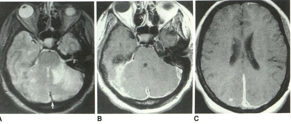
involve-ment, which was not apparent on the unenhanced scans (Fig.
1 ). Seven of 12 patients had intraaxial areas of high signal
intensity in the cerebellum, internal capsule, and gray-white
junction, on T2-weighted images, which did not enhance after
contrast administration (Fig. 2). Eight patients had enhancing
lesions that were not apparent, or were poorly seen on
unenhanced scans; these included one patient with optic
nerve enhancement (Fig. 3), two with parenchymal and one
with ependymal lesions (Fig. 4), and three with enhancing
hypothalamic lesions (Fig. 5). Three patients had enhancing
extraaxial masses mimicking meningiomas on postcontrast
T1-weighted images (Figs. 3 and 6). On unenhanced
T2-weighted images, these lesions were of very low intensity.
One patient presented with dementia and had hydrocephalus
(Fig. 7). In addition, two patients had vasogenic edema (Fig.
6). Of the two patients with spinal sarcoidosis, one had
enlargement of the cervical cord noted on the unenhanced
scan with enhancement of the cord on postcontrast images (Fig. 8); the other had plaquelike enhancing thickening of the
pia at the level of the conus, with no definite abnormality
identified on the unenhanced study. Superior sagittal sinus
thrombosis, a rare complication of sarcoidosis [5], was noted
in one case (Fig. 2).
Discussion
Sarcoidosis is a systemic granulomatous disease of
un-known origin that affects the CNS in 5% of cases [1-3].
Manifestations of the disease on unenhanced MR images
have been reported [9, 1 0]. Recently, several reports have
emphasized the sensitivity of MR imaging to the detection of meningeal disease (Ross MR et al., paper presented at the annual meeting of the Radiological Society of North America,
Chicago, November 1989, and [12]). Since meningeal
involve-ment in sarcoidosis is common, we hypothesized that the
addition of gadopentetate dimeglumine would greatly
en-hance the sensitivity of MR in the detection of CNS
sarcoid-osis. Our study confirmed this hypothesis. Indeed, eight of
our 12 patients with intracranial sarcoidosis and one of the
two patients with spinal sarcoidosis had meningeal
involve-ment that was not apparent on the unenhanced scans. The
detection of meningeal enhancement immediately narrows
the differential diagnosis to a few conditions, including
bac-terial, viral, lymphomatous, or carcinomatous meningitis,
many of which may be diagnosed by lumbar puncture. The
1230 SELTZER ET AL. AJNR:12, November/December 1991
Fig. 1.-Case 4: 29-year-old man with suspected encephalitis.
A, Axial unenhanced T2-weighted image shows nonspecific foci of high signal intensity at gray-white junction (arrows).
B, Axial unenhanced T1-weighted image reveals prominent suprasellar soft tissue.
C, Axial postcontrast T1-weighted image shows diffuse marked enhancement of leptomeninges.
A
B
c
Fig. 2.-Case 3: 42-year-old woman with altered mental status.
A, Axial unenhanced T2-weighted image shows mass with increased signal intensity in left cerebellum. Note lack of flow void and flow artifact in
superior sagittal sinus (arrow).
B, Axial postcontrast T1-weighted image shows marked enhancement of meninges. No flow in superior sagittal or transverse sinus. Note lack of
enhancement of left cerebellar hemisphere.
C, Axial postcontrast T1-weighted image shows enhancing intraaxiallesion and meningeal enhancement in interhemispheric fissure. Repeat
contrast-enhanced imaging showed resolution of the enhancing extraaxiallesion after a course of steroid therapy.
of meningeal enhancement, are nonspecific and tissue biopsy
is necessary for definitive diagnosis. This can usually be obtained from sites such as the lacrimal gland, peripheral
lymph nodes, or, rarely, the mediastinum or meninges.
Occasionally, meningeal enhancement may be due to hy-peremia associated with dural sinus thrombosis (Ross MR et
al., paper presented at RSNA, November 1988). We believe
this to be the case in our patient with superior sagittal sinus
thrombosis, since results of the biopsies of the meninges
were negative while results of the cerebellar biopsy revealed
noncaseating granulomata.
While the characteristic pathologic changes of sarcoidosis consist of noncaseating granulomata containing
[image:4.612.55.563.78.303.2] [image:4.612.59.561.358.572.2]necro-AJNR:12, November/December 1991 MR OF CNS SARCOIDOSIS 1231
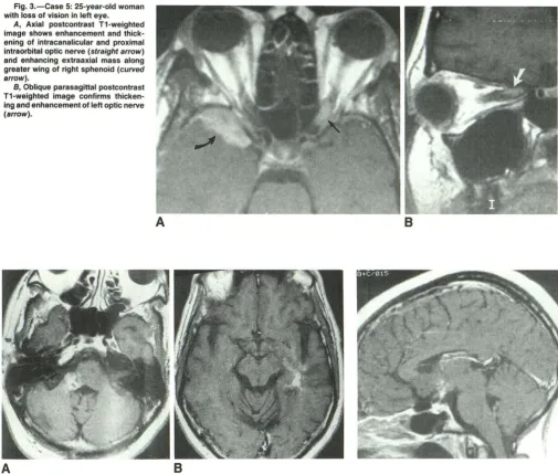
Fig. 3.-Case 5: 25-year-old woman with loss of vision in left eye.
A, Axial postcontrast T1-weighted
image shows enhancement and thick-ening of intracanalicular and proximal
intraorbital optic nerve (straight arrow) and enhancing extraaxial mass along
greater wing of right sphenoid (curved arrow).
B, Oblique parasagittal postcontrast
T1-weighted image confirms thicken-ing and enhancement of left optic nerve (arrow).
A
8
A
8
Fig. 4.-Case 9: 57-year-old man with hearing loss and tinnitus in right ear.
A, Axial postcontrast T1-weighted image shows enhancing 1-cm lesion in right middle cerebellar peduncle bulging into right cerebellopontine angle at the emergence of cranial nerve
VIII.
Fig. 5.-Case 1: 18-year-old woman with amenor-rhea. Sagittal postcontrast T1-weighted image shows punctate enhancement of hypothalamus.
B, Axial postcontrast T1-weighted image shows enhancement of ependyma of left temporal
horn.
sis can mimic caseation, presenting problems in diagnosis. Such a problem was encountered in case 5, a patient in whom biopsy results of an extraaxial right temporal mass revealed necrotizing granulomas consistent with tuberculosis or sar-coidosis. All cultures and special stains were negative for acid-fast bacillus, and the patient improved on steroids.
The presence of abnormal areas of high signal intensity on T2-weighted images at the gray-white junction, as seen in seven of our patients, is a nonspecific finding that by itself is not characteristic of CNS sarcoidosis. The combination of such findings with meningeal enhancement or with lesions in the hypothalamus should suggest the diagnosis of sarcoido-sis. In our series, enhancement of intraaxial lesions was
uncommon (two of 12). In one patient in whom a biopsy of high-intensity nonenhancing lesion was done, noncaseating granulomata characteristic of sarcoidosis were encountered. Thus, while meningeal involvement with sarcoidosis often results in enhancement, the intraparenchymal lesions en-hance more rarely.
Involvement of the hypothalamus and the pituitary stalk is characteristic of CNS sarcoidosis. Thickening of the hypo-thalamus may be apparent on the unenhanced study, but the lesion is more obvious on the postcontrast study when en-hancement of the hypothalamus is seen.
1232 SELTZER ET AL. AJNR:12, November/December 1991
Fig. 6.-Case 7: 41-year-old man with speech problems.
A, Axial postcontrast T1-weighted image shows enhancing mass in left cavernous sinus. Note previous left-sided craniotomy for biopsy.
8, Axial unenhanced T2-weighted image shows mass of low signal intensity in cavernous sinus. Note extensive vasogenic edema.
c, Axial postcontrast T1-weighted image obtained after steroid treatment. Note marked decrease in size of extraaxiallesion. The edema also showed a marked decrease on T2-weighted images (not shown).
A
B
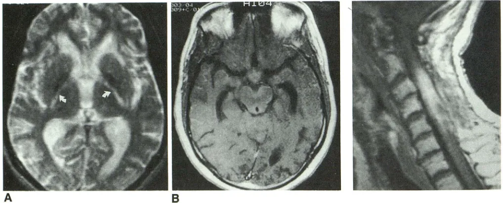
Fig. 7.-Case 6: 32-year-old man with dementia.
A, Axial unenhanced T2-weighted image shows slight enlargement of ventricles and increased signal intensity of both internal capsules (arrows).
Fig. 8.-Case 14: 56-year-old woman with quadriparesis. Sagittal postcontrast T1-weighted image shows enhancing intramedul-lary mass mimicking a neoplasm.
8, Axial postcontrast T1-weighted image confirms hydrocephalus. Note enhancement of hypothalamus and pia around brainstem. Sagittal postcontrast T1-weighted image (not shown) confirmed enhancement of hypothalamus and leptomeninges.
meningiomas, lymphoma, metastasis, or syphilis. However,
our study suggests that these lesions may have a character-istic, very low signal intensity on T2-weighted images, which may suggest the correct diagnosis. Heavily calcified menin-giomas, of course, could have a similar appearance.
Even though it was retrospective, our study provides some information about the response of CNS sarcoidosis to treat-ment. Four patients in our series had repeat studies after steroid therapy. Corticosteroids constitute the most common treatment for sarcoidosis. The disease often responds initially
[image:6.612.58.559.81.271.2] [image:6.612.52.561.331.537.2]cyclophospha-AJNR:12, November/December 1991 MR OF CNS SARCOIDOSIS 1233
mide treatment. Simultaneously, infiltration of the optic nerve dramatically diminished and a contralateral extraaxial mass also almost completely resolved. Thus, MR imaging offered an objective measure of the patient's response to therapy.
Involvement of the cranial nerves is a rare complication of CNS sarcoidosis, with peripheral involvement of the 7th nerve the most common site. The optic nerve and the chiasm can also be involved. Cavernous sinus involvement may second-arily affect the cranial nerves coursing in the cavernous sinus. Contrast-enhanced MR imaging can provide, for the first time, evidence of sarcoid infiltration of the optic nerve (by demon-strating enhancement of the optic nerve) in the absence of morphologic changes. However, enhancement of the optic nerve is not specific for sarcoidosis and can be seen in optic neuritis with or without other findings of multiple sclerosis, syphilis, cryptococcosis, and radiation-induced optic neuritis [12]. Hearing loss in patients with CNS sarcoidosis can result from infiltration of the meninges in the internal auditory canal or from intraaxial lesions in the brainstem. MR imaging can easily differentiate between these two conditions (Mark AS et al., paper presented at the annual meeting of the Radiolog-ical Society of North America, Chicago, November 1990).
In this series, we found that the addition of contrast agent increased the sensitivity of MR imaging in the detection of CNS sarcoid lesions, permitting the detection of meningeal, parenchymal, optic nerve, and ependymal sarcoid lesions not seen on unenhanced MR scans. If the diagnosis of sarcoidosis can be made on the basis of clinical and imaging information, a CNS biopsy may be unnecessary. For cases in which a CNS biopsy cannot be avoided, contrast-enhanced MR im-aging may indicate an optimal site for biopsy by identifying a region of active parenchymal or meningeal granulomata. MR
imaging with and without contrast medium may also provide a noninvasive means of monitoring patients' response to therapy. We recommend that contrast agent be used routinely in patients in whom CNS sarcoidosis is suspected, especially if the unenhanced studies are normal or nonspecific.
REFERENCES
1. Delaney P. Neurologic manifestations in sarcoidosis: review of the lit
era-ture, with a report of 23 cases. Ann Intern Med 1977; 87:336-345
2. Stern BJ, Krumholz A, Johns C, Scott P, Nissim J. Sarcoidosis and its
neurological manifestations. Arch Neurol1985; 42:909-917
3. Matthews WB. Neurologic manifestations of sarcoidosis. In: Asbury AK, McKhann GM, McDonald WI, eds. Diseases of the nervous system: clinical neurobiology. London: Heinemann, 1986:1563-1570
4. Kendall BE, Taller GLV. Radiological findings in neurosarcoidosis. Br J Radio/1978; 51:81-92
5. Leeds NE, Zimmerman RD, Elkin CM, Nussbaum M, LeVan AM. Neuro-sarcoidosis of the brain and meninges. Semin Roentgenol 1985; 20: 387-392
6. Widerholt we, Siekert RG. Neurological manifestations of sarcoidosis.
Neurology 1965; 15:1147-1154
7. Anderson WAD. Pathology, 6th ed. St. Louis: Mosby, 1971:939-942 8. Nesbit GM, Miller GM, Baker HL Jr, Ebersold MJ, Scheithauer BW. Spinal
cord sarcoidosis: a new finding at MR imaging with Gd-DTPA
enhance-ment. Radiology 1987; 173:839-843
9. Hayes WS, Sherman JL, Stern BJ, Citrin CM, Pulaski PD. MR and CT evaluation of intracranial sarcoidosis. AJR 1987; 149: 1 043-1049 10. Miller DH, Kendall BE, BarterS, et al. Magnetic resonance imaging in
central nervous system sarcoidosis. Neurology 1988; 38:378-383 11. Brooks BS, El Gamma! T, Hungerford GD, Acker J, Trevor RP, Russell W.
Radiologic evaluation neurosarcoidosis: role of computed tomography.
AJNR 1982; 3:513-521
12. Guy T, Mancuso A, Quisling RG, Beck R, Moster M.
Gadolinium-DTPA-enhanced magnetic resonance imaging in optic neuropathies.