Bernard Gero 1 Gordon Sze1 Hassan Sharif2
Received October 10, 1990; revision requested December 20, 1990; revision received March 8,
1991; accepted March 11, 1991.
' Department of Diagnostic Radiology, Yale
Uni-versity School of Medicine, 333 Cedar St., New
Haven, CT 06510. Address reprint requests to G.
Sze.
2 Department of Radiology, Riyadh Armed
Forces Hospital, Riyadh, Saudi Arabia.
0195-6108/91/1205-1009
© American Society of Neuroradiology
MR Imagi
n
g
of I
n
tradural
Inflam
matory Diseases of the
Spine
1009
Twenty-eight patients with intradural inflammatory disease of the spine were studied in order to characterize the MR imaging findings of infectious and inflammatory condi-tions. Patients were categorized according to the spinal compartment involved. Among the 12 patients in the intradural extramedullary group, unenhanced scans were either normal or nonspecific while contrast-enhanced scans were helpful in visualizing and localizing the lesion. Nevertheless, contrast-enhanced MR studies were unable to differentiate infection and inflammation from tumor in this compartment. Among 16 patients with intramedullary lesions, four had granulomatous disease and 12 had nongranulomatous disease. The granulomatous lesions resembled tumors and displayed MR characteristics of a focal lesion with large nodular enhancement. The patients with nongranulomatous intramedullary lesions exhibited two subsets of MR findings. In the first subset of nine patients, diffuse cord swelling and high signal were seen on long TR images, combined with either no enhancement or peripheral, diffuse, or speckled enhancement of the spinal cord on contrast-enhanced short TR images. In the second subset of three patients, minimal or no spinal cord swelling was displayed despite the visualization of high signal on long TR scans and nodular enhancement with contrast administration on short TR scans. Both subsets were sufficiently unique that nongranu-lomatous myelitis could usually be differentiated from spinal cord tumors.
AJNR 12:1009-1019, September/October 1991
Intradural inflammatory diseases of the spine include meningitis and myelitis.
There have been no previous reports of MR imaging in cases of spinal meningitis. Previous reports of MR imaging of myelitis have described numerous findings,
including fusiform swelling of the spinal cord [1-7], high signal in the cord on T2
-weighted images [1-3, 5, 6, 8, 9], high signal in the cord with no swelling or very slight cord swelling [8-1 0], or normal signal in the spinal cord on T2-weighted images despite spinal cord swelling [4]. Myelitis has been reported to enhance with contrast administration [1, 5, 7], but enhancement patterns have not been c har-acterized. The purpose of this study is to see if MR imaging shows any abnormality in patients with intradural inflammatory disease and, if so, to then characterize these lesions.
Materials and Methods
The MR images of 28 patients with intradural inflammatory disease of the spine were reviewed. The patients were divided into two groups, those with intradural extramedullary lesions (12 patients) and those with intramedullary lesions (16 patients). The patients with intramedullary lesions were further divided into two groups, those with granulomatous disease and those with nongranulomatous disease. The diagnoses were confirmed by a variety of methods, including CSF analysis (organism isolation, antigen specific titers) (five patients:
four intradural extramedullary, one intramedullary), open biopsy (six patients: three intradural
extramedullary, three intramedullary), and, in patients with tuberculosis, dramatic response
1010 GERO ET AL. AJNR:12, September/October 1991
intramedullary). The diagnosis was strongly presumed when the disease manifested first in an extraspinal location followed by direct time-related causal involvement of the spine (five patients, all intra-medullary) and was considered immune-mediated when the proper clinical history and findings were present (one patient, intradural extramedullary, presumed antigen-antibody reaction to the adminis-tration of gamma globulin) or idiopathic when exhaustive CSF and clinical analyses were unable to identify a specific origin (five patients: one intradural extramedullary, four intramedullary). Patients with known multiple sclerosis were excluded.
The patients were imaged on four different MR imaging systems. Eighteen patients were imaged on a 1.5-T magnet, eight on a 0.5-T system, and one each on a 1.0-T and a 2.0-T system. Both short TR (450-650) and long TR (1200-2500) spin-echo sequences were used. Gradient-moment nulling and cardiac gating were used where appro-priate. In five patients, all with intradural extramedullary inflammatory disease, long TR sequences were not obtained.
All patients were imaged in the sagittal plane. Axial images were obtained in 23 cases and coronal images were obtained in four cases. A single patient was imaged in both the axial and coronal planes. In the majority of cases the slice thickness varied between 3 and 5 mm, although 7 mm was employed in some instances in the lower-field magnet. Eleven patients were imaged more than once.
Twenty-one of the 28 patients received IV gadopentetate dimeglu-mine (0.1 mmoljkg), and postcontrast short TR sagittal scans were acquired. The images were reviewed and the results tabulated.
Results
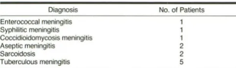
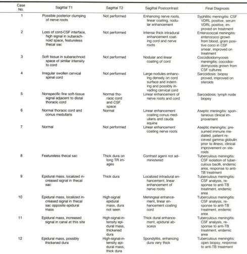
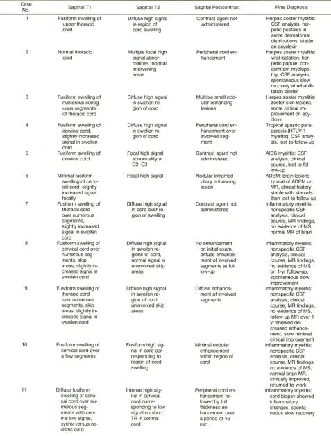
The number of patients in each diagnostic group are tabu-lated in Tables 1 and 2. The MR findings and associated diagnoses are tabulated in Tables 3 and 4.
Intradural Extramedullary Disease
Twelve patients were found to have intradural extramed-ullary disease. In these patients, unenhanced scans were usually unrevealing. In two cases no abnormality was identi-fied; in 10 cases there was a questionable abnormality of a nonspecific nature. These nonspecific findings varied from
TABLE 1: Intradural Extramedullary Spinal Lesions
Diagnosis
Enterococcal meningitis
Syphilitic meningitis
Coccidioidomycosis meningitis Aseptic meningitis
Sarcoidosis
Tuberculous meningitis
No. of Patients
1 1 1 2 2 5
TABLE 2: Intramedullary Granulomatous and Nongranulomatous Spinal Lesions
Sarcoidosis
AIDS myelitis
Diagnosis
Tropical spastic paraparesis (HTLV-1)
Acute disseminated encephalomyelitis
Herpes zoster myelitis
Tuberculosis
Inflammatory myelitis of uncertain origin
No. of Patients
1
3 3
6
linear strands of soft-tissue intensity in the spinal canal to a complete obliteration of the normal canal space. The location of the lesion was indeterminate and the relationship to the cord or nerve roots unclear. Long TR images were obtained in seven of the 12 patients with intradural extramedullary inflammatory disease. In three of the seven patients, the long TR images masked the intradural abnormality. The dura was
considered thickened in four of the seven cases.
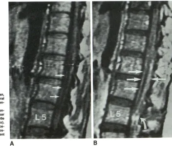
Eleven of the 12 patients with intradural extramedullary disease received gadopentetate dimeglumine. The adminis-tration of contrast agent proved to be useful in visualizing the lesion in two patients in whom unenhanced scans were normal and in defining the lesion in the remaining nine patients (Fig. 1 ). After the administration of contrast material, the lesion could be accurately located in the intradural extramedullary compartment by visualization of the intact spinal cord or nerve roots surrounded by enhancing tissue, creating an impression of a filling defect.
Three patterns of enhancement of the intradural extramed-ullary lesions were noted. The first pattern was linear en-hancement on the surface of the spinal cord andjor nerve roots and was present in nine patients (Fig. 1 ). The enhance-ment was delicate and smooth and outlined the underlying nerve roots and spinal cord.
The second mode of enhancement was nodular in nature. This was present in three patients (Fig. 1 ), and consisted of discrete foci of enhancement on the surface of the spinal cord andjor nerve roots. In two patients there was a combination of nodular and linear enhancement within the intradural com-partment.
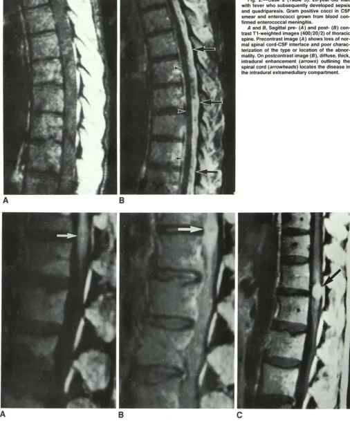
The third pattern, seen in two patients, consisted of diffuse, thick, intradural enhancement of the leptomeninges, com-pletely surrounding the spinal cord and appearing as an intradural filling defect (Fig. 2). No correlation was found between the pattern of enhancement and the severity of the presenting neurologic symptoms.
Intramedullary Disease
The 16 patients with intramedullary disease were divided into two groups, those with granulomatous disease and those with nongranulomatous disease. In the group of patients with intramedullary granulomatous disease (four patients), each showed the same MR characteristics. Focal spinal cord swell-ing was identified on the short and long TR images (Figs. 3A and 38). There was localized high signal intensity on the long TR images in the region of cord swelling. After contrast administration, nodular enhancement within the cord was seen at the site of the suspected abnormality (Fig. 3C). On follow-up MR after 6 months of antituberculous therapy in the patients with tuberculomas, the tuberculomas disappeared
and the spinal cord returned to normal.
In the 12 patients with nongranulomatous intramedullary
inflammatory disease, a spectrum of MR findings was noted. First, the amount of cord swelling varied from none to diffuse. In the cases in which the spinal cord was diffusely swollen,
the swelling extended over numerous spinal segments. In
[image:2.614.55.296.535.604.2] [image:2.614.53.297.651.730.2]AJNR:12, September/October 1991 MR OF SPINAL INTRADURAL INFLAMMATORY DISEASE 1011
TABLE 3: MR Findings and Final Diagnoses of Patients with Intradural Extramedullary Lesions
Case
Sagittal T1 Sagittal T2 No.
Possible posterior clumping Not performed of nerve roots
2 Loss of cord-CSF interface, Not performed high signal in
subarach-noid space, featureless thecal sac
3 Soft tissue in subarachnoid Not performed space of similar intensity
to cord
4 Irregular swollen cervical Not performed
spinal cord
5 Nonspecific fine soft-tissue Normal tho-signal adjacent to distal racic cord thoracic cord and CSF
space 6 Normal thoracic cord and Normal
conus medullaris
7 Normal Not performed
8 Featureless thecal sac Thick dura on
long TRim-ages
9 Epidural mass, localized in- Thick dura creased signal in thecal
sac
10 Epidural mass, localized in- High-signal creased signal in thecal epidural
sac opposite epidural mass, dura
mass not seen
11 Epidural mass, increased
High-signal-in-signal in canal at this site tensity epi-dural mass, thickened dura 12 Epidural mass, possibly
High-signal-in-thickened dura tensity epi-dural mass,
thick dura
Second, the signal characteristics within the cord on unen-hanced scans varied. On unenhanced short TR scans, only once case showed diffuse low signal in the cord. In the other 11 cases, the cord was of normal signal intensity or slightly increased signal intensity on short TR images. The faintly increased signal intensity was only seen in those cases as-sociated with cord swelling. In the two patients in whom there was no cord swelling, no signal abnormality could be identified on the short TR sequence. In all cases, high signal intensity was seen on long TR sequences in a focal or diffuse location
Sagittal Postcontrast Final Diagnosis
Enhancing nerve roots, Syphilitic meningitis: CSF linear coating, nodu- VORL positive, serum lar enhancement VORL positive,
im-proved on treatment Intense thick intradural Enterococcal meningitis:
enhancement coat- enterococci grown
ing cord and nerve from blood, gram
posi-roots live cocci in CSF
smear, improved on treatment Nodular and linear Coccidioidomycosis
coating of cord meningitis:
coccidioi-domycosis grown from CSF cultures Large nodules enhanc- Sarcoidosis: biopsy
ing densely on cord proved, improved on
surface and indent- steroids
ing and possibly in-vading cervical cord
Linear enhancement of Sarcoidosis: lymph node nerve roots and cord biopsy
Linear enhancement Aseptic meningitis:
span-coating conus med- taneous clinical
im-ullaris and cauda provement equina
Linear enhancement Aseptic meningitis:
pre-coating nerve roots sumed immune
me-diated, patient re-ceived gamma globulin prior to illness, clinical improvement on ste-raids
Contrast agent not ad- Tuberculous meningitis:
ministered CSF isolation of tuber-culous bacilli, endemic area, response to anti-TB treatment Localized intradural en- Tuberculous meningitis:
hancement, linear CSF analysis,
re-enhancement of sponse to anti-TB
nerve roots treatment, endemic area
Meningeal enhance- Tuberculous meningitis:
ment, linear en- CSF analysis, re-hancement coating sponse to anti-TB
cord treatment, endemic
area
Thick dural enhance- Tuberculous meningitis:
ment, epidural ab- CSF analysis,
re-scess sponse to anti-TB
treatment, endemic area
Spondylitis, enhancing Tuberculous meningitis:
dura very thick open biopsy, response to anti-TB treatment
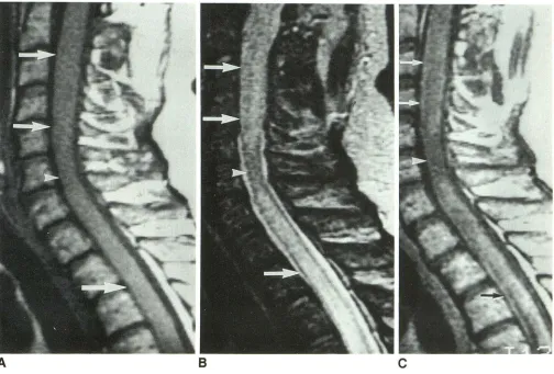
(Figs. 4 and 5). There was normal signal intensity in the cord in the apparently uninvolved skip areas (Fig. 4).
Contrast-enhanced scans were available in eight patients with nongranulomatous inflammatory intramedullary lesions. Three patterns of enhancement were seen. More than one enhancement pattern was seen in two patients.
[image:3.617.56.546.93.596.2]1012 GERO ET AL. AJNR:12, September/October 1991
TABLE 4: MR Findings and Final Diagnoses of Patients with Intramedullary Lesions
Case
Sagittal T1 Sagittal T2 Sagittal Postcontrast Final Diagnosis
No.
Fusiform swelling of Diffuse high signal Contrast agent not Herpes zoster myelitis: upper thoracic in region of administered CSF analysis,
her-cord cord swelling petic pustules in
same dermatomal distributions, stable on acyclovir 2 Normal thoracic Multiple focal high Peripheral cord en- Herpes zoster myelitis:
cord signal abnor- hancement viral isolation, her
-malities, normal petic papule,
con-intervening comitant
myelopa-areas thy; CSF analysis;
spontaneous slow recovery at rehabili-tation center 3 Fusiform swelling of Diffuse high signal Multiple small nod- Herpes zoster myelitis:
numerous contig- in swollen re- ular enhancing zoster skin lesions,
uous segments gion of cord lesions some clinical
im-of thoracic cord provement on
acy-clovir
4 Fusiform swelling of Diffuse high signal Peripheral cord en- Tropical spastic
para-cervical cord, in swollen re- hancement over paresis (HTLV-1 slightly increased gion of cord involved seg- myelitis): CSF
analy-signal in swollen ment sis, lost to follow-up
cord
5 Fusiform swelling of Focal high signal Contrast agent not AIDS myelitis: CSF cervical cord abnormality at administered analysis, clinical
C2-C3 course, lost to
fol-low-up
6 Minimal fusiform Focal high signal Nodular intramed- ADEM: brain lesions
swelling of cervi- ullary enhancing typical of ADEM on
cal cord, slightly lesion MR, clinical history,
increased signal stable with steroids
focally then lost to follow-up
7 Fusiform swelling of Diffuse high signal Contrast agent not Inflammatory myelitis:
thoracic cord in cord over re- administered nonspecific CSF over numerous gion of swelling analysis, clinical
segments, course, MR findings,
slightly increased no evidence of MS,
signal in swollen normal MR of brain
cord
8 Fusiform swelling of Diffuse high signal No enhancement Inflammatory myelitis: cervical cord over in swollen re- on initial exam, nonspecific CSF numerous seg- gions of cord, diffuse enhance- analysis, clinical ments, skip normal signal in ment of involved course, MR findings,
areas, slightly in- uninvolved skip segments at fol- no evidence of MS
creased signal in areas low-up on 1-yr follow-up,
swollen cord spontaneous slow
improvement
9 Fusiform swelling of Diffuse high signal Diffuse enhance- Inflammatory myelitis:
thoracic cord in swollen re- ment of involved nonspecific CSF over numerous gion of cord, segments analysis, clinical
segments, skip uninvolved skip course, MR findings,
areas, slightly in- areas no evidence of MS,
creased signal in follow-up MR over 1
swollen cord yr showed
de-creased enhance-ment, slow minimal clinical improvement 10 Fusiform swelling of Fusiform high sig- Minimal nodular Inflammatory myelitis:
cervical cord over nal in cord cor- enhancement nonspecific CSF
a few segments responding to within region of analysis, clinical region of cord cord course, MR findings,
swelling no evidence of MS,
normal brain MR, clinically improved, returned to work 11 Diffuse fusiform Intense high sig- Peripheral cord en- Inflammatory myelitis:
swelling of cervi- nal in cervical hancement fol- cord biopsy showed
cal cord over nu- cord corre- lowed by full inflammatory
merous seg- sponding to low thickness en- changes, sponta
-ments with cen- signal on short hancement over neous slow recovery trallow signal, TR in central a period of 45
syrinx versus ne- cord min
[image:4.612.63.540.93.721.2]AJNR:12, September/October 1991 MR OF SPINAL INTRADURAL INFLAMMATORY DISEASE 1013
TABLE 4: Continued
Case No.
12
13
14
15
16
Sagittal T1
Normal thoracic
cord
Fusiform swelling over numerous
segments with
diffuse low signal
Focal enlargement
of
cervicomedul-lary junction, low
signal focally
Low-intensity signal
focally at
cervico-medullary junc-tion
Focal enlargement of conus,
low-in-tensity signal fo -cally
Sagittal T2
Diffuse high signal
in upper
tho-racic cord
Focal high signal
in swollen re-gion of cord
Focal high signal with low inten-sity centrally
Focal high signal
with low int en-sity centrally
Focal high signal
with low inten-sity centrally
[image:5.614.53.551.92.341.2]patients with skip areas, the spinal cord enhanced over the entire length of the enlarged segments but failed to enhance in areas of normal intervening cord. In two cases, the initial contrast-enhanced MR study showed little or no spinal cord enhancement despite fusiform swelling and associated diffuse
Fig. 1.-Case 1 (Table 3): 33-year-old woman, 3 months postpartum, who developed bilateral leg weakness and radiculopathy. Syphilitic men-ingitis proved by CSF VORL.
A and 8, Sagittal pre- (A) and post-(8) con-trast T1-weighted images (600/20/4) of lumbar
spine. Precontrast image (A) shows subtle
clumping of nerve roots posteriorly (arrows). Postcontrast image (8) shows linear enhance-ment (straight arrows) of nerve roots and
pos-sibly the dura, as well as nodular enhancement distally (curved arrow).
A
Sagittal Postcontrast
Multiple enhancing
nodules of
var-ious size
Solid nodular
en-hancement over two segments
Contrast agent not administered
Thick rim
enhance-ment with cen-tral low signal
Thick rim enhance-ment with cen-tral low signal
Final Diagnosis
Inflammatory myelitis: cord biopsy showed inflammatory changes, AIDS, lost to follow-up Sarcoidosis: biopsy,
improved on steroids
Tuberculoma: endemic area, marked re-sponse to anti-TB therapy, complete resolution on follow-up MR
Tuberculoma: endemic area, marked re-sponse to anti-TB therapy, complete resolution on follow-up MR
Tuberculoma: endemic area, marked re-sponse to anti-TB
therapy, complete resolution on follow -up MR
high signal over numerous segments on long TR scans. On
subsequent follow-up MR scans several weeks later, the
contrast-enhanced scans showed clear diffuse enhancement
in the spinal cord in the abnormally enlarged areas.
In two patients peripheral enhancement within the cord was
[image:5.614.204.556.421.721.2]1014
A
8
A
8
GERO ET AL. AJNR:12, September/October 1991
Fig. 2.-Case 2 (Table 3): 26-year-old man with fever who subsequently developed sepsis and quadriparesis. Gram positive cocci in CSF smear and enterococci grown from blood con-firmed enterococcal meningitis.
A and 8, Sagittal pre- (A) and post-(8) con-trast T1-weighted images (400/20/2) of thoracic spine. Precontrast image (A) shows loss of nor-mal spinal cord-CSF interface and poor charac-terization of the type or location of the abnor-mality. On postcontrast image (8), diffuse, thick, intradural enhancement (arrows) outlining the spinal cord (arrowheads) locates the disease in the intradural extramedullary compartment.
c
Fig. 3.-Case 16 (Table 4): Conus medullaris tuberculoma in male patient who presented with progressive urinary retention and leg weakness.
A, Sagittal precontrast T1-weighted image (450/30/6) shows focal enlargement of conus (arrow).
8, Precontrast T2-weighted image (1800/50/2) shows a nodular high-signal-intensity lesion in conus with surrounding poorly defined higher signal (arrow).
[image:6.613.53.559.82.691.2]AJNR:12, September/October 1991 MR OF SPINAL INTRADURAL INFLAMMATORY DISEASE 1015
[image:7.612.57.561.74.415.2]A
B
c
Fig. 4.-Case 8 (Table 4): Inflammatory myelitis in 45-year-old man who presented with chest and abdominal numbness.
A, Sagittal precontrast T1-weighted image (600/20/4) shows fusiform swelling of noncontiguous segments of cervical cord (arrows), which are separated by a short normal intervening skip segment (arrowhead) of cord.
8 and C, Swollen areas of spinal cord demonstrate diffuse high signal ort precontrast T2-weighted image (2200/80/2) (arrows in 8) and diffuse enhancement after contrast administration (arrows in C) with a nonenhanc;in9 sl!ip {!rea (arrowhead). This pattern is unlike that usually seen in tllmor.
seen (Fig. 6). The unenhanced short and long TR MR scans showed fusiform swelling of the spinal cord over numerous
segments and diffuse high signal on the long TR images. The
immediate postcontrast scans showed peripheral enhance-ment but in one of these cases a delayed scan was performed,
which showed progressive enhancement of the spinal cord
toward the center (Fig. 7).
Finally, a nodular enhancement pattern within the spinal
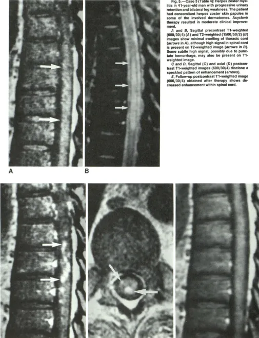
cord was identified in four cases (Fig. 5). The amount of cord
swelling varied from none to diffuse. In one case of acute
disseminated encephalomyelitis, the unenhanced short TR
scan was normal despite a focal nodular high-signal-intensity intramedullary lesion on the long TR scan. This focal area demonstrated intense enhancement after contrast
administra-tion. In the remaining three cases the enhancement consisted
of multitudinous small foci of varying size, all smaller than 4
mm in diameter. No dominant nodule was seen. The
enhance-ment occurred within the cord in the region of cord swelling
or signal abnormality only. One patient in this group had three
follow-up MR examinations over a 14-month period, all of
which showed a decrease in the enhancement in the spinal
cord (Fig. 5) associated with clinical improvement.
Discussion
Intradural inflammatory diseases of the spine include
men-ingitis and myelitis of diverse origin. Patients who present
with isolated acute or subacute neurologic symptoms refera
-ble to the spinal cord require prompt attention. Initial
investi-gation is aimed at identifying surgical from nonsurgical lesions
[11], and imaging studies, especially MR, assist in making
this distinction. The administration of contrast material allows
the definitive identification of lesions and subsequent
char-acterization.
Intradural Extramedullary Disease
In patients with inflammatory intradural extramedullary
le-sions, unenhanced MR scans were either normal or unable
to help localize the abnormality to one of the three spinal
compartments. Contrast-enhanced MR imaging was essential
in some cases (two patients) for visualizing the abnormality
and in others for localizing the inflammation to the intradural
extramedullary space and for defining the extent of the dis
1016 GERO ET AL.
A
B
AJNR:12, September/October 1991
Fig. 5.-Case 3 (Table 4): Herpes zoster mye-litis in 41-year-old man with progressive urinary retention and bilateral leg weakness. The patient had concomitant herpes zoster skin papules in some of the involved dermatomes. Acyclovir therapy resulted in moderate clinical improve-ment.
A and 8, Sagittal precontrast T1-weighted (600/30/4) (A) and T2-weighted (1500/50/2) (8) images show minimal swelling of thoracic cord (arrows in A), although high signal in spinal cord is present on T2-weighted image (arrows in 8). Some subtle high signal, possibly due to
punc-tate hemorrhage, may also be present on T1-weighted image.
C and D, Sagittal (C) and axial (0) postcon-trast T1-weighted images (600/30/4) disclose a
speckled pattern of enhancement (arrows).
[image:8.618.54.559.78.737.2]AJNR:12, September/October 1991 MR OF SPINAL INTRADURAL INFLAMMATORY DISEASE 1017
[image:9.617.56.299.77.389.2]A
B
Fig. 6.-Case 4 (Table 4): Tropical spastic paraparesis (HTLV-1 myelop-athy) in 34-year-old man from Barbados who presented with progressive quadriparesis, proved by CSF titers.
A, Sagittal precontrast T1-weighted image (600/20/4) shows diffuse
swelling of cervical spinal cord (arrows).
8, Sagittal postcontrast T1-weighted image (600/20/4) shows peripheral enhancement of cord (arrows).
The appearance and location of the enhancement in the cases of intradural extramedullary disease make it likely that the enhancing structures were inflamed meninges, similar in appearance to intracranial meningeal enhancement [12]. The three patterns of enhancement identified (linear enhancement coating the nerve roots and spinal cord, nodular enhancement on the surface of the spinal cord, or nerve roots and thick intradural enhancement completely filling the subarachnoid space) most likely reflect the degree and nature of the men-ingeal inflammation. Cranial menmen-ingeal enhancement has been shown to correlate well with severe meningeal inflam-mation on histopathologic sections; areas of less intense meningeal inflammation histologically did not exhibit enhance-ment [12]. While this trend may be the same in the meninges of the spine, in this series no correlation was found between the patterns of enhancement-that is, linear, nodular, or thick intradural-and the initial severity of neurologic symptoms.
All the enhancement patterns noted in the patients with intradural extramedullary inflammatory changes have been seen in patients with leptomeningeal spread of tumor; the MR findings alone could not differentiate infection from tumor [13].
Although the IV administration of gadopentetate dimeglumine greatly increased the sensitivity of MR in detecting an abnor-mality and localizing it to the intradural extramedullary
com-partment, it did not improve the diagnostic specificity. In addition, no enhancement pattern characterized a specific inflammatory origin. Fine linear enhancement on the cord surface was seen in patients with lesions varying from sar-coidosis to bacterial meningitis.
Intramedullary Disease
In the subgroup of patients with intramedullary disease of granulomatous origin, the MR characteristics were focal cord swelling, high signal on long TR images, and nodular enhance-ment with contrast administration. On both the long TR images and the contrast-enhanced scans, differentiation of granulomatous disease from an intramedullary tumor could not be made.
In the subgroup of patients, however, with nongranuloma-tous intramedullary lesions, the MR characteristics were often sufficiently unique to suggest the probability of an inflamma-tory intramedullary lesion and to make consideration of an intramedullary tumor less likely. Of course, the clinical pres-entation alone is often suggestive. Patients with inflammatory myelitis usually present acutely in contradistinction to the usual insidious onset of symptoms associated with spinal cord tumors, although exceptions in both instances do occur.
MR, however, can further help in the differentiation of the two. Cord tumors are generally characterized by a significant amount of cord swelling, which is usually of low intensity on short TR images and high intensity on long TR images. If hemorrhage is present, it is usually focal. After the adminis-tration of contrast agent, focal nodular enhancement is usually seen. Of the 55 reported gliomas of the cord studied with contrast material, 54 enhanced [14-19]. Nonenhancing gliomas appear to be comparatively unusual. In addition,
gliomas that enhance in a peripheral, diffuse, or speckled pattern have not been described.
In our study, nongranulomatous myelitis was characterized on MR by two sets of findings. In the first set, there was diffuse swelling of the spinal cord over numerous segments, sometimes with skip areas, and a subtle increase in the signal of the cord on short TR scans, possibly due to petechial hemorrhage. This was accompanied by a diffuse high signal on long TR scans in the enlarged areas of the spinal cord. In all cases except one, diffuse, peripheral, or speckled enhance-ment was seen. Any of these enhancement patterns would be most unusual in cases of intramedullary tumor where large nodular enhancement is generally seen. The presence of skip areas would also be atypical. In this number of cases, if these were tumors, one might also have expected to see cases with definite cyst formation.
In the second set of findings that characterized nongranu-lomatous myelitis, the cord did not show enlargement on the short TR scans. Focal areas of high signal intensity were seen on the long TR scans and nodular enhancement after contrast administration was noted in these regions. Tumor was con-sidered less likely in these cases owing to the fact that there was no swelling despite the relatively large focal lesion.
Pathologically, patients with myelitis exhibit perivascular inflammation, primarily perivenular lymphocytic infiltration [20,
inflam-1018
A
D
GERO ET AL. AJNR:12, September/October 1991
8
c
Fig. 7.-Case 11 (Table 4): Inflammatory myelitis in 28-year-old woman with acutely progres-sive arm and truncal numbness, inability to walk, urinary and fecal incontinence. Open biopsy demonstrated inflammatory myelitis without a syrinx.
A, Sagittal precontrast T1-weighted image (500/11/4) shows fusiform swelling of cervical cord with central cord hypointensity.
B, Immediate postcontrast sagittal T1-weighted image (500/11/4) shows some subtle periph-eral enhancement.
C and 0, Sagittal (C) and axial (0) T1-weighted images (500/11/4) obtained 1 hr after initial
injection of contrast agent show progressive peripheral and central enhancement.
mation show degradation with relative sparing of the axons.
An inflammatory exudate bathes this process. Ischemia may
occur in association with arterial spasm in the pia or other
spinal arteries and may result in myelomalacia [5, 22, 23]. In
the chronic stage, the inflammatory exudate is replaced by
fibrous gliosis. Depending on the severity of the inflammation
and the time from onset of the disease, cord swelling may or
may not be seen.
These pathologic findings correlate with the MR findings
encountered in this study. Diffuse swelling of the cord or no
swelling of the cord was seen on short TR images. High signal
intensity seen on T2-weighted images most likely reflected
the inflammatory exudate, edema, and ischemia in the acute
and subacute cases or areas of gliosis in the more chronic examples.
[image:10.612.56.562.78.609.2]AJNR:12, September/October 1991 MR OF SPINAL INTRADURAL INFLAMMATORY DISEASE 1019
minimal in two patients despite marked abnormalities on the unenhanced sequences. Follow-up scans, however, within a few weeks depicted marked enhancement. Thus, findings in patients with myelitis may progress fairly rapidly, unlike the usual appearance of tumors. In addition, delayed scans may also be of use. The immediate postcontrast image may dem-onstrate only peripheral cord enhancement, with progressive central enhancement seen later (Fig. 7).
The identification of a causative agent producing myelitis remains difficult. CSF analysis, specifically viral antigen titers and viral cultures, are helpful if positive but often take weeks to return. Even open biopsy, as shown in two of our cases in which the preoperative diagnosis was intramedullary tumor,
demonstrated nonspecific changes consistent with myelitis; isolation of the causative agent was unsuccessful.
One of the dilemmas facing clinicians involves the determi-nation of whether an acute myelitis may be the initial pres-entation of multiple sclerosis (MS). MS as described by Poser and colleagues [24] is a multiphasic disease. In as many as one third of patients, the initial presentation of MS, however, may be an isolated spinal cord syndrome [25]. Hence, an acute myelitis may herald the onset of MS. On the basis of spinal MR findings alone, myelitis resulting from MS could not be differentiated from myelitis of an infectious or inflammatory origin in patients presenting with isolated spinal cord syn-drome. This is not surprising considering that MS also induces an inflammatory condition. The presence of coterminous white matter lesions in the brain, however, at the time of initial presentation is known to increase the likelihood of MS as the eventual diagnosis [26].
Of our 16 patients with inflammatory cord lesions, 1 0 were given a specific diagnosis. Of the remaining six, five showed no evidence of MS on clinical follow-up a year later. Their myelopathic symptoms were stable or improved and they developed no symptoms referable to previously uninvolved portions of the CNS. In addition, two had negative MR studies of the brain. One was lost to follow-up.
In conclusion, contrast-enhanced MR images proved es-sential for visualization, characterization, and localization of intradural extramedullary inflammatory diseases; however, the enhancement patterns were nonspecific. In patients with granulomatous cord lesions, MR disclosed focal lesions in the
spinal cord but differentiation from tumor was difficult. In patients with nongranulomatous cord lesions, two sets of MR findings that were sufficiently characteristic to suggest infec-tion or inflammation rather than tumor were identified.
ACKNOWLEDGMENT
We thank Helmut Gahbauer for contributing one of the cases.
REFERENCES
1. Nesbit GM, Miller GM, Baker HL Jr, Ebersold MJ, Scheithauer BW. Spinal cord sarcoidosis: a new finding at MR imaging with Gd-DTPA
enhance-ment. Radiology 1989;173:839-843
2. Awerbuck G, Feinberg WM, Ferry P, Komar NN, Clements J. Demonstr
a-tion of acute post-viral myelitis with magnetic resonance imaging. Pediatr
Neuro/1987;3:367-369
3. Larson EN, Holtas S, Cronqvist S. Emergency magnetic resonance ex-amination of patients with spinal cord symptoms. Acta Radio/ 1988;29:
69-75
4. Merine F, Wang H, Kumar AJ, Zinreich SJ, Rosenbaum AE. CT
myelog-raphy and MR imaging of acute transverse myelitis. J Comput Assist
Tomogr 1987;11 :606-608
5. Appel B, Moens E, Lowenthal A. MRI of the spine and spinal cord: infectious and inflammatory pathology. J Neuroradio/1988;15:325-334
6. Kesselring J, Miller DH, Robb SA, et al. Acute disseminated encephalo-myelitis. Brain 1990;113:291-302
7. Tashiro K, Moriwaka F, Sudo K, Akino M, Abe H. Syphylitic myelitis with its magnetic resonance imaging (MRI) verification and successful
treat-ment. Jpn J Psychiatry Neuro/1987;41 :269-272
8. Kenik JG, Krohn K, Kelly RB, Bierman M, Hammeke MD, Hurley JA. Transverse myelitis and optic neuritis in systemic lupus erythematosus: a case report with magnetic resonance imaging findings. Arthritis Rheum
1987;30:947-950
9. Shabas D, Gerard G, Cunha B, Malhotra V, Leeds N. MR imaging of AIDS
myelitis. AJNR 1989;10:S51-S52
10. Miller DH, McDonald WI, Blumhardt LD, et al. Magnetic resonance imaging
in isolated noncompressive spinal cord syndromes. Ann Neural
1987;22:714-723
11. Roper AH, Poskanzer DC. The prognosis of acute and subacute transverse
myelopathy based on early signs and symptoms. Ann Neural 1978;4:
51-59
12. Matthews V, Kuharik MA, Edwards MK, D'Amour PG, Azzarelli B, Dreesen RG. Gd-DTPA-enhanced MR imaging of experimental bacterial meningitis:
evaluation and comparison with CT. AJNR 1988;9: 1045-1050
13. Sze G, Abramson A, Krol G, et al. Gadolinium-DTPA in the evaluation of intradural extramedullary spinal disease. AJNR 1988;9: 153-163
14. Breger RK, Williams AL, Daniels DL, et al. Contrast enhancement in spinal
MR imaging. AJNR 1989;1 0:633-637
15. Bydder G, Brown J, Niendorf HP, et al. Enhancement of cervical intraspinal
tumors in MR imaging with intravenous gadolinium-DTPA. J Comput Assist
Tomogr 1985;9:847-851
16. Dillon WP, NormanD, Newton TH, et al. Intradural spinal cord lesions: Gd
-DTPA-enhanced MR imaging. Radiology 1989;170:229-237
17. Parizel PM, Baleriaux D, Rodesch G, et al. Gadolinium-DTPA-enhanced
MR imaging of spinal tumors. AJNR 1989;10:249-258
18. Sze G, Krol G, Zimmerman RD, et al. Intramedullary disease of the spine:
diagnosis using gadolinium-DTPA enhanced MR imaging. AJNR
1988;9:847-858,AJR 1988;151 :1193-1204
19. Valk J. Gadolinium-DTPA in MR of spinal lesions. AJNR 1988;9:345-350
20. Russell DS. The neurological unity of acute hemorrhagic leucoencephalitis and acute disseminated encephalomyelitis. Brain 1955;78:369-382 21. Waksman BH, Adams RD. Infectious leucoencephalitis: a critical comp
ari-son of certain experimental and naturally occurring leucoencephalitides. J Neuropathol Exp Neuro/1962;21 :491-518
22. Singer HD. The pathology of so called acute myelitis. Brain 1902;25: 332-340
23. Lipton H, Teasdall RD. Acute transverse myelopathy in adults. Arch Neural
1973;28:252-257
24. Poser CM, Paty DW, Scheinberg L, et al. New diagnostic criteria for
multiple sclerosis: guidelines for research protocols. Ann Neural 1983;
13:227-231
25. Shibasaki H, McDonald WI, Kuroiwa Y. Racial modification of the clinical
picture of multiple sclerosis: comparison between British and Japanese patients. J Neural Sci 1981 ;49: 253-271
26. Miller DH, Ormerod IEC, Rudge P, Kendall BE, Moseley IF, McDonald WI.