Original Article
Effects of mannitol on hypoxic-ischemic brain
edema and aquaporin-4 expression in neonatal rats
Rujuan Chen1, Li Liu1, Mi Xiao1, Shuzhen Jiao2, Xiaoxia Qiao3, Xiumei Wu1, Yong Liu4, Haixia Lv4, Xinlin Chen4,
Jianxin Liu4
1Department of Neonatology, The First Affiliated Hospital of Xi’an Jiaotong University, Xi’an, China; 2Department of Neonatology, The First Affiliated Hospital of Xi’an Medical University, Xi’an, Shaanxi, China; 3Department of NICU, Xi’an Children’s Hospital, Xi’an, China; 4Institute of Neurobiology of Xi’an Jiaotong University School of Medicine, Xi’an, China
Received June 20, 2016; Accepted August 15, 2016; Epub November 15, 2016; Published November 30, 2016
Abstract: Brain edema after hypoxic-ischemic brain damage (HIBD) is a serious problem in neonates. Mannitol can mitigate edema; however, the molecular mechanism has not been fully elucidated. It has been reported that aqua-porin-4 (AQP4), a member of the water channel protein family, plays a vital role in the development of brain edema. We therefore investigated whether mannitol might alleviate brain edema by regulating the expression of AQP4. In the present study, we used an established neonatal rat model of HIBD. A blood-brain barrier (BBB) permeability test revealed BBB leakage following HIBD, suggesting the vasogenic edema contributed to brain swelling during HIBD. Brain water content analysis indicated that mannitol could relief brain edema induced by HIBD after 6 h. In addition, qPCR showed that mannitol upregulated the expression of AQP4 in the first 48 h following HIBD induction. In conclu-sion, this study suggests that mannitol could alleviate brain edema after HIBD via upregulating the expression of AQP4. It may provide a novel insight into the treatment of HIBD.
Keywords: AQP4, brain edema, hypoxic-ischemic brain damage, mannitol, neonate
Introduction
Brain edema is a vital characteristic in the pathophysiological progress of hypoxic-isch-emic brain damage (HIBD). Igor Klatzo classi-fied brain edema into cytotoxic edema and vasogenic edema according to whether dam-age in the blood-brain barrier (BBB) happens [1]. Both types may result in acute intracra- nial hypertension and aggravate brain damage [2]. Therefore, it is essential to maintain nor- mal brain water homeostasis after HIBD. The molecular mechanism of brain edema during HIBD as a hot area of research has not been fully elucidated.
Recent studies have shown that the aquapo-rin-4 (AQP4) is an important member of water channel proteins family and plays a key role in brain water homeostasis [3]. AQP4 is abun- dant in the central nervous system (CNS), espe-cially in astrocyte foot processes and capillary endothelial cells on the BBB [4], a diffusion
membrane which exerts a vital role in inhibit- ing unwanted substances entering into the brain. However, the roles of AQP4 in brain water homeostasis and brain edema are complic- ated and often depend on the form of edema. Several researches have proved that in the model of cytotoxic edema, AQP4 deletion could reduce brain swelling by limiting water entry into cells [5]. Accordingly, overexpression of AQP4 was found to accelerate cytotoxic brain edema [6]. While in vasogenic edema, AQP4 deletion promoted the formation of brain ede- ma by reducing brain water clearance [7]. Although there are numerous literatures have reported the important roles of AQP4 in brain edema, little is known about the relationship between brain edema after HIBD and the dyn- amic changes of AQP4.
rela-tionship among mannitol, brain edema and AQP4. In this study, the HIBD animal model was successfully built to investigate the effect of mannitol on brain edema and AQP4 expression. Brain water contents at different time points were examined to assess the level of brain edema. qRT-PCR was performed to analyze the expression of AQP4. The results revealed that mannitol alleviated brain edema caused by HIBD via regulating the expression of AQP4. This study investigated the effect of mannitol on brain edema and AQP4 expression in HIBD animal model. Furthermore, it will provide a novel insight into the treatment of HIBD in neonates.
Materials and methods
Animals
A total of 216 neonatal Sprague-Dawley (SD) rats (10-20 g) at the age of 7 days were obt- ained from the Animal Experiments Center of Xi’an Jiaotong University. The rats were hou- sed in a specific pathogen free environment with free access to water and chow. The SD rats were randomly divided into 3 groups: sham operation group (n=72), the right common car- otid artery (CCA) was separated without liga- ted; HIBD group (n=72), the HIBD animal model was constructed; mannitol group (n=72), after HIBD animal model was completed, the rats received an intraperitoneal injection of 20% mannitol at a dose of 1 g/kg. Each group was divided into 6 subgroups according to diffe- rent time points of their termination (0 h, 6 h, 12 h, 24 h, 48 h, 72 h).
The experimental procedures and animals us- ed in this study were approved by the Ethics Committee of the First Affiliated Hospital of Xi’an Jiaotong University. All efforts were made to minimize suffering.
Construction of HIBD animal model
HIBD animal model was constructed as our previous study [9]. Briefly, the rats were
anes-thetized with chloral hydrate, right CCA was carefully separated and ligated with 7-0 surgi-cal silk. After which the animals were kept in normoxic environment for 1 h, then exposed to 1 hour period of hypoxia (mixture of 92% nitrogen and 8% oxygen). Then the rats were returned to their dams until sacrificed.
Detection of BBB permeability
BBB permeability was detected by using Evans Blue (EB, 2%) as a tracer. Rats were injected with 2 ml/kg EB and physiological saline solution to heart cavity 2 hours before decapitated. Brain tissues were obtained imm- ediately and homogenized with 50% trichlo- roacetic acid. After centrifuging at 15,000 rpm for 20 min, the EB content was detected by a spectrophotometer at 600 nm. The cal- culation of EB was based on the standard curve and the data were expressed in µg/g wet brain tissue.
Determination of brain water content
The rats were sacrificed by decapitation, fresh brain tissues were removed and weighed im- mediately to measure wet weight. Then dried these tissues in an oven at 100°C for 24 h and reweighed to measure dry weight. Water con-tent of brain tissue was calculated as the formula: [(wet weight-dry weight)]/wet weight ×100%.
qRT-PCR
Total RNA was isolated using Trizol reagent (Invitrogen, CA, USA) and reverse transcribed into complementary DNA (cDNA) using Prime- Script® 1st Strand cDNA Synthesis Kit (Takara,
Dalian, China). Realtime quantitative PCR ana- lysis was performed applying SYBR Premix Ex TaqTM II (Takara, Dalian, China) according
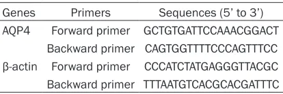
to manufacturer’s instruction. The expression level of AQP4 was analyzed with melting curve analysis and normalized to internal β-actin. The primers for AQP4 and β-actin were provided by Takara (Table 1).
Statistical analysis
[image:2.612.91.298.83.152.2]Data were expressed as mean ± standard deviation (SD), statistical analysis was per-formed applying SPSS 17.0 (SPSS Inc). The dif-ferences between two groups were analyzed using independent 2-sample t test. The differ-ences among multiple groups were analyzed Table 1. The primers for AQP4 and β-actin
Genes Primers Sequences (5’ to 3’) AQP4 Forward primer GCTGTGATTCCAAACGGACT
Figure 1. EB content in the three groups. The EB content in sham operation group, HIBD group and mannitol group at different time points after treat-ments was detected. The sham operation group showed no significant differ-ence at each time point (P>0.05). The HIBD group showed significantly higher EB level than the sham operation group at each time point (P<0.05). The mannitol markedly decreased EB content except for 0 h (P<0.05).
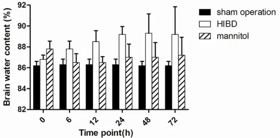
Figure 2. Brain water content in the three groups. The level of brain water content in the sham operation group had no significant difference at each time point (P>0.05). HIBD group showed significantly higher water content than the sham operation group at each time point (P<0.05). The mannitol decreased the water content almost to baseline.
[image:3.612.92.379.320.462.2]While in the mannitol group, the EB content decreased from 0 h to 6 h and then increased slightly. The level at each time point was signi- ficantly lower than that of HIBD group except for 0 h (Figure 1).
Brain water content
Brain wet-dry weight ratios were calculated to determine water content in different groups. The level of brain water content in the sham operation group had no sig-nificant difference at each time point (P>0.05). Water content in HIBD group rose sharply from 0 h to 48 h, and decreased at 72 h. The level was significantly higher than the sham operation group at each time point (P<0.05). Conversely, in the mannitol group, the brain water con-tent at each time point was significantly lower than that of HIBD group except for 0 h, almost reached to base-line. In the comparison bet- ween the sham operation and mannitol group, no sig-nificant difference was fo- und (P>0.05) except for 0 h (Figure 2).
AQP4 mRNA expression
using one-way ANOVA analysis. P value <0.05 was considered statistical significance.
Results
Detection of BBB permeability
To determine the effects of HIBD and mannitol on BBB, we detected BBB permeability by EB staining. As shown in Figure 1, The EB con-tent in sham operation group showed no signi- ficant difference at each time point (P>0.05). However, in HIBD group, the EB content inc- reased from 0 to 48 h, and then decreased. The level was significantly higher than the sham operation group at each time point (P<0.05).
groups, the expression was significantly higher at each time point except at 72 h (P<0.05). Discussion
In the present study, we used a HIBD animal model to investigate the effect of mannitol on brain edema after HIBD and its mechanisms via AQP4. Since the roles of AQP4 are totally different in cytotoxic edema and vasogenic edema, it is important to find out the major type of brain edema during HIBD. It has been reported that cytotoxic edema without obvi- ous BBB damage is the major form in the early period of hypoxia or ischemia. With the BBB damaging, vasogenic edema occurred in the later period of hypoxia or ischemia [10]. Our research showed that hypoxia-ischemia (HI) increased brain water content and BBB perme-ability from 0 h, which proved that in the begin-ning stage of HI, the vasogenic edema had begun to form and continued to the later peri-od. The possible reason may be the simult- aneous occurrence of hypoxia and ischemia in our model aggravated the damage of BBB thus accelerated the progress of vasogenic edema. In HIBD group, we found that the changing trends of the brain water content and BBB per-meability at different time points from 0 h to 72 h were similar, the peak level were at 48 h. It is indicated that hypoxia and ischemia induce brain edema by damaging the BBB. However,
we speculate that the decrease of AQP4 after HI limited the water clearance thus acceler- ated the progress of brain edema. The follow- ing upregulation of AQP4 may be explained as a self-protective reaction to eliminate excess water in brain and prevent the development of brain edema.
[image:4.612.93.378.83.243.2]While in the mannitol group, the results sho- wed that mannitol had a significant effect on decreasing BBB permeability and alleviating edema induced by HI, especially at 6 h after operation, the brain water content almost reached to baseline. However, in the early stage of HI, mannitol seemed to increase the BBB permeability and thus aggravated edema. It is indicated that mannitol was capable of open- ing BBB transitorily and accelerated the early period of brain edema. From the analysis of AQP4, we found that mannitol markedly upre- gulated the expression of AQP4 but downregu-lated the brain water content, suggesting that the alleviation of brain edema was at least in part associated with the high expression of AQP4. It can also be explained as the AQP4 contributed to water clearance in vasogenic edema. However, the mechanism for the regu-lating effect of mannitol on AQP4 is not fully understood. Recent studies have demonstr- ated that hyperosmotic stress could increase the expression level of AQPs. For instance, Hajime Arima et al found that mannitol was Figure 3. AQP4 expression in the three groups. No distinct difference at each
time point in the sham operation group was observed (P<0.05). The HIBD group showed significantly higher AQP4 level than sham operation group at 0 h, 6 h and 72 h (P<0.05), but significantly lower at the other three time points. In mannitol group, the AQP4 expression was significantly higher than sham operation and HIBD group except at 72 h (P<0.05).
able to increase the expression of AQP4 and AQP9 in cultured rat astrocytes and brain cor-tex. Besides, Hajime Arima also confirmed that the neuroprotection of mannitol was associ- ated with the activation of p38 MAPK pathway in astrocytes [11]. However, little research has been done on the relationship between manni-tol and AQP4 in neonatal brain.
In conclusion, the present study proves that mannitol relieves brain edema after HIBD by upregulating the expression of AQP4. More functional research of AQP4 is needed to fur-ther illustrate the mechanism after HIBD. Acknowledgements
This work was supported by a grant from the National Natural Science Foundation of China (No. 810705390).
Disclosure of conflict of interest
None.
Address correspondence to: Li Liu, Department of Neonatology, The First Affiliated Hospital of Xi’an Jiaotong University, 277 West Yanta Road, Xi’an 710061, Shaanxi, China. Tel: 86-029-85323829; Fax: 86-029-85323829; E-mail: liuli918@163.com
References
[1] Klatzo I. Presidential address. Neuropathologi-cal aspects of brain edema. J Neuropathol Exp Neurol 1967; 26: 1-14.
[2] Yu LS, Yi JP, Ye GH, Zheng YY, Song ZJ, Yang YM, Song YL, Wang ZY and Bao QY. Effects of curcumin on levels of nitric oxide synthase and AQP-4 in a rat model of hypoxia-ischemic brain damage. Brain Res 2012; 1475: 88-95.
[3] Fu XM, Li QP, Feng ZH and Mu DZ. The roles of aquaporin-4 in brain edema following neonatal hypoxia ischemia and reoxygenation in a cul-tured rat astrocyte model. Glia 2007; 55: 935-941.
[4] Li XM, Gao JY, Ding J, Hu G and Xiao M. Aqua-porin-4 expression contributes to decreases in brain water content during mouse postnatal development. Brain Res Bull 2013; 94: 49-55. [5] Saadoun S, Papadopoulos MC, Watanabe H,
Yan DH, Manley GT and Verkman AS. Involve-ment of aquaporin-4 in astroglial cell migration and glial scar formation. J Cell Sci 2005; 118: 5691-5697.
[6] Yang B, Zador Z and Verkman AS. Glial cell aquaporin-4 overexpression in transgenic mice accelerates cytotoxic brain swelling. J Biol Chem 2008; 283: 15280-15286.
[7] Papadopoulos MC, Manley GT, Krishna S and Verkman AS. Aquaporin-4 facilitates reabsorp-tion of excess fluid in vasogenic brain edema. FASEB J 2004; 18: 1291-1293.
[8] Grande PO and Romner B. Osmotherapy in Brain Edema: A Questionable Therapy. J Neu-rosurg Anesth 2012; 24: 407-412.
[9] Yu L, Fan SJ, Liu L, Xiao M, Lin XJ, Liu Y, Lv HX, Chen XL and Liu JX. Effect of ischemic post- conditioning on cerebral edema and the AQP4 expression following hypoxic-eschemic brain damage in neonatal rats. World J Pediatr 2015; 11: 165-170.
[10] Papadopoulos MC and Verkman AS. Aquapo-rin-4 and brain edema. Pediatr Nephrol 2007; 22: 778-784.