Original Article
Targeting Notch-1 reverses cisplatin chemosensitivity in
ovarian cancer cells by upregulation of PUMA
Jin Qu1, Yun Wang1, Yonglin Yang2, Jiacun Liu2,3
1Department of Gynaecology, People’s Hospital of Jining, Jining, China; 2Department of Clinical Laboratory,
People’s Hospital of Laiwu, Jinan, China; 3Department of Clinical Laboratory, The People’s Hospital of Weifang,
Weifang, China
Received October 28, 2016; Accepted March 17, 2017; Epub May 15, 2017; Published May 30, 2017
Abstract: Aim: Cisplatin (DDP) is the first-line chemotherapeutic therapeutics for ovarian cancer treatment. Its ear -ly response is usual-ly effective, the majority of patients will ultimate-ly recur with chemotherapy-resistant cancer. However, the mechanisms of DDP chemoresistance remain unclear. The aim of this work is to explore the poten -tial mechanism for development of chemoresistance in ovarian cancer. Methods: SKOV3 and SKOV3DDP cells was treated with 3 μg/ml of cisplatin for 24-72 h. Using siRNAs and inhibitors to examine the relation between p53, Notch-1 and PUMA expression exposure to cisplatin, and the effect on cisplatin chemosensitivity of ovarian cancer cells. A subcutaneously implanted tumor model of SKOV3 and SKOV3DDP cells in nude mouse was used to observe the effects of Notch-1 knockdown in combination with cisplatin on tumorigenesis. Results: Our data revealed that p53, Notch-1 and PUMA protein expression were induced by DDP. In the early, DDP-induced p53-dependent PUMA expression contributes to DDP-induced apoptosis of SKOV3 cells. In the later, DDP-induced p53 enhanced expres -sion of Notch1, further downregulated PUMA expres-sion, and weaken p53-induced apoptosis and cell growth inhi -bition. Knockdown of Notch-1 expression sensitized SKOV3 and SKOV3DDP cells to DDP treatment in cultured cell lines and xenograft nude mice cell model. Furthermore, our data demonstrated that Notch1 was overexpressed in SKOV3DDP cells, and the expression of Notch1 correlated with chemoresistance of SKOV3 cells. Conclusions: P53/ Notch-1/PUMA axis has an important role in the development of chemoresistance in ovarian cancer. Notch1 has an anti-cancer role in ovarian cancer. Thus, inhibition of Notch1 function reverses cisplatin chemosensitivity in ovarian cancer cells, which provides a logical approach for effective cancer therapy.
Keywords: Ovarian cancer, chemoresistance, cisplatin, P53, Notch-1, PUMA
Introduction
Epithelial ovarian carcinoma (EOC) remains the most lethal gynecologic malignancy [1]. Currently, chemotherapy in combination with
surgical debulking is the preferred treatment option and derivatives of cisplatin (DDP) are first-line chemotherapeutic therapeutics [2]. While early response is usually effective, the majority of patients will ultimately recur with
chemotherapy-resistant cancer and succumb
to disease [2]. Therefore, the control of drug-resistance against DDP is one of the important issues in the improved treatment of ovarian cancer. To date, the mechanisms by which
tumor cells develop resistance to DDP remain incompletely understood.
p53 is a short lived protein, which is activated
(phosphorylation) by DNA damage signal. The
activated p53 in turn activates its downstream signals, and regulates cell cycle progression, DNA repair and apoptosis. p53 can regulate cis-platin induced cell death by several
mecha-nisms like: Degradation of flice-like inhibitory protein (FLIP), direct binding and counteracting the antiapoptotic function of B-cell lymphoma-extra-large (Bcl-xL), over expression of phos
-phatase and tensin homolog (PTEN) and inhibi
-tion of AMPK [3]. PUMA (p53 upregulated mo-dulator of apoptosis), a BH3-only Bcl-2 family
protein, plays an essential role in p53-depen-dent and p53-indepenp53-depen-dent pathway induced apoptosis by various stimuli [4, 5]. Cisplatin
could activate p53 dependent PUMA upregula -tion in ovarian cancer cells and act on the
mitochondria and cause the release of mito -chondrial death proteins [6-8]. Although DDP induced apoptosis in EOC cells, only low levels
following DDP treatment [8]. This might be due to simultaneous induction of the antiapoptotic protein in addition to p53 and PUMA.
The Notch signaling pathway, a highly evolution -arily conserved pathway, which is activated by its ligands, in both invertebrate and vertebrate
development, plays a key role in cell differentia
-tion, survival, and proliferation [9]. When the
Notch receptor is activated, the receptor-ligand then triggers a second Notch extracellular domain cleavage by a metalloproteinase ADAM, which in turn downregulates ligand activity [10].
Up to date, only four Notch genes have been identified in mammals (Notch-1 to 4) [11]. All
the Notch receptors are very similar in
struc-tures, although there are some subtle differ -ences in their cytoplasmic domains and extra-cellular [9].
The function of Notch signaling in tumorigene
-sis could be either oncogenic or anti-prolifera
-tive, and the function could be context depen -dent. Notch signaling has been shown to be
anti-proliferative in a limited number of tumor types, including skin cancer, human hepatocel -lular carcinoma, medullary thyroid, cervical cancer, and small cell lung cancer [12-16].
However, most of the studies have shown the
Notch pathway has been shown to be activated in multiple tumors, including EOC [17], and as
aoncogenic function of Notch in many human
carcinomas [18-20]. Recently, Notch pathway has been reported to be involved in intrinsic resistance and acquired drug-resistance, and
knockdown of Notch-1 could reverse chemo
-sensitivity [21]. The mechanism for this poten
-tiation of chemosensitivity reversion by Notch inhibition may be related to downregulation of pro-survival pathways and upregulation of
anti-survival pathways [22].
Recently, it has found that activated p53 could upregulate the expression of Notch1 inhuman cancer cell lines. Moreover, inhibition of Notch1 activity after genotoxic stress by an inhibitor of Notch signaling increased susceptibility to
apoptosis [23], suggesting that Notch-1 plays
anti-apoptosis effect. However, the molecular
processes that mediate antiapoptotic activity
of Notch-1 are not completely understood. Meurette et al. has found that activation of
Notch-1 signalling in breast cancer cells caused resistance to p53-dependent apoptosis in-
duced by DNA damage, followed by decreased
pro-apoptotic PUMA and NOXA accumulation [24], suggesting that activation of p53-depen
-dent Notch-1 signalling could result in PUMA
inactivation.
In the present study, we demonstrate that
knockdown of Notch-1 could induce apoptosis
and reverses cisplatin chemosensitivity in ovar-ian cancer cells via a direct interaction
amon-gp53/Notch-1/PUMA axis. Our results suggest
that DDP treatment early activates
p53-depen-dent PUMA upregulation and induces cell apop -tosis. DDP treatment later activates
p53-depen-dent Notch-1 signal, resulting in PUMA
inac-tivation and apoptosis inhibition.
Materials and methods
Ethics statement
All studies involving mice were approved by the
Institutional Animal Care and Treatment Com-mittee of People’s hospital of Laiwu, Shandong.
Cell lines and culture
The human ovarian cancer cell line SKOV3 were purchased from the Cell Bank of the Chinese Academy of Science (Shanghai, China). Cisplatin (DDP)-resistant SKOV3 cell (SKOV3DDP)
were purchased from shybio.biomart (ATCC, Shanghai, China). Both cell lines were cultured
in RPMI-1640 medium supplemented with 10%
fetal bovine serum (FBS) in 5% CO2/95% air at 37°C. In vitro assays were performed at 60-70%
cell density.
Antibodies and agents
Cisplatin [cis-diammine-dichloroplatinum (II)]
was acquired from Sigma-Aldrich. Anti-Notch-1, Anti-PUMA, Anti-P53 and Anti-β-actin antibod
-ies were purchased from Santa Cruz Biotech-nology (Shanghai, China). Notch-1 siRNA,
PU-MAsiRNAs, p53 siRNA and Notch 1 shRNA Plasmid (h) were purchased from Santa Cruz Biotechnology (Guangzhou, China).
Transfection with siRNAs
Lipofectamine 2000 (Invitrogen) kits were used for transfections of siRNAs, respectively, ac-cording to the manufacturer’s instructions. Briefly, SKOV3 and SKOV3DDP cells were seeded in six-well plates (density 2.5 × 10⁵ cells/well)
50 nM Notch-1 siRNAs or PUMAsiRNAs or p53
siRNA or scramble siRNA or Notch 1
shRNA-plasmid in serum-free medium for 5-8 h. The culture medium was then replaced with fresh medium for 48 h. Cells were then treated accordingly. For stable siRNA transfection, 24 hs after Notch-1 siRNAs or Notch 1 shRNA transfection, the cells were split into 96-well plates and subjected to the G418 (1 mg/ml) selection for 2-3 weeks.
Cisplatin treatment
The length of treatment for all experiments con
-sisted of 24-72 h. SKOV3 and SKOV3DDP cells
were first plated and cultured with 10% FBS in RPMI 1640 media for 24 hours before initiation of treatment to allow cells to attach to the plate. The next day, the media was replaced with 0.1% FBS in RPMI 1640 containing DDP 3 µM concentrations for 24-72 h. To determine the effect of Notch-1, PUMA and p53 on DDP-induced apoptosis and growth inhibition, SK-OV3 and SKSK-OV3DDP cells were transfected with
siRNA targeting Notch-1 or PUMA or p53 24 h before DDP treatment about. Cells were then
treated accordingly.
Cell survival assay
After exposure to various treatments, cells were seeded in 96-well plates at a density of 3
× 103 cells/well in DMEM containing 10% FBS.
After they had adhered, cells were assessed for growth using an MTT assay. Briefly, 20 μl of MTT solution (5 mg/ml in PBS) was added into triplicate wells and cells were incubated for 4-6
h in an incubator. Absorbance at 490 nm was read with a microplate reader.
Apoptosis assay
Apoptosis induction was quantified by Annexin V/PI double staining followed by flow cytometry. Annexin V/PI double staining was performed using an apoptosis detection kit (Biovision, Mountain view, CA) following the manufactur
-er’s instruction. Briefly, after exposure to vari -ous treatments, cells were gently detached by
brief trypsinization, and then washed with ice cold PBS. After another wash with binding buf
-fer, cells were suspended in 300 µL binding buffer containing Annexin V and propidium iodide, and incubated for 5 min at room tem
-perature. Early apoptotic cells were identified
as Annexin V positive/PI negative cells, while late apoptotic/necrotic cells were identified as Annexin V positive/PI positive cells using a BD LSR II cell analyzer.
Western blotting
Cell lysates were homogenized by sample buf
-fer (100 mM Tris-HCl (pH 6.8), 2% Sodium Dodecyl Sulfate (SDS), 0.002% bromophenol blue, 20% glycerol, 10% β-mercaptoethanol (all from Sigma-Aldrich). Those were subjected to SDS-PAGE and transferred onto nitrocellu
-lose membranes (Amersham Biosciences). The following primary antibodies were used for immunodetection: anti-β-actin, Notch-1, P53 and PUMA and the Western Blot Substrate kit (Pierce) were used to detect
chemilumine-scence.
Xenograft models
Immunodeficient female mice, 4 to 6 weeks old, were purchased from the Shanghai
Ani-mal Center. Stable Notch-1 shRNA or control
shRNA transfected SKOV3 and SKOV3DDP cells (5 × 10⁶) were injected subcutaneously into
6-week-old female athymic nude mice. On days 6-8 and 16-18, cisplatin (3 mg/kg/d) was injected intraperitoneally into the mice. Tumor
growth was monitored every 3 days with cali-pers to calculate tumor volume according to
the formula [length × width2]/2. Six weeks later,
primary tumor masses were excised, fixed in 4% paraformaldehyde, and embedded in paraf
-fin. Sections (5 μm thick) were prepared and
stained with hematoxylin and eosin.
Terminal deoxynucleotidyl-transferase-mediat-ed dUTP nick end labeling (TUNEL) analysis
TUNEL was performed with an In situ Cell Death Detection Kit (Roche). Cell apoptosis was quan
-tified by determining the percentage of posi
-tively stained cells for all of the nuclei in 20 ran
-domly chosen fields/section at 200× magni-fication. Slides of the apoptosis studies were quantified in a blind manner by two indepen
-dent reviewers two different times.
Immunohistochemistrical staining
Tissue sections were deparaffinized and rehy
blocked in 5% BSA in PBS at RT for 1 h. The sec -tions were incubated with primary antibody
(PUMA and Notch-1) overnight at 4°C followed
by incubation with biotin-conjugated secon-
[image:4.612.91.517.73.562.2]dary antibodies for 1 h at RT. The sections were incubated with an Avidin-Biotin complex for 45 min, followed by diaminobenzidine (DAB) staining. The DAB-stained preparations were
visualized with a TE-2000U bright-field optical
microscope. Statistics
Data was expressed as means ± S.E. Statistical
analysis was performed by ANOVA or Student’s t-test (two-tailed) using SPSS 17.0 software. A
P-value of <0.05 was considered to be statisti
-cally significant.
Results
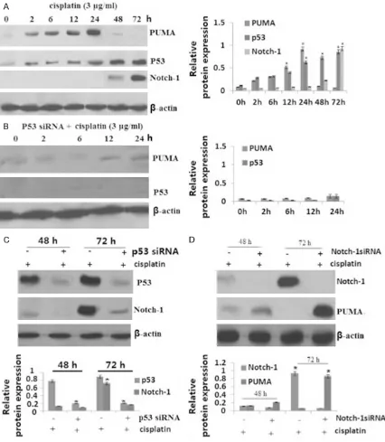
Early p53-dependent induction of PUMA by cisplatin treatment
SKOV3 ovarian cancer cells was treated with 3 μg/ml of cisplatin. PUMA protein was induced by cisplatinat 2 h, with the peak level of PUMA
protein at 24 h. After treatment of cisplatin for 24 h, PUMA protein expression was decreased, and undetectable after 48 h (Figure 1A). We next detected p53 protein expression in the
SKOV3 cells after cisplatin treatment. The
re-sults showed that P53 protein was induced gradually with cisplatin treatment, and reach
the peak at 48-72 h (Figure 1A).
We also detected Notch-1 protein expression in
the SKOV3 cells after cisplatin treatment. The
results showed that Notch-1 protein began to
induce by cisplatinat 48 h, and reach the peak
at 72 h (Figure 1A).
To determine whether p53 is involved in PUMA induction, SKOV3 cells were transfected with
p53 siRNA to inhibit p53 expression, then
treat-ed with 3 μg/ml of cisplatin for 24 h, only to find that induction of PUMA protein was blocked in the present of cisplatin (Figure 1B).
p53-dependent induction of Notch-1 by cispla-tin
SKOV3 ovarian cancer cells was treated with 3 μg/ml of cisplatin for 24-72 h. Notch-1 protein
was induced by cisplatinat 48 h, and reached
the peak level of PUMA protein at 72 h (Figure 1A). To determine whether p53 is involved in Notch-1 induction, SKOV3 cells were transfect -ed with p53 siRNA to inhibit p53 expression,
[image:5.612.93.517.77.347.2]then treated with 3 μg/ml of cisplatin for 48-72 h, only to find that induction of Notch-1 protein was blocked in the present of cisplatin (Figure 1C).
Activated P53/Notch-1 signal inhibits cisplatin-induced PUMA upregulation
To determine whether PUMA was inhibited by cisplatin-activated Notch-1, SKOV3 cells were transfected with 1 siRNA to inhibit Notch-1 expression, then treated with 3 μg/ml of cisplatin for 72 h, only to find that PUMA protein was significantly induction in the pres
-ent of cisplatin (Figure 1D). The above data col -lectively indicate that p53-dependent induction
of PUMA by cisplatinis blocked by
cisplatin-induced Notch-1 activation.
Knockdown of Notch-1 induced PUMA-depen-dent growth inhibition and apoptosisin SKOV3 cells
18% of apoptosis was detected in SKOV3 cells following cisplatin treatment for 24 h-72 h
(Figure 2A). Knockdown of Notch-1 by siRNA
alone did not affect cell apoptosis (P<0.01,
Figure 2A) and cell growth (P<0.01, Figure 2B)
of SKOV3 cells, but led to a significant increase
in cisplatin-induced apoptosis and growth inhi-bition at 48-72 hs (P<0.01, Figure 2A, 2B).
However, the effect of Notch-1 knockdown on
cisplatin-induced apoptosis was much reduced and growth inhibitionin was decreased in the
PUMA siRNA/SKOV3 cells (P<0.01, Figure 2A, 2B). The control siRNA has no effect on cisplat -in-induced apoptosis at 48-72 hs (data not
shown). In addition, knockdown of p53 by siRNA inhibited cisplatin-induced apoptosis at 24 h
(P<0.05), and led to a significant increase in
cisplatin-induced apoptosis and growth inhibi-tion at 48-72 hs (P<0.01, Figure 2A, 2B).
However, the effect of p53 knockdown on cispl -atin-induced cell apoptosis and growth was
much reduced in the PUMA siRNA/SKOV3 cells
(P<0.01, Figure 2A, 2B). These results suggest that PUMA induction represents a novel mech -anism mediating cisplatin-induced apoptosis
and growth inhibition, and that activation of
p53-dependent Notch-1 signal can
compro-mise cisplatin-induced and PUMA-mediated
apoptosis and growth inhibition. Knockdown of Notch-1 induced growth inhibition and PUMA-dependent apoptosisin SKOV3DDP cells
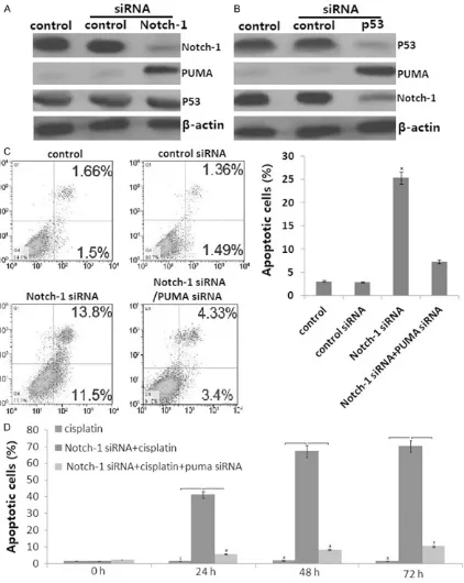
Notch-1 and p53 protein was overexpressed,
and PUMA was less expressed in the SKOV3DDP
cells by western blot assay. Knockdown of
Notch-1 (Figure 3A) or p53 (Figure 3B) by
siRNA induced PUMA protein expression in the SKOV3DDP cells. In addition, knockdown of p53
blocked Notch-1 protein in SKOV3DDP cells (Figure 3B). However, knockdown of Notch-1 did not affect p53 protein in SKOV3DDP cells (Figure 3A). The above data indicate that PUMA
was p53/Notch-1 dependent regulation in the
SKOV3DDP cells.
Knockdown of Notch-1 reverses cisplatin che-mosensitivity of SKOV3DDP cells by upregulation
of PUMA
SKOV3DDP cells were transfected with Notch-1
siRNA or control siRNA for 48 h, then treated with 3 μg/ml of cisplatin for 24-72 h. Analysis of apoptosis using annexin V/propidium iodide (PI) staining showed that knockdown of Notch-1 alone induced apoptosis of SKOV3DDP cells (P<0.05, Figure 3C). However, when PUMA pro
-tein was knockdown by siRNA (Figure 3C),
knockdown of Notch-1 did not affect apoptosis of SKOV3DDP cells (Figure 3C).
Although SKOV3DDP cells were resistant to
cis-platin treatment, knockdown of Notch-1 signifi -cantly enhanced cisplatin-induced apoptosis (Figure 3D). However, when PUMA protein was knockdown by siRNA, knockdown of Notch-1 blocked cisplatin-induced apoptosis (Figure 3D). MTT assay also showed that knockdown of Notch-1 significantly enhanced
cisplatin-induced cell growth inhibition (data not shown).
However, when PUMA protein was knockdown by siRNA, knockdown of Notch-1 blocked cispl -atin-induced cell growth inhibition (data not
shown). The above data indicate that knock
-down of Notch-1 reverses cisplatin chemosen
-sitivity in SKOV3DDP cells by upregulation of
PUMA.
Knockdown of siRNA in combination cisplatin yields significantly better antitumor efficacy compared with the single-agent treatment
Next, to run a pilot efficacy study to combine
the Notch-1 sliencing and cisplatin treatment,
mice bearing SKOV3DDP or SKOV3 cells (Notch-1 siRNA or control siRNA) tumors were treated
with PBS or cisplatin. There was tumor growth
inhibition observed in mice that had Notch-1 siRNA alone (~35%) or cisplatin alone (~8%) or
inhi-bition observed in mice that had Notch-1 siRNA alone (~10%) or cisplatin alone (~20%) or the
combination treatment (~45%) in SKOV3 cells.
[image:7.612.95.517.72.602.2]The combination group showed significantly better growth inhibition compared with PBS or either of the single-agent treatment on day 42
in both of the cells (P<0.01, respectively)
(Figure 4A, 4B).
Immunohistochemistrical staining assay sho-
wed that Notch-1 was knockdown and PUMA was induced in the groups of combination treat
-ment in the SKOV3DDP cells. (Figure 4C, 4D).
Analyzing tissue sections from tumors revealed that combination treatment had significantly
increased apoptosis in the tumors assessed by
TUNEL staining in both of the cells (P<0.01,
respectively) (Figure 4E). Notch-1 siRNA has
the same results as that to Notch-1 and PUMA expression, as well as cell apoptosis in SKOV3
cells (data not show).
Discussion
Inhibition of cell proliferation and inducing
apoptosis by p53 is largely attributable to its ability to transcriptionally activate the
expres-sion of genes that encode proteins, which determine cell fate [25, 26]. Depending on cel -lular context, wild-type p53 limits cell proli-
feration in response to DNA damage and other
cellular stresses by inducing cell cycle arrest, apoptosis, or senescence [27, 28].
In our study, we found that treatment of SKOV3
cells with DDP induced apoptosis and growth
inhibition of SKOV3 cells at 24 h. Furthermore, p53 and PUMA expression was significantly increased. Knockdown of PUMA blocked DDP
induced cell apoptosis at 24 h. In addition,
knockdown of p53 blocked DDP induced cell apoptosis and PUMA upregulation at 24 h. We therefore suggested that DDP treatment acti
-vated p53 dependent PUMA, which plays an
important role in DDP-induced apoptosis and
growth inhibition of SKOV3 cells. DDP induced apoptosis (<20%) in SKOV3 cells at 24 h, no sig
-nificant change was found at 48-72 h compared to the apoptosis at 24 h. Furthermore, PUMA was significantly increased at 24 h and unde
-tectable at 48 h following DDP treatment.
We suggested that DDP might simultaneously
induce some antiapoptotic protein after 24 h in addition to p53 and PUMA.
Notch-1 signaling plays a pivotal role in cell
survival, in a manner that can deeply influence the final outcome in tumor development. Re-cently, Notch1 gene has been identified as a direct transcriptional target of p53 [29]. In pri
-mary human keratinocytes, knock-down of p53
results in down-modulation of Notch1 expres -sion [30]. Conversely, increased p53 levels
leads to Notch1 up-regulation in normal kerati -nocytes and, to a substantial greater extent, in
SCC cells [31]. In the present study, we found that Notch-1 was induced at 48 h after DDP treatment, and reached the peak at 72 h. knockdown of p53 expression in SKOV3 cells reduced the expression levels of Notch1 pro -tein in response to DDP treatment. In addition,
knockdown of Notch-1 increased susceptibi-lity to apoptosis induced by DDP treatment of cells. These observations provide support for the idea that p53-mediated upregulation of
Notch1 expression in SKOV3 cells counteracts p53-mediated proapoptotic functions.
It has recently found that activation of Notch-1 signalling decreased pro-apoptotic PUMA
ex-pression and resulted in resistance to p53- dependent apoptosis induced by DNA damage
[24]. In the present study, Notch-1 was signifi
-cantly increased and PUMA was signifi-cantly decreased at 72 h after DDP treatment. Kn-ockdown of Notch-1 expression in SKOV3 cells enhanced the expression levels of PUMA pro -tein in response to DDP treatment. In addition,
knockdown of PUMA decreased susceptibility to apoptosis of Notch-1 silenced SKOV3 cells. Based on our observation and accumulating
evidence, we could concluded that Notch1
gene is atranscriptional target of p53, and PUMA is atranscriptional target of Notch-1. Activation of Notch1 expression inhibited
PU-MA activation, and decreased p53-dependent apoptosis.
We further observed that Notch-1 was overex
-pressed in the SKOV3DDP cells. Knockdown of
Notch-1 restored the susceptibility of SKOV3DDP
cells to DDP treatment via PUMA upregulation.
In addition, p53 was also overexpressed in the
SKOV3DDP cells. Knockdown of p53 decreased
Notch-1 expression and increased the PUMA expression, and restored the susceptibility of SKOV3DDP cells to DDP treatment. We there-
fore suggested that inhibition of Notch1 func -tion could reverse cisplatin chemosensitivity in ovarian cancer cells.
In the present study, we found that early che
-mosensitivity of ovarian cancer cells to cisplat
-in is associated with p53-dependent PUMA upregulation. Later chemoresistance of ovarian
p53-dependent Notch-1 activation, resulting in
PUMA inactivation and counteracting p53-medi
-ated proapoptotic functions. Overexpression of Notch-1 is associated with chemoresistance of ovarian cancer cells to cisplatin. Thus, inhibi
-tion of Notch1 func-tion reverses cisplatin che -mosensitivity in ovarian cancer cells, which
pro-vides a logical approach for effective cancer
therapy.
Acknowledgements
This study was supported Jining nature scien
-tific research fund (No: 2014JWM201).
Disclosure of conflict of interest
None.
Address correspondence to: Yun Wang, Depart- ment of Gynaecology, People’s Hospital of Jining, Jining, China. E-mail: wangyunliuzi@163.com
References
[1] Siegel R, Naishadham D, Jemal A. Cancer sta-tistics, 2012. CA Cancer J Clin 2012; 62: 10-29.
[2] Kartalou M, Essigmann JM. Mechanisms of re -sistance to cisplatin. Mutat Res 2001; 478: 23-43.
[3] Basu A, Krishnamurthy S. Cellular responses to cisplatin-induced DNA damage. J Nucleic Ac-ids 2010; 2010.
[4] Yu J, Zhang L, Hwang PM, Kinzler KW, Vogel -stein B. PUMAinduces the rapid apoptosis of
colorectal cancer cells.Mol Cell2001; 7: 673-682.
[5] Wu WS, Heinrichs S,Xu D, Garrison SP, Zam
-betti GP, Adams JM, Look AT. Slug antagonizes p53-mediated apoptosis of hematopoietic pro -genitors by repressing puma. Cell 2005; 123: 641-653.
[6] Maurmann L, Belkacemi L, Adams NR, Majmu -dar PM, Moghaddas S, Bose RN. A novel cispl-atinmediated apoptosis pathway is associated
with acid sphingomyelinase and FAS proapop -totic protein activation inovarian cancer. Apop-tosis 2015; 20: 960-974.
[7] Gu J, Tang Y, Liu Y, Guo H, Wang Y, Cai L, Li Y, Wang B. Murine double minute 2 siRNA and wild-type p53 gene therapy enhances sensitiv-ity of the SKOV3/DDP ovarian cancercell line tocisplatinchemotherapy in vitro and in vivo. CancerLett 2014; 343: 200-209.
[8] Fraser M, Bai T, Tsang BK. Akt promotes cispla-tinresistance in human ovarian cancercells through inhibition of p53 phosphorylation and
nuclear function. Int JCancer2008; 122: 534-546.
[9] Miele L. Notch signaling. Clin Cancer Res 2006; 12: 1074-1079.
[10] Zolkiewska A. ADAM proteases: ligand pro -cessing and modulation of the Notch pathway. Cell Mol Life Sci 2008; 65: 2056-2068. [11] Bolós V, Grego-Bessa J, de la Pompa JL. Notch
signaling in development and cancer. Endocr Rev 2007; 28: 339-363.
[12] Wang M, Xue L, Cao Q, Lin Y, Ding Y, Yang P, Che L. Expression of Notch1, Jagged1 and be
-ta-catenin and their clinicopathological signifi -cance in hepatocellular carcinoma.
Neoplas-ma2009; 56: 533-541.
[13] Gao J, Chen Y, Wu KC, Liu J, Zhao YQ, Pan YL, Du R, Zheng GR, Xiong YM, Xu HL, Fan DM. RUNX3 directly interacts with intracellular do -main of Notch1 and suppresses notch signal -ing in hepatocellular carcinoma cells.Exp Cell Res2010; 316: 149-157.
[14] Wang C, Qi R, Li N, Wang Z, An H, Zhang Q, Yu Y, Cao X. Notch1 signaling sensitizes tumor ne -crosis factor-related apoptosis-inducing ligand-induced apoptosis in human hepatocellular carcinoma cells by inhibiting Akt/Hdm2-medi -ated p53 degradation and up-regulating p53-dependent DR5 expression. J Biol Chem 2009; 284: 16183-16190.
[15] Dotto GP. Notch tumor suppressor function. Oncogene2008; 27: 5115-5123.
[16] Nicolas M, Wolfer A, Raj K, Kummer JA, Mill P, van Noort M, Hui CC, Clevers H, Dotto GP, Radtke F. Notch1 functions as a tumor sup -pressor in mouse skin. Nat Genet 2003; 33: 416-421.
[17] Groeneweg JW, Foster R, Growdon WB, Verhei -jen RH, Rueda BR. Notchsignaling in serous ovariancancer. JOvarianRes2014;7: 95. [18] Rizzo P, Miao H, D’Souza G, Osipo C, Song LL,
Yun J, Zhao H, Mascarenhas J, Wyatt D, Antico G, Hao L, Yao K, Rajan P, Hicks C, Siziopikou K, Selvaggi S, Bashir A, Bhandari D, Marchese A, Lendahl U, Qin JZ, Tonetti DA, Albain K, Nickol -off BJ, Miele L. Cross-talk between notch and the estrogen receptor in breast cancer sug-gests novel therapeutic approaches. Cancer Res2008; 68: 5226-5235.
[19] Song LL, Peng Y, Yun J, Rizzo P, Chaturvedi V, Weijzen S, Kast WM, Stone PJ, Santos L, Lore -do A, Lendahl U, Sonenshein G, Osborne B, Qin JZ, Pannuti A, Nickoloff BJ, Miele L. Notch-1 as -sociates with IKKalpha and regulates IKK ac -tivity in cervical cancer cells.Oncogene 2008; 27: 5833-5844.
induces apoptosis via inactivation of Akt, mTOR, and NF-kappaB signaling pathways. J Cell Biochem 2010; 109: 726-736.
[21] Gottesman MM. Mechanisms of cancer drug resistance. Annu Rev Med 2002; 53: 615-627. [22] Wang Z, Li Y, Ahmad A, Azmi AS, Banerjee S,
Kong D, Sarkar FH. TargetingNotchsignaling pathwaytoovercomedrug resistancefor can-cer therapy. Biochim Biophys Acta 2010; 1806: 258-267.
[23] Alimirah F,Panchanathan R, Davis FJ,Chen J, Choubey D. Restorationof p53expressionin human cancer celllines upregulates the ex-pression of Notch1: implications for cancer cell fate determination after genotoxicstress. Neoplasia2007; 9: 427-34.
[24] Meurette O, Stylianou S, Collu GM, Gilmore AP, Brennan K. Inhibition of apoptosis byNotch signalling inbreastepithelial cells. Breast Can -cerRes2008;10: P20.
[25] Vousden KH and Lu X. Live or let die: the cell’s response to p53. Nat Rev Cancer 2002; 2: 594-604.
[26] McKay BC and Ljungman M. Role for p53 in the recovery of transcription and protection against apoptosis induced by ultraviolet light. Neoplasia 1999; 1: 276-284.
[27] Ljungman M. Dial 9-1-1 for p53: mechanisms of p53 activation by cellular stress. Neoplasia 2000; 2: 208-225.
[28] Pucci B, Kasten M, Giordano A. Cell cycle and apoptosis. Cell 2010; 2: 291-299.
[29] Wei CL, Wu Q, Vega VB, Chiu KP, Ng P, Zhang T, Shahab A, Yong HC, Fu Y, Weng Z, Liu J, Zhao XD, Chew JL, Lee YL, Kuznetsov VA, Sung WK, Miller LD, Lim B, Liu ET, Yu Q, Ng HH, Ruan Y. A global map of p53 transcription-factor binding sites in the human genome. Cell 2006; 124: 207-219.
[30] Mandinova A, Lefort K, Tommasi VA, Stonely W, Ostano P, Chiorino G, Iwaki H, Nakanishi J, Dotto GP. The FoxO3a gene is a key negative target of canonical notch signalling in the kera -tinocyte UVB response. EMBO J 2008; 27: 1243-54.