Original Article
Protective effect and mechanism of hydrochloride
dexmedetomidine on the severe craniocerebral
trauma in rats
Xingzhi Liao1*, Yufang Liu1*, Likun Yang2, Wenhua Wu1, Maitao Zhou1
Departments of 1Anesthesiology, 2Neurosurgery, PLA 101 Hospital, Wuxi 214044, China. *Equal contributors.
Received November 3, 2016; Accepted March 14, 2017; Epub May 15, 2017; Published May 30, 2017
Abstract: To investigate the cerebral protection effect of dexmedetomidine (DEX) in severe craniocerebral injury rat model. A total of 120 male SD rats were equally randomly divided into DEX group, sham group, and control. Rat acute severe craniocerebral injury model was established using hydraulic blow method. The rats received
seda-tive treatment at 2 h after injury. Rat behavior changes before and after modeling were observed. Serum S-100β, neuron specific enolase (NSE), and myelin basic protein (MBP) levels on pre-operation (T1), day 1 post operation
(POD1, T2), day 3 post operation (POD3, T3), day 5 post operation (T4), and day 7 post operation (T5) were de-tected by ELISA. Rat hippocampus was extracted for pathological section on T2, T3, T4, and T5. Neuronal cell death
was observed upon Nissl’s staining. NF-κB and Caspase-3 expression in hippocampus were tested by immuno
-histochemistry. Serum S-100β, NSE, and MBP levels significantly decreased on T2, T3, T4, and T5 in DEX group compared with control group (P < 0.05). Neuronal cell death, NF-κB, and Caspase-3 expression markedly reduced on T2, T3 (except NF-κB), T4, and T5 in DEX group compared with control group (P < 0.05). DEX can protect brain
upon improving cerebral oxygen metabolism and alleviating cerebral ischemia hypoxia in severe craniocerebral injury.
Keywords: Severe craniocerebral injury, dexmedetomidine, cerebral metabolism, cerebral protection
Introduction
Following the development of economy and traffic transportation industry, the incidence of craniocerebral trauma shows rising trend year by year. As one of the more common car acci-dent injuries in clinic, craniocerebral trauma and its complications has a serious influence on the central nervous system [1]. According to the Glasgow scoring system, patients with cra-niocerebral trauma can be grouped into mild (13-15 points), medium (9-12 points), heavy (6-8 points), and severe (3-5 points). Among them, patients in heavy and severe types have the most severe injury and complex treatment because of the interaction of injury factors, treatment methods, and individual differences, leading to the total fatality rate remains at 30%-50% [2, 3]. Heavy and severe craniocerebral injury may cause cerebral edema, nonphysical brain blood oxygenation and perfusion, local
cerebral ischemia, cerebral embolism, and inflammatory cascade reaction, which can induce central nervous system damage [4]. Thus, application of sedative with central ner-vous system protection can alleviate cerebral function damage after severe craniocerebral injury, thus has a practical value and clinical significance.
seda-tive treatment after severe craniocerebral inju-ry. This study intends to explore the cerebral protection effect of DEX in severe craniocere-bral injury through the establishment of rat severe craniocerebral injury model.
Materials and methods
Experimental animals
Male SD rats at 6-week old and weighted 250~300 g were purchased from laboratory animal center of Wuxi Medical College.
Rats were used for all experiments, and all pro-cedures were approved by the Animal Ethics Committee of PLA 101 Hospital.
Main reagents and instruments
Serum S-100β, neuron-specific enolase (NSE), and myelin basic protein (MBP) ELISA kits were got from R&D. Bio-Rad 680 microplate reader was bought from Bio-Rad. Claudin 5 antibody was got from Abcam. CD31 antibody was pur-chased from Invitrogen (USA). Fluorescence microscope was from Olympus.
Rat acute severe craniocerebral injury model preparation
The rats in DEX group and control were anes-thetized by 10% chloral hydrate (3.0 ml/kg) intraperitoneal injection. The head was fixed on stereotactic frame and disinfected. A straight incision was made on the top to expose parietal bone. A bone window with diameter at 4 mm was opened on 3 mm after bregma and 2 mm on the right side of sagittal suture. The endo-cranium was kept integrity. The skull connect-ing pipe was adhered to bone window and ster-ile saline solution was injected through the lumen. After consciousness recovery, the hy- draulic blow apparatus was connected to the connecting pipe to perform hydraulic blow with the striking force at 3.2~3.5 atm. The rat in sham group received same procedure without Control operation with before, but no hydraulic blow. The rats were kept in single cage after spontaneous breathing recovery.
Experimental animals grouping and treatment
A total of 120 male SD rats were equally ran-domly divided into DEX group, sham group, and control. Serum S-100β, neuron-specific eno -lase (NSE), and myelin basic protein (MBP) lev
-els on pre-operation (T1), POD1 (T2), POD3 (T3), POD5 (T4), and POD7 (T5) were detected by ELISA. Rat hippocampus was extracted for pathological section on T2, T3, T4, and T5. Neuronal cell death was observed upon Nissl’s staining. The rats in DEX group received 30 ug/ kg DEX intraperitoneal injection at 12 h after modeling, while the rats in control received equal amount of normal saline.
Blood sample collection
A total of 2 ml peripheral venous blood was extracted in EDTA anticoagulated tube and cen-trifuged to extract supernatant. The serum was stored at -80°C.
Serum S-100β, NSE, and MBP detection
Serum S-100β, NSE, and MBP levels were de-tected according to the ELISA manual.
Hippocampus pathological examination
The rats were killed on POD1 and POD7 after hydraulic injury. The brain tissue was fixed in 4% paraformaldehyde and then incubated in 30% sucrose solution. After embedding, the tis-sue was coronary serial sectioned at 10 μm thickness from 0.7~2.3 mm after bregma for immunofluorescent staining. Classical immuno -fluorescent staining was applied for CD31 and Claudin 5 staining. After washed by PBS for three times, the slice was blocked by normal rabbit or mouse serum for 30 min. Next, the slice was incubated in rabbit anti rat Claudin 5 antibody (1:500) and mouse anti rat CD31 anti-body (1:500) at 4°C overnight. After washed by PBS for three times, the slice was further incu -bated in Alexa Fluor 488-conjugated goat anti rabbit antibody (1:1000) and Mexa Fluor 568-conjugated goat anti mouse antibody (1:1000) at 20-37°C for 1 h. At last, the slice was stained by DAPI avoid of light for 5 min. After sealed by anti-fluorescence quenching agent, the slice was observed under the fluo -rescence microscope. The data was analyzed by the Image J software (National Institutes of Health, Bethesda, MD, USA).
Statistical analysis
com-pared by chi-square test. Correlation analysis was adopted upon Spearman rank analysis. Multivariate analysis was applied using Logistic regression analysis. P < 0.05 was presented as statistical significance.
Results
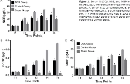
Serum S-100β, NSE, and MBP levels compari-son
Serum S-100β, NSE, and MBP levels in T1 showed no statistical significance among three groups (P > 0.05). Serum S-100β, NSE, and MBP levels in T2, T3, T4, and T5 were obviously
higher than T1 in DEX group and control (P < 0.05). Serum S-100β, NSE, and MBP levels sig -nificantly increased on T2, T3, T4, and T5 in DEX group and control compared with sham group (P < 0.05). Their levels in DEX group were obviously lower than the control (P < 0.05) (Table 1; Figure 1).
Hippocampal neuron cell death comparison
Cellular morphology in hippocampus was nor-mal in sham group. The cells in control group arranged disorder, appeared edema and num-ber reduce on T2, and showed Nissl body disin-Table 1. Serum S-100β, NSE, and MBP levels comparison (x±s, µg/L)
Group n Index T1 T2 T3 T4 T5
Control 40 S-100β 0.072±0.002 0.245±0.055*,# 0.334±0.002*,# 0.567±0.050*,# 0.735±0.015*,#
MBP 7.34±0.02 12.45±2.15*,# 21.45±1.15*,# 23.56±0.15*,# 36.45±3.78*,#
NSE 8.35±0.45 21.46±2.55*,# 30.84±5.25*,# 34.56±5.74*,# 36.45±3.78*,#
Sham 40 S-100β 0.073±0.001 0.066±0.001 0.056±0.154 0.056±0.001 0.045±0.005
MBP 7.35±0.05 8.31±0.03 10.15±0.05 11.56±1.05 19.55±3.56
NSE 8.23±0.55 11.45±2.55 16.56±3.46 17.45±5.67 19.55±3.56
DEX 40 S-100β 0.071±0.001 0.197±0.056*,#,& 0.234±0.002*,#,& 0.324±0.001*,#,& 0.335±0.004*,#,&
MBP 7.35±0.05 10.15±0.15*,#,& 17.35±1.05*,#,& 18.45±0.07*,#,& 21.45±0.15*,#,&
NSE 8.33±0.46 16.34±2.45*,#,& 20.84±1.13*,#,& 26.45±3.45*,#,&
*P < 0.05, compared with T1; #P < 0.05, compared with sham group; &P < 0.05, compared with control group. T1:
[image:3.612.91.526.82.217.2]pre-opera-tion; T2: 1 day post the operapre-opera-tion; T3: 3 day post the operapre-opera-tion; T4: 5 day post the operapre-opera-tion; T5: 7 day post the operation.
Figure 1. Serum S-100β, NSE, and MBP lev
-els (x±s, µg/L) comparison among all of three groups. A. Serum S-100β comparison; B. Se
-rum MBP comparison; C. Se-rum NSE compari
-son. *P < 0.05 represents the NSE, S-100β or
[image:3.612.90.517.266.542.2]tegration and lost, together with karyopyknosis, karyorrhexis, and karyolysis on T5. Cell damage in DEX group were obviously slighter than that in control at the same time point (Figure 2).
NF-κB expression in hippocampal neuron cells
Western blot revealed that NF-κB showed no statistical difference in sham group among
[image:4.612.90.523.73.196.2]sion in T2 was lower in DEX compared with sham group (P < 0.01). It gradually elevated and reached top in T4, which was obviously higher than that in sham group (P < 0.01). It then reduced, which was lower than that of sham group in T5 (P < 0.01). Caspase-3 level in T2 was lower in DEX group compared with sham and control (P < 0.05). Caspase-3 kept Figure 2. Hippocampal neuron cell death comparison in all groups. A. Sham group; B. Control group; C. DEX group.
[image:4.612.92.372.251.374.2]The arrows represent the Nissl body in the tissues.
Figure 3. Hippocampal neuron cells NF-κB expression comparison in all
groups. *P < 0.05 represents the NF-κB levels in Control group or Sham
group compared to the DEX group.
different time points (P > 0.05). NF-κB expression in T2 was lower in DEX compared with sham group (P < 0.01). It gradually elevated and re- ached top in T3, which was obviously higher than that in sham group (P < 0.01). It then reduced, which was lower than that of sham group in T4 and T5 (P < 0.01). NF-κB level in T2 was lower in DEX group compared with sham and control (P < 0.05). It showed no statistical difference in T3 compared with control while higher than that in sham group. NF-κB kept in relative low level in T4 and T5 at DEX group, which was signifi -cantly lower than that in sham and control (P < 0.01) (Figure 3).
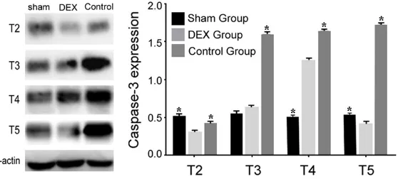
Caspase-3 expression in hip-pocampal neuron cells
Western blot revealed that Caspase-3 showed no statis-tical difference in sham group among different time points (P > 0.05). Caspase-3 expres-Figure 4. Hippocampal neuron cells Caspase-3 expression comparison in all
groups. *P < 0.05 represents the caspase-3 levels in control group or sham
[image:4.612.97.376.438.564.2]in relative low level in T5 at DEX group, which was significantly lower than that in sham and control (P < 0.01) (Figure 4).
Discussion
Heavy and severe craniocerebral injury induced nonphysical brain blood oxygenation and per-fusion, local cerebral ischemia, cerebral embo-lism, and inflammatory cascade reaction can trigger central nervous system damage [9-11]. Searching for correct cerebral protective mea-sures can alleviate cerebral function damage after severe craniocerebral injury.
Ehsan Z showed that 1 μg/kg DEX infusion before vein anesthesia induction and 0.4 μg/ kg DEX injection till finish operation can reduce the perioperative inflammatory response in patients with acute craniocerebral injury [12]. Therefore, this study selected DEX for investi-gation. S-100β, MBP, and NSE proteins are mainly located in glial cells of the central ner-vous system. They can be detected in blood during glial cell damage and blood brain barrier dysfunction [13, 14]. Their protein concentra-tions in the blood reflect the particular sensi -tive index of brain damage. This study applied the classical ELISA method to detect serum S-100β, MBP, and NSE protein concentrations to ensure the examination accuracy. Our results demonstrated that serum S-100β, MBP, and NSE levels decreased in DEX group, suggesting that DEX produced cerebral protection after severe craniocerebral injury.
It was revealed that cell apoptosis is the major type of delayed neuron death caused by cere-bral hypoxia ischemia, involving the mecha-nisms of (1) energy metabolism disorder; (2) intracellular calcium overload; (3) lipid peroxi-dation; (4) the toxic effects of excitatory amino acids; (5) free radical damage; (6) inflammatory response [15-18]. It was found that excitatory amino acids play a critical role in the pathogen-esis of cell death induced by neuron toxic effect [19]. Glu is the major part of excitatory amino acids that acts the excitatory neurotransmitter effect to maintain normal brain function. However, cerebral ischemia hypoxia induces a large number of substances, such as reactive oxygen species, leading to GLT concentration at 10 times in synaptic cleft and extracellular glutamate. Excessive glutamate releases to be a potential neurotoxin. Once the glutamate
content in synaptic cleft exceeds the GLT adjusting range and breaks the physiological balance between excitatory and inhibitory amino acids, it may over stimulate ionic gluta-mate receptor on cell membrane, such as N-methyl-D-aspartic acid (NMDA) receptor. It further leads to a series of diseases, including early Na+, Cl-, H
2O internal flow induced cell edema and Ca2+ overload, and late apoptosis induced secondary nerve injury, leading to “exi-totoxicity” [20].
In this study, Western blot revealed that NF-κB expression was lower in DEX compared with sham group. It gradually elevated and reached top in T3 and then reduced, which was lower than that of sham group and Control group. NF-κB level in T2 was lower in DEX group compared with sham and control. NF-κB kept in relative low level in T4 and T5 at DEX group, which was significantly lower than that in sham and control. It suggested that DEX may enhance excitatory amino acids Glu transport, alleviate nerve excitability toxicity effect, improve brain hippocampus tissue pathology, and reduce neuron apoptosis through down-regulating NF-κB expression, thus to play an important role in neuroprotection.
excitability toxicity effect, improve brain hippo-campus tissue pathology, and reduce neuron apoptosis via downregulating Caspase-3 ex- pression.
Conclusion
DEX can protect brain upon improving cerebral oxygen metabolism and alleviating cerebral ischemia hypoxia in severe craniocerebral injury through downregulating NF-κB and Caspase-3 levels.
Acknowledgements
This work was supported by Medical innovation foundation of Nanjing military region (NO. 14MS011).
Disclosure of conflict of interest
None.
Address correspondence to: Dr. Maitao Zhou, De- partment of Anesthesiology, PLA 101 Hospital, Wuxi 214044, China. Tel: +86-0510-85142312; Fax: +86-0510-85142312; E-mail: MaitaoZhouzxc@163. com
References
[1] Patel MB, McKenna JW, Alvarez JM, Sugiura A,
Jenkins JM, Guillamondegui OD, Pandhari-pande PP. Decreasing adrenergic or sympa-thetic hyperactivity after severe traumatic brain injury using propranolol and clonidine
(DASH After TBI Study): study protocol for a
randomized controlled trial. Trials 2012; 13: 177.
[2] Gu JW, Yang T, Kuang YQ, Huang HD, Kong B,
Shu HF, Yu SX, Zhang JH. Comparison of the
safety and efficacy of propofol with midazolam
for sedation of patients with severe traumatic brain injury: a meta-analysis. J Crit Care 2014; 29: 287-290.
[3] Flower O, Hellings S. Sedation in traumatic brain injury. Emerg Med Int 2012; 2012: 637171.
[4] James ML, Olson DM, Graffagnino C. A pilot study of cerebral and haemodynamic physio-logical changes during sedation with dexme-detomidine or propofol in patients with acute brain injury. Anaesth Intensive Care 2012; 40: 949-957.
[5] Yardan T, Erenler AK, Baydin A, Aydin K, Cokluk C. Usefulness of S100B protein in neurological
disorders. J Pak Med Assoc 2011; 61: 276-281.
[6] Dunn LK, Durieux ME, Nemergut EC. Non-opi -oid analgesics: novel approaches to
periopera-tive analgesia for major spine surgery. Best
Pract Res Clin Anaesthesiol 2016; 30: 79-89. [7] Thomas A, Miller JL, Couloures K, Johnson PN.
Non-intravenous sedatives and analgesics for procedural sedation for imaging procedures in pediatric patients. J Pediatr Pharmacol Ther 2015; 20: 418-430.
[8] Zamani MM, Keshavarz-Fathi M, Fakhri-Bafghi MS, Hirbod-Mobarakeh A, Rezaei N, Bahrami A, Nader ND. Survival benefits of dexmedeto -midine used for sedating septic patients in in-tensive care setting: a systematic review. J Crit Care 2016; 32: 93-100.
[9] Lundblad M, Trifa M, Kaabachi O, Ben Khalifa
S, Fekih Hassen A, Engelhardt T, Eksborg S, Lonnqvist PA. Alpha-2 adrenoceptor agonists as adjuncts to peripheral nerve blocks in chil-dren: a meta-analysis. Paediatr Anaesth 2016; 26: 232-238.
[10] Zhong WG, Ge XY, Zhu H, Liang X, Gong HX, Zhong M, Xiao X. Dexmedetomidine for anti-emesis in gynecologic surgery: a meta-analysis of randomized controlled trials. Int J Clin Exp Med 2015; 8: 14566-14576.
[11] Pan W, Wang Y, Lin L, Zhou G, Hua X, Mo L. Outcomes of dexmedetomidine treatment in pediatric patients undergoing congenital heart disease surgery: a meta-analysis. Paediatr An-aesth 2016; 26: 239-248.
[12] Ehsan Z, Mahmoud M, Shott SR, Amin RS, Ish-man SL. The effects of anesthesia and opioids on the upper airway: a systematic review. La-ryngoscope 2016; 126: 270-284.
[13] Serafim RB, Bozza FA, Soares M, do Brasil PE,
Tura BR, Ely EW, Salluh JI. Pharmacologic pre -vention and treatment of delirium in intensive care patients: a systematic review. J Crit Care 2015; 30: 799-807.
[14] Ford AH, Almeida OP. Pharmacological inter-ventions for preventing delirium in the elderly. Maturitas 2015; 81: 287-292.
[15] Gulabani M, Gurha P, Dass P, Kulshreshtha N.
Comparative analysis of efficacy of lignocaine
1.5 mg/kg and two different doses of dexme-detomidine (0.5 mug/kg and 1 mug/kg) in at-tenuating the hemodynamic pressure re-sponse to laryngoscopy and intubation. Anesth Essays Res 2015; 9: 5-14.
[16] Liu ZX, Xu FY, Liang X, Zhou M, Wu L, Wu JR, Xia
JH, Zou Z. Efficacy of dexmedetomidine on
postoperative shivering: a meta-analysis of clinical trials. Can J Anaesth 2015; 62: 816-829.
[18] Darnobid JA. The pharmacology of total intra-venous anesthesia. Int Anesthesiol Clin 2015; 53: 13-27.
[19] Richards JR, Albertson TE, Derlet RW, Lange
RA, Olson KR, Horowitz BZ. Treatment of toxic -ity from amphetamines, related derivatives, and analogues: a systematic clinical review. Drug Alcohol Depend 2015; 150: 1-13. [20] Longrois D, Conti G, Mantz J, Faltlhauser A,
Aantaa R, Tonner P. Sedation in non-invasive ventilation: do we know what to do (and why)? Multidiscip Respir Med 2014; 9: 56.
[21] Page VJ, McAuley DF. Sedation/drugs used in intensive care sedation. Curr Opin Anaesthesi-ol 2015; 28: 139-144.
[22] Piva S, McCreadie VA, Latronico N.
Neuroin-flammation in sepsis: sepsis associated deliri -um. Cardiovasc Hematol Disord Drug Targets 2015; 15: 10-18.
[23] Sheahan CG, Mathews DM. Monitoring and
de-livery of sedation. Br J Anaesth 2014; 113