Original Article
Changes and significance of peripheral blood helper
T cell 9 and interleukin-9 in patients with chronic
obstructive pulmonary disease
Xujun Ye, Lizheng Liang, Jinmeng Xiong, Kexi Mao
Department of Geriatrics, Zhongnan Hospital of Wuhan University, Wuhan, China
Received May 5, 2017; Accepted August 3, 2017; Epub September 15, 2017; Published September 30, 2017
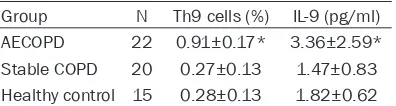
Abstract: Objective: To observe the changes of peripheral blood helper T cell 9 (Th9) and interleukin-9 (IL-9) in the stable and acute exacerbation phases of chronic obstructive pulmonary disease (COPD) so as to investigate its effects and clinical significance in the pathogenesis of COPD. Methods: A total of 42 patients (22 patients in the acute exacerbation chronic obstructive pulmonary disease (AECOPD) and 20 patients in the stable chronic obstruc-tive pulmonary disease (SCOPD) and 15 healthy controls were enrolled in this study to test the Th9 content by flow cytometry (FCM) and the serum IL-9 level by enzyme linked immunosorbent assay (ELISA). Results:The levels of Th9 and IL-9 in Group AECOPD were significantly higher than those in Group SCOPD (0.91±0.17 vs 0.27±0.13 and 0.28±0.13, P<0.05; 3.36±2.59 vs 1.47±0.83 and 1.82±0.62, P<0.05), and the IL-9 level and the Th9 ratio in Group AECOPD showed slight positive correlation (r=0.436, P<0.05). There was no significant difference in the IL-9 level and Th9 ratio in the AECOPD and SCOPD (P>0.05). Conclusions:The Th9 cells may be involved in the patho-genesis of AECOPD by secreting IL-9.
Keywords: Chronic obstructive pulmonary disease, Th9, IL-9, pulmonary function
Introduction
Chronic obstructive pulmonary disease (COPD) is a common disease characterized by
persis-tent airflow limitation. The degree of airflow
limitation is not completely reversible and pro-gressively develops, and its acute exacerbation and systemic complications may affect the severity and prognosis of the disease [1]. The morbidity and mortality of COPD increases year by year, and now it ranks the third place of glob-al death causes [2]; in 2020, COPD will become
the fifth of world’s disease-caused economic
burden. The pathogenesis of COPD has not been clear yet, and certain scholars thought that COPD may be a smoking triggered autoim-mune disease [3]. Previous studies have shown that COPD is characterized by the increase of macrophages, activated neutrophils, and lym-phocytes in the peripheral airway, lung paren-chyma, or pulmonary vessels. Some patients may also appear the increase of eosinophils, Th2 cells, or type 2 intrinsic lymphocytes (ILC2).
The above inflammatory cells, epithelial cells,
and other structural cells co-release a variety
of inflammatory mediators and thus are
invol-ved in the pathogenesis of COPD [4]. Different studies all consider that the immune system
plays an important role in the inflammatory
response and immune response of COPD. Stu-
dies aiming the specific immune function in
COPD patients mainly investigate the changes of CD3+T cells, CD4+T cells, CD8+T cells, and CD4+/CD8+T cells, and consider the decreased cell ratio of CD4+/CD8+T as an important indi-cator of immune function decrease. The helper T cells are a class of CD4+T cell subset that are
functionally classified according to different
cytokines they secrete. Studies targeting the helper T cells and COPD mainly concentrate on the balance disorder among the Th1/Th2, Th17, and regulatory T cells (Treg). The Th9 cells are a class of CD4+T lymphocyte subset newly dis-covered in recent years, and mainly secrete IL-9 thus participating in the processes of allergic
neutrophils. The Th9 cells and IL-9 may be involved in allergic asthma-caused chronic
air-way inflammation in patients with allergic asth -ma [7], and anti-IL-9 antibodies can reduce air-way hyperresponsiveness, goblet cell
metapla-sia, and other inflammatory responses in rat
allergic asthma model. Previously, IL-9 has been investigated the expression from the sputum and exhaled condensate gas in COPD rat model and COPD crowd. The IL-9 levels in smoking-induced COPD rat model, rat alveolar
lavage fluid, peripheral blood, and lung tissue are significantly higher than that in normal rats,
and positively correlated with the number of white blood cells, macrophages, or neutrophils [8]. Certain studies also reported higher IL-9 in the induced sputum of COPD patients, and pos-itively related to the macrophages and IL-8, so
IL-9 may be involved in the COPD airway inflam -mation and injury through macrophages [9, 10]. There is no report about the relationships of Th9 and IL-9 with COPD. The aim of this study is to investigate the changes of Th9 and IL-9 in the AECOPD and SCOPD patients so as to
explore their roles and clinical significance in
the pathogenesis of COPD. Materials and methods
General information
The study subjects were divided into AECOPD, SCOPD, and Control, and all selected from the inpatient or outpatient COPD patients from February to April 2017 in our hospital; the AECOPD and SCOPD met the standards of Global Initiative for Chronic Obstructive Lung Disease (GOLD) in 2017 [3] and had a history of
more than 10 years of smoking. Group SCOPD had 20 cases, including 18 males and 2
females, aging (75.3±9.17) years old, with sta -ble condition for 3 months or more; Group AECOPD had 22 cases, including 19 males and
3 females, aging (77.32±9.09) years old.
Mean-while, 15 healthy subjects were also selected as Group Control, including 11 males and 4
females, aging (72.5±6.04) years old, with no
history of respiratory infection for nearly 1 month. All the selected subjects should be excluded from the combination of allergic asth-ma, autoimmune diseases, blood system dis-eases, tumors, or recent application of hor-mones or immunosuppressive therapy. The age, sex, smoking history, previous history of illness, and other basic information of all the subjects were collected. This study was appro- ved by the Ethics Committee of Central South Hospital, and all the subjects signed the infor- med consent.
Comparison of Th9 cell ratio
The human peripheral blood mononuclear phocytes were extracted using the human lym-phocyte isolation solution (Sigma, St. Louis, USA), cultured in 1640 medium containing phorbol ester PMA (Sigma, St. Louis, USA), monensin (eBioscience, San Diego, USA), and ionomycin (Sigma, St. Louis, USA) for 5 hours; 5
μl of PE-C y7-CD3 (eBioscience, San Diego, USA)
and FITC-CD8a (eBioscience, San Diego, USA) were added, respectively, and the mixture was then incubated in darkness at 4°C for 30 min, followed by phosphate buffered saline (PBS) rinsing, two-time centrifugation, 30-min
incu-bation using one fix&perm kit at 4°C, and
two-time rinsing and centrifugation using the
fix&perm kit. 5 μl of PE-IL9 antibody (eBiosci -ence, San Diego, USA) was then added together with PE-IgG2a K as the isotype control for 30- min incubation at 4°C in darkness; after rinsed
using PBS, centrifuged twice, and fixed in 4%
paraformaldehyde, the mixture was then pre-served in darkness at 4°C for the detection.
The Th9 cells were defined as CD9+CD8-IL9+, and detected the ratio by FC500 flow cytometer
(Beckman, CA, USA). Comparison of IL-9
[image:2.612.92.288.83.138.2]4 ml of fasting peripheral blood was collected from each subject in the morning, and after centrifuged at 2000 r/min for 6 min, the upper Table 1. Three groups in general
Groups N General (m/f) Ages
AECOPD 22 19/3 77.32±9.09
Stable COPD 20 18/2 75.3±9.17 Healthy control 15 11/4 72.5±6.04
Table 2. Three groups Th9 cells and the level of IL-9 (_x±s)
[image:2.612.92.290.184.237.2]blood serum was tested the serum level of IL-9 by IL-9 ELISA kit (eBioscience, San Diego, USA). Statistical analysis
The general data such as sex, age, and smok-ing history of all the subjects were archived using EXCEL and analyzed using SPSS16.0.
The data were expressed as mean ± standard
deviation (_x±SD), and performed one-way
ANOVA after tested the homogeneity of vari-ance. The intragroup data correlation was ana-lyzed using the Pearson correlation analysis,
with P<0.05 considered as statistical
signifi-cance. Results
General information
There was no significant difference in the age
and sex among the three groups (P>0.05) (Table 1).
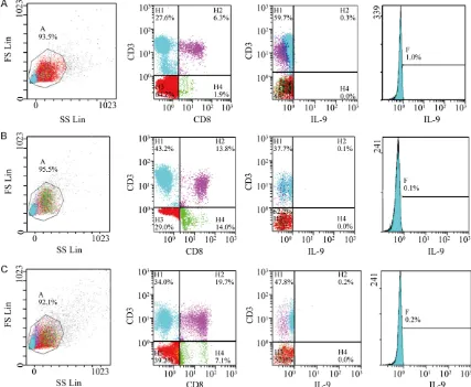
Comparison of Th9 cell ratio
The Th9 cell ratio in Group AECOPD was signifi -cantly higher than those in the other two groups
(P<0.05), while there was no significant differ -ence in the Th9 cell ratio between Group SCOPD and Control (P>0.05) (Table 2; Figure 1). Comparison of IL-9
The IL-9 level in Group AECOPD was significant -ly higher than those in the other two groups
(P<0.05). There was no significant difference in
the IL-9 level and Th9 cell ratio between Group COPD and Control (P>0.05) (Table 2; Figure 2). Correlation analysis
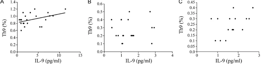
The IL-9 level and the Th9 cell ratio in Group
AECOPD were slightly correlated (r=0.436, P<
[image:3.612.91.518.76.427.2]0.05). In contrast, there were no relationship between the IL-9 level and the Th9 cell ratio in Group SCOPD and Control group (Figure 3).
Discussion
Chronic airway inflammation and emphysema
are the main pathological features of COPD,
and many immune cells and inflammatory fac -tors are involved in. Certain studies have shown that COPD patients exist immune dysfunction or disorder, and mainly manifest the imbalance of Th1/Th2 and Th17/Treg cells at the cellular
level. The Th1 and Th17 cells secrete proinflam
-matory cytokines, thus aggravating local inflam -matory response and causing airway injury; however, the decrease of Th2 and Treg cell
functions will cause the insufficient secretion of inhibitory inflammatory factors, thus resulting in proinflammatory - anti-inflammatory function imbalance, so it’s closely related to the severi -ty, disease progression, and prognosis of COPD. The Th9 cells are derived from the initial CD4+T
lymphocytes under the co-stimulation of TGF-β
and IL-4, which highly secrete IL-9, and IL-9 then combines with IL-9 receptor (IL-9R) and plays its various biological effects, such as par-ticipating in the pathogenesis of bronchial asth-ma, autoimmune diseases, or tumors. IL-9R is widely distributed in the lymphocytes, neutro-phils, respiratory epithelial cells and respiratory
smooth muscle cells. The Th9 cells and IL-9
level are significantly elevated in the peripheral
blood of patients with allergic asthma, and the apoptosis of neutrophils is negatively correlat-ed with the IL-9 level. Th9 and IL-9 may be involved in allergens caused chronic airway
inflammation in allergic asthma patients [11];
in rat allergic asthma model, anti-IL-9 antibod-ies can reduce airway hyperresponsiveness,
goblet cell metaplasia, and other inflammatory
responses. In the mouse model with experi-mental autoimmune encephalomyelitis, IL-9 or
its receptor deficiency is found to be associat -ed with the severity of this disease [12]. The results of this study show that the Th9 ratio and the IL-9 level in the peripheral blood of
Group AECOPD are significantly higher than
those in Group SCOPD and Control (Figure 2A, 2B), in contrast, the IL-9 level and the Th9 ratio
[image:4.612.92.521.73.210.2]in Group SCOPD show no statistical signifi -cance when compared with Control (Figure 2A, 2B). And the IL-9 level and the Th9 ratio in Group AECOPD show slight positive correlation (Figure 3A). However, there were no relation-ship between the IL-9 level and the Th9 cell ratio in Group SCOPD and Control group (Figure
Figure 2. Three groups the comparison of peripheral blood IL-9 levels and Th9 percentage comparison. Contents of peripheral blood Th9 and IL-9 AECOPD up from SCOPD group and Control group significantly (P<0.05).
[image:4.612.91.520.261.367.2]3B, 3C). Previous studies have found that the IL-9 level in the induced sputum of COPD
patients is significantly elevated, especially in
the cytoplasm of macrophages, and IL-9 may
be involved in COPD airway inflammation and
injury through the macrophages [13]. IL-9 can promote the proliferation of mast cells in immune response, induce the degranulation of mast cells, and release histamine, IL-13, or
IL-1β, thus participating in the body allergic and inflammatory responses [14]. In addition, IL-9
can also stimulate the secretion of IL-17 by stimulating the Th17 cells, thus enhancing
inflammatory responses and co-participating
in the pathogenesis of autoimmune diseases
[15]. The Th9 cells and IL-9 are significantly
increased in the peripheral blood of the patients in Group AECOPD, we speculate the possible mechanisms may be: AECOPD may
amplify chronic lung inflammation due to acute
respiratory infection, which may even develop
into systemic inflammatory response; under
the stimulation generated by micro-environ-mental cytokines, the Th9 cells can differenti-ate, proliferdifferenti-ate, and secrete IL-9, and IL-9 can
further participate in inflammatory response
and airway injury through combining with IL-9R (on the surface of macrophages) or directly through the macrophages. IL-9 can also stimu-late the neutrophils and the Th17 cells to
secrete proinflammatory cytokines, so that more inflammatory cells and cytokines may gather together, thus enhancing local inflam -matory response; when IL-9 stimulates the
mast cells to secrete TGF-β and VEGF, it can
promote airway remodeling and neovascular-ization; IL-9 can also promote the proliferation of the mast cells in the immune response and induce the degranulation of mast cells, release
histamine, IL-13, IL-1β, and other media, thus participating in the body allergic and inflamma -tory responses [13]. When IL-9 binds with the receptors on the surface of smooth muscle cells, it can cause airway smooth muscle con-traction; IL-9 can also stimulate the Th17 cells
to secrete IL-17, thus enhancing inflammatory
response and co-participating in the pathology of autoimmune diseases; when IL-9 acts on the airway epithelial cells, it can make it secrete excessive mucus [16]. Asthma-COPD overlap syndrome (ACOS) has both asthma and COPD-related characteristics. Some scholars have suggested that ACOS may be the overlap
air-way inflammation co-mediated by the eosino
-phils and neutro-phils. Combined with the re- sults of this study, we hypothesize that in such populations, IL-9 may be involved in
eosinophil-mediated inflammatory response by promoting
the maturation of eosinophils and inhibiting their apoptosis.
Currently, smoking is widely recognized as a risk factor for COPD, but only some smokers eventually develop into COPD, and airway
inflammation in most COPD patients continues
when they stop smoking; furthermore, the degree of cigarette exposure determines the anti-trypsin antibody concentration in COPD patients [17]. A multi-center longitudinal study in the US and Canada has shown that the decline rate of lung function in COPD patients who discontinue smoking is faster than before [18]. Nunez et al. [19] also found that the titers of anti-nuclear antibodies, anti-tissue antibod-ies, and anti-lung epithelial cell antibodies in nearly 1/3 of patients with stable COPD increa-
se, and 21% of COPD patients exhibit correla -tions of the titers of peripheral blood anti-tissue antibodies with airway limit severity and injury. Feghali-Bostwick et al. [20] reported that the IgG autoantibodies in COPD patients increase
and show high affinity to the lung epithelial
cells, so they may be involved in regulating the oxidative stress response, and the adaptive immune response of autoimmune response may be involved in the pathological processes of COPD. Autoimmune response may be invol- ved in the pathogenesis of COPD, and the Th9 cells and IL-9 are both upregulated in some autoimmune diseases; the results of this study show that the Th9 and IL-9 levels in Group
AECOPD are significantly increased than Group SCOPD and Control, and whether it’s involved
by Th9 and IL-9-mediated autoimmune respon- se still needs further studies.
The Th9 and IL-9 levels in Group SCOPD and
Control show no significant difference. AECOPD mainly appears systemic inflammation and
increased expression of IL-32 in the superna-tant of lung tissue than in the peripheral blood,
suggesting the existence of pulmonary inflam
-mation-based systemic inflammation in COPD, and local inflammation is more severe [21]. Combined with previous studies, we find that the peripheral blood-related to proinflammato -ry immune cells and cytokines in SCOPD are reduced than those in AECOPD, so we presume
confined to the lungs, and the lab indexes in the peripheral blood can’t fully respond to the real pulmonary inflammatory responses. The rela -tionships among various types of immune cells and cytokines are intertwined and complex,
and we’ll analyze whether the Th9 cells and IL-9 participate in the pulmonary chronic inflamma -tory process in SCOPD by multi-layer analysis,
such as bronchial lavage fluid, bronchoscopy,
or tissue biopsy, when certain conditions are adequate.
Patients with COPD generally suffer from im- mune system dysfunction or disorder, as well as imbalance of lymphocyte subsets and cyto-kine network, and these changes are directly or indirectly involved in pulmonary structural dam-age and disease evolution of COPD. The Th9 cells are one auxiliary T lymphocyte subset newly discovered in recent years, and their dif-ferentiation, development, and IL-9 secretion are affected by a variety of factors. This study suggests that the Th9 cells and IL-9 may be involved in the pathogenesis of AECOPD, while
the specific pathways and regulatory mecha
-nisms in this disease are not clarified yet. With
the deepening of immunological research,
more immune cells and inflammatory media -tors will be discovered. Exploring the mecha-nisms among these immune cells and cyto-kines in diseases, investigating disease-specif-ic biomarkers, and further studying methods to reconstruct the in vivo immune balance and to make the immune system play its normal immune defense function, as well as to block
or reduce inflammation, oxidative stress, etc.,
through the body immune functions and to effectively improve the prognosis and natural progress of COPD, will be our further investiga-tion steps and direcinvestiga-tions.
Acknowledgements
This project was supported by grants from Natural Science Foundation of Hubei Province of China (Grant number: 2016CFB680).
Disclosure of conflict of interest
None.
Address correspondence to: Xujun Ye, Department of Geriatrics, Zhongnan Hospital of Wuhan Univer- sity, Wuhan 430071, China. Tel: +86 27 67813449; Fax: +86 27 67812892; E-mail: cnxujunye@163. com
References
[1] Global initiative for chronic obstructive lung disease. Global strategy for the diagnosis, management, and prevention of chronic ob-structive pulmonary disease 2017 report [EB/ OL]. (2016-11-16) [2016-12-09]. http://www. goldcopd.org.
[2] WHO. The top 10 causes of death. May, 2014. http://www.Who.Int/mediacentre/factsheets/ fs310/en/.
[3] Cosio MG, Saetta M and Agusti A. Immunologic aspects of chronic obstructive pulmonary dis-ease. N Engl J Med 2009; 360: 2445-2454. [4] Barnes PJ. Inflammatory mechanisms in pa
-tients with chronic obstructive pulmonary dis-ease. J Allergy Clin Immunol 2016; 138: 16-27. [5] Sehra S, Yao W, Nguyen ET, Glosson-Byers NL,
Akhtar N, Zhou B and Kaplan MH. TH9 cells are required for tissue mast cell accumulation during allergic inflammation. J Allergy Clin Im -munol 2015; 136: 433-440.
[6] Wang Y, Bi Y, Chen X, Li C, Li Y, Zhang Z, Wang J, Lu Y, Yu Q, Su H, Yang H and Liu G. Histone deacetylase SIRT1 negatively regulates the dif-ferentiation of interleukin-9-producing CD4(+) T cells. Immunity 2016; 44: 1337-1349. [7] Koch S, Graser A, Mirzakhani H, Zimmermann
T, Melichar VO, Wölfel M, Croteau-Chonka DC, Raby BA, Weiss ST and Finotto S. Increased ex-pression of nuclear factor of activated T cells 1 drives IL-9-mediated allergic asthma. J Allergy Clin Immunol 2016; 137: 1898-1902, e7. [8] Zhang M, Xie Y, Yan R, Shan H, Tang J, Cai Y, Yin
J, Chen M, Zhang J, Yang X, Zhang Q and Li Y. Curcumin ameliorates alveolar epithelial injury in a rat model of chronic obstructive pulmo-nary disease. Life Sci 2016; 164: 1-8.
[9] Nguyen HQ, Herting JR, Pike KC, Gharib SA, Matute-Bello G, Borson S, Kohen R, Adams SG and Fan VS. Symptom profiles and inflamma -tory markers in moderate to severe COPD. BMC Pulm Med 2016; 16: 173.
[10] Jin Y, Wan Y, Chen G, Chen L, Zhang MQ, Deng L, Zhang JC, Xiong XZ and Xin JB. Treg/IL-17 ratio and Treg differentiation in patients with COPD. PLoS One 2014; 9: e111044.
[11] Kaplan MH, Hufford MM and Olson MR. The Development and in vivo function of TH9 cells. Nat Rev Immunol 2015; 15: 295-307.
[12] Hoppenot D, Malakauskas K, Lavinskienė S, Bajoriūnienė I, Kalinauskaitė V and Sakalaus -kas R. Peripheral blood Th9 cells and eosino-phil apoptosis in asthma patients. Medicina (Kaunas) 2015; 51: 10-17.
[14] Koch S, Sopel N and Finotto S. Th9 and other IL-9-producing cells in allergic asthma. Semin Immunopathol 2017; 9: 55-68.
[15] Elyaman W, Bradshaw EM, Uyttenhove C, Dar-dalhon V, Awasthi A, Imitola J, Bettelli E, Oukka M, van Snick J, Renauld JC, Kuchroo VK and Khoury SJ. IL-9 induces differentiation of TH17 cells and enhances function of FoxP3+ natural regulatory T cells. Proc Natl Acad Sci U S A 2009; 106: 12885-12890.
[16] Singh TP, Schön MP, Wallbrecht K, Gruber-Wackernagel A, Wang XJ and Wolf P. Involve-ment of IL-9 in Th17-associated inflammation and angiogenesis of psoriasis. PLoS One 2013; 8: e51752.
[17] Alam S, Li Z, Atkinson C, Jonigk D, Jan-ciauskiene S and Mahadeva R. Zα1-antitrypsin confers a proinflammatory phenotype that con -tributes to chronic obstructive pulmonary dis-ease. Am J Respir Crit Care Med 2014; 189: 909-931.
[18] Anthonisen NR, Connett JE and Murray RP. Smoking and lung function of lung health study participants after 11 years. Am J Respir Crit Care Med 2002; 166: 675-679.
[19] Núñez B, Sauleda J, Antó JM, Julià MR, Orozco M, Monsó E, Noguera A, Gómez FP, Garcia-Aymerich J and Agustí A. Anti-tissue antibodies are related to lung function in chronic obstruc-tive pulmonary disease. Am J Respir Crit Care Med 2011; 183: 1025-1031.
[20] Feghali-Bostwick CA, Gadgil AS, Otterbein LE, Pilewski JM, Stoner MW, Csizmadia E, Zhang Y, Sciurba FC and Duncan SR. Autoantibodies in patients with chronic obstructive pulmonary disease. Am J Respir Crit Care Med 2008; 177: 156-163.