Original Article
Tailoring the binding properties of SpA
Ig binding domains by
in vitro
molecular evolution
Lili Wang1*, Yingying Ding1*, Ziyu Lin2*, Jie Bai1, Chunyan Zhao1, Caixia Gao1, Jiaojiao Feng1, Jinhong Wang1,
Bing Rui1, Xiangyu Li1, Hao Wu3, Baohua Qian4, Wei Pan1
1Department of Medical Microbiology and Parasitology, School of Basic Medicine, Second Military Medical
Univer-sity, Shanghai City, China; 2Department of Pathology, The Second People’s Hospital of Lu’an City, Anhui Province,
China; 3Department of Infectious Diseases, Beijing Youan Hospital, Capital Medical University, Beijing City, China; 4Department of Blood Transfusion, Changhai Hospital, Second Military Medical University, Shanghai City, China. *Equal contributors and co-first authors.
Received July 12, 2017; Accepted August 22, 2017; Epub October 15, 2017; Published October 30, 2017
Abstract: Background: Staphylococcus aureus protein A (SpA) is a bacterial immunoglobulin (Ig)-binding protein (IBP) and has fundamental applications in medical and biological sciences associated with IgG. In addition to its
high affinity for IgG Fc, SpA also has a low affinity for the VH3 regions of IgG, IgM and IgA Fab, which may complicate its IgG applications. Methods: To diminish its VH3 binding potential and preserve the Fc binding potential, the amino acids which are involved in the interaction with VH3 at positions 29 and 30 in SpA A domain and at positions 36
and 37 in SpA C domain were randomly mutated respectively, meanwhile, a combinatorial phage library displaying randomly-rearranged mutated A and C domains of SpA was constructed. Then, a combination of AL29I30-AV29K30was generated by in vitro molecular evolution using human IgG as bait. Results: The binding assays demonstrated that comparable to that of the its equivalent (A-A), AL29I30-AV29K30 exhibited affinity for IgG, but diminished affinity for IgM
and IgA. Horse-radish peroxidase (HRP) conjugated AL29I30-AV29K30 presented enhanced IgG binding potential and
improved effects for antibody detection against HIV core antigen in serum. Further, AL29I30-AV29K30 affinity chromatog
-raphy recovered purified IgG in amount comparable to that for SpA but recovered no detectable IgM and IgA, thus
demonstrating the former’s application advantages. Conclusions: This study demonstrates a successful example of functional protein engineering via in vitro molecular evolution and provides a useful approach to remold the Ig binding property of SpA for application purposes.
Keywords: NEIBM, IgG, phage-based molecular evolution, antibody detection, antibody purification
Introduction
Bacterial immunoglobulin (Ig)-binding proteins
(IBPs) specifically bind to immunoglobulins (Igs)
and play important roles in the pathogenicity of bacteria [1]. Protein A of Staphylococcus aure-us (SpA) [2], protein L of Finegoldia magna, for-merly Peptostreptococcus magnus (PpL) [3], and protein G of groups C, G streptococci (SpG)
[4], are three well-defined IBPs. Among them,
SpA with a molecular weight of 57 kDa com- prises 524 amino acid residues [5]. SpA has a
tandem repeat sequence that containing five
highly homologous IgG-binding domains, which named (from the N terminus) E, D, A, B and C. Each domain comprises of approximately 58 amino acid residues with a characteristic sec-ondary structure consisting of three up-down
α-helixes that mediate the binding with IgG
Fc region [6]. In addition, SpA also binds to a fraction of Ig molecules other than IgG Fc, for
instance, the VH3 heavy chains in Fab region of
Igs [7-9]. Crystal structure studies have shown that SpA B domain binding with Fc mainly involves residues in helix I and less involves in helix II [10, 11], whereas D domain interacting
with VH3 of Fab involves residues in helix II and
helix III [5].
SpA and other IBPs have fundamental applica-tions in medical and biological area, e.g.
diag-nostic antibody detection, antibody purification,
research reagents. SpA-based purification, as a key purification step, is the main part of the
raw-material costs [16, 17].In addition to the
specific interaction with the Fc region of IgG during antibody purification, SpA may also bind to the Fab domain of the VH3-clan of IgG, IgM and IgA. Therefore, this VH3 binding potential might disturb the purification of recombinant
monoclonal antibodies and lead to an
inaccu-rate specific detection of the IgG antibody
responses against pathogens by false detec-tion of the IgM antibodies. Developing SpA-derived IgG binding proteins with preserved IgG
Fc binding potential and diminished VH3 bind -ing potential remains an interest-ing research topic.
Numerous novel combinations of Ig-binding domains of SpA, SpG and PpL that do not exist in natural bacterial IBPs were generated by in vitro molecular evolution. These molecules are named as newly evolved Ig-binding molecules (NEIBM) and exhibit novel Ig-binding properties [18-22]. For example, NEIBM LD5 and LD3 not only could interact with human Ig Fab in the
VH3 and Vκ regions via a double-binding me-thod, but also show a high affinity for human
IgM [19]. Additionally, the horse-radish peroxi-dase (HRP) conjugated LD5 has better IgM detection sensitivity in the ELISA assay for
HCV and improves antibody detection in anti-HIV and anti-EV71 VP1 ELISA assays [20, 23,
24]. Additionally, NEIBM D-C-G3, with potential simultaneous tri-site binding for some IgG sub-classes, is generated through in vitro molecular evolution as well [21].
In the present study, the amino acids which are
involved in the interaction with VH3 at positions
29 and 30 of SpA A domain and those at posi-tions 36 and 37 of SpA C domain, were random-ly mutated, and a combinatorial phage library displaying randomly-rearranged mutated A and C domains of SpA was constructed. A NEIBM combination, AL29I30-AV29K30, was obtained by in vitro molecular evolution using human IgG as bait, which preserved the IgG Fc binding poten-tial and diminished the IgM and IgA binding potential.
Materials and methods
Ethical approval
All aspects of the study on the forty
anti-HIV-positive human serum samples were approved
by the Ethics Committee of Beijing You An Hospital, Capital Medical University, China; the written informed consent was provided by all subjects in the study [23]. And all aspects of
the study on the forty anti-HIV-negative human
serum samples were approved by the Ethics Committee of Changhai Hospital, Shanghai, China; oral informed consent was provided by all subjects in the study.
Vectors, reagents and serum samples
The phagemid vector pCANTAB5S was con-structed in advance in our laboratory. The pro-karyotic expression plasmid, pET-32a (+), and
E. coli host strains Top10, were purchased from Novagen Company (Darmstadt, Germany), and
E. coli TG1 was obtained from Stratagene Com- pany (Cambridge, England). All the primers in this study were synthesized by the Sangon Biotech (Shanghai, China). The horseradish pe- roxidase (HRP)-conjugated anti-M13 antibody
and helper phage M13K07 were obtained from
Pharmacia Biotech (Uppsala, Sweden). Human IgG (hIgG), human IgM (hIgM) and human IgA (hIgA) were purchased from Sigma (St. Louis, MO, USA).
Forty anti-HIV-positive human serum samples
were collected from the AIDS high-risk cohort at YouAn Hospital in Beijing, China [23]. Forty
anti-HIV-negative human serum samples were
collected from healthy blood donors at Chang- hai Hospital, Shanghai, China. Upon receipt, all samples were aliquoted and stored at -80°C. Referring to the related study, the seropositive
status of the subjects was confirmed using Western blotting (HIV Blot 2.2 WB, MP Bio-medicals Asia Pacific Pte. Ltd., Singapore) and ELISA (Diagnostic Kit for Antibody to HIV (ELI-SA), Shanghai Kehua Bio-Engineering Co., LTD.,
China) [23].
Construction of combinatorial phage libraries
mutat-ed phage library was constructmutat-ed by digesting the PCR products with Xba I and ligating into the phagemid pCANTAB5S on the Xba I site (library 1, Table 1).
Gene fragments with similarly Xba I restriction sites that encode the SpA A and C domains were respectively synthesized by PCR with primers listed in Table S2. Then, the combinato-rial phage library displaying various randomly-rearranged A and C domains was built with above mentioned method (library 2, Table 1).
After amplification in E. coli Top10, the recombi-nant phagemids were transformed into the host bacterial strain TG1. Helper phages M13-
K07 (approximately 1.8*1011 transformation
unit (TU)) were added when the optical density (OD) value at 600 nm was about 0.5, and cul-tured in the shaker at 230 rpm for 1 h at
37°C. Subsequently, kanamycin (15 μg/ml)
was added and the cells kept culturing over-night. After the centrifugation at 5,000 g for 10 min, the supernatant was collected and the
phages were harvested by a 0.22 μm filter.
Then, the combinatorial phage display libraries displaying the randomly-rearranged A and C domains of SpA were obtained (Table 1). The primer pairs pCANTAB5S-1 (5’-CAACGTGAAAA- AATTATTATTCGC-3’) and pCANTAB5S-6 (5’-GTA- AATGAATTTTCTGTATGAGG-3’) were applied for
the amplification and sequencing analysis of
inserted fragments for the positive phages [22].
In vitro molecular evolution of the libraries by hIgG molecule
Dilute the hIgG to the concentration of 10
μg/ml in 0.1 M NaHCO3 (pH9.6), then add to 96-well ELISA plates for incubating at 37°C for 3 h. Then, block the plates with 0.01 M phos-phate buffered saline (PBS) containing 10% of skimmed milk, 0.1% of Tween 20 and 0.2% of
mercurothiolate (blocking buffer) for 3 h. Add the constructed phage-displaying libraries into the plates and incubate at 37°C for 2 h. Wash each well with PBS containing 0.1% of Tween 20 for 10 times to remove the unbound phag-es. When the OD at 600 nm was about 0.5,
100 μl of the E. coli TG1 was added and the plates were incubated at 37°C for 1 h. Take 10 ml infected TG1 cells to culture in tryptone-yeast extract (2X) plates containing ampicillin
(100 μg/ml) and then, calculate the number of
colony forming units. After growing with helper
phages M13K07 (approximately 1.8*1011 TU)
overnight, the residual cells were cultured in 2X tryptone-yeast extract medium (8 ml)
contain-ing ampicillin (100 μg/ml). After the centrifuga -tion at 5,000 g for 10 min, the supernatant was collected and the phages were harvested by a
0.22 μm filter for the next selection round with
the hIgG as well. The selection was performed 3-6 rounds totally.
A total of 22 phage clones that randomly picked from each primary library and each round of the post-selection libraries were cultured with 2X tryptone-yeast extract medium (1 ml) at 37°C overnight. The primers pCANTAB5S-1 and
pCANTAB5S-6 were applied for PCR amplifica -tion and analysis of the inserted fragments of phages. The empty plasmid pCANTAB5S was used as negative control for the evolution [22].
Phage ELISA test
Screening the hIgG binding phages in post-selection libraries by ELISA: The recombinant phagemids in the post-selection library 1 and library 2 were transformed into the E. coli TG1, respectively. Subsequently, select ninety clones from each library randomly and then cul-ture them by shaking at 230 rpm at 37°C
over-night. With the helper phages M13K07 (about
1.8*1011 TU), monoclonal phages were
select-ed out from the last round from each
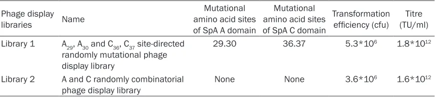
post-Table 1. The names, mutational sites and evaluation of the combinatorial phage libraries
Phage display
libraries Name
Mutational amino acid sites of SpA A domain
Mutational amino acid sites of SpA C domain
Transformation
efficiency (cfu) (TU/ml)Titre Library 1 A29, A30 and C36, C37 site-directed
randomly mutational phage display library
29.30 36.37 5.3*106 1.8*1012
Library 2 A and C randomly combinatorial
phage display library None None 3.6*10
[image:3.612.92.522.84.181.2]selection library and added to plates after
coating with 10 μg/ml of hIgG as mentioned
above, and the plates were incubated at 37°C for 2 h. After washing with the solution conta- ining 0.25% of Tris, 0.05% of Tween 20, HRP-conjugated anti-M13 phage antibody was ap- plied to detect the bound phages. Then, 3, 3’, 5, 5’-tetramethylbenzidine (TMB) (Sigma, St. Lou- is, MO, USA) and H2O2 were adopted as the substrate for HRP, and the absorbance at 450 nm was measured with an ELISA Reader. Pla- smid pCANTAB5S-phage was applied as a neg-ative control [22].
Sequence analyses: A total of 10 positive phage clones with the highest OD450nm were sequenced with the primers pCANTAB5S-1 and pCANTAB5S-6 by JIE LI Biology Company (Shanghai China). The sequences of target amino acid were deduced on the basis of DNA sequencing result, and the DNASTAR software package was adopt of the multiple sequence alignment analysis.
Prokaryotic expression, purification, and bind-ing analysis of AL29I30-AV29K30 and A-A
Expression and purification of AL29I30-AV29K30 and A-A: With the primer pairs U-AA-BamH and D-AA-Sal, the representative phagemids, including AL29I30-AV29K30 and A-A, were used as templates to amplify DNA fragments by PCR using primers in Table S3 respectively. After the
amplification, the target DNA was inserted into
the plasmid pET-32a (+) and the result was examined via DNA sequencing. The proteins expression of AL29I30-AV29K30 and A-A was medi-ated by isopropyl-beta-D-thiogalactopyranoside (1 mmol/L) in E. coli BL21 (DE3). After sonica-tion for 30 min, the proteins were collected
and purified by a Ni-NTA column (Amersham
Pharmacia Biotech).
ELISA test: The ELISA test was conducted as
previously described [22]. Briefly, the immuno -assay strips (Nunc, Rochester, NY, USA) were
coated with the purified AL29I30-AV29K30 and A-A at
a concentration of 1 μg per well that diluted by
0.1M NaHCO3 (pH9.6) and incubated at 37°C for 3h. After blocking with blocking buffer for 3h, a fold-dilution of biotin-labeled, IgG, IgM and IgA (0.1 mg/ml) was added and incubated at 37°C for 45 min respectively. The reactive compounds were detected with HRP-conjugat- ed streptavidin (2 µg/ml, Sigma, St. Louis, MO,
USA), and the absorbance at 450 nm was mea-sured with an ELISA Reader.
Application of AL29I30-AV29K30 and A-A in anti-HIV ELISA test
HRP labeling: Using sodium periodate, HRP-labelled AL29I30-AV29K30 (HRP-AL29I30-AV29K30) and HRP-labelled A-A (HRP-A-A) were prepared as
follows [22]. Briefly, a mixture with 5 mg/ml of HRP and 200 μl of 0.1 M sodium periodate
was stirred about 20 min in the dark surround-ing and dialyzed against 1 mM sodium acetate buffer (pH4.4) at 4°C overnight. Then 1 ml of 5 mg/ml AL29I30-AV29K30 and A-A in PBS (pH7.2) was added immediately after adding 20 ul of
0.2 M carbonate buffer (pH9.5). Keep gently
stirring for 2 h in the dark surrounding, 100 ul of sodium borohydride solution (4 mg/ml) was added and reacted for 3h at 4°C. Finally, the HRP-AL29I30-AV29K30 and HRP-A-A were dialyzed against 0.01M PBS (pH7.2) at 4°C overnight and stored at -20°C [22].
Test of binding activity of HRP-AL29I30-AV29K30 and HRP-A-A: The ELISA plates were coated with
hIgG, hIgM and hIgA respectively (1 μg per well)
using 0.1 M NaHCO3 (pH9.6) and were incubat-ed at 37°C for 3 h. After blocking with skimmincubat-ed milk, 100 µl of serial double-diluting solutions of HRP-AL29I30-AV29K30 and HRP-A-A (1 mg/ml) were added to each well which was then incu-bated at 37°C for 45 min. The plates were developed by TMB and the absorbance at 450 nm was measured by ELISA Reader.
Detection of anti-HIV antibody: To detect the
anti-HIV antibody using HRP-AL29I30-AV29K30 and HRP-A-A, the immunoassay strips were coated
with 1 ug of the recombinant HIV core antigen (CA, GenBank accession No. AAC82593.1). HIV
CA was inserted into the cloning sites of a pET-32a (+) vector under the control of the T7 pro-moter. A His tag was added at the N-terminus of the target protein to form a fusion protein. The strips were incubated at 37°C for 3 h, and
then blocked as above. Then, 100 μl of 10-fold dilutions of the forty anti-HIV-positive human
serum samples were added for binding reac-tion at 37°C for 45 min. Meanwhile, the forty
anti-HIV-negative human serum samples were
set as negative control. After washing four
times with washing buffer, 100 μl of a
37°C for 45 min. The strips were developed with TMB and the absorbance at 450 nm was measured by ELISA Reader.
Biosensor analyses
The binding properties of AL29I30-AV29K30 and A-A to hIgG, hIgM, and hIgA were studied by sur-face plasmon resonance (SPR) using the Bio-Rad XPR36 protein interaction array system.
Briefly, hIgG, hIgM and hIgA (diluted in 10 mM
sodium acetate, pH4.5) were coupled to a GLC ProteOn sensor chip using amine-coupling chemistry according to the manufacturer’s instructions. The association and disassocia-tion condidisassocia-tion were measured with serial 1:5 dilutions of AL29I30-AV29K30 and A-A with the con-centrations of 1.6 nM, 8 nM, 40 nM, 200 nM,
1 µM and 5 µM. The flow rate was set as 100
µl/min using PBST (pH7.4, 0.005% Tween 20)
as flow buffer. The sensor-chip surfaces were
regenerated by using 10 mM Glycine-HCl (pH-
2.0). KA (affinity constant) = ka (association
rate constant)/kd (dissociation rate constant) [21].
Affinity chromatography
The AL29I30-AV29K30 coupled-column was made by immobilizing 30 mg of the AL29I30-AV29K30 fusion protein onto 3 ml sepharose column (Amers- ham, Uppsala, Sweden) according to the manu-facturer’s instructions. Then, 2 ml of human serum was diluted in PBS (1:8) and then appli-
ed on the 3 ml AL29I30-AV29K30 coupled-column or SpA coupled-column (Amersham Pharmacia Biotech AB, Uppsala, Sweden) respectively at room temperature. Subsequently, wash the col-umns with extensive PBS (pH7.0), elute the bound proteins using 100 mM sodium acetate (pH3.0) and collect the approximately 4 ml of the eluted proteins respectively, and dialyze the proteins against PBS (pH7.0). Ten-microliter of the eluted proteins were analyzed by 12% of SDS-PAGE using the purchased hIgG, hIgM, and hIgA as control [21].
Data analysis
All the experiments were repeat no less than three times in each triplicate (n = 9) and per -formed independently. The data were analyzed
with one-way ANOVA or Student’s t-test. P value
< 0.05 was considered significant.
Results
In vitro molecular evolution of the combinato-rial phage library displaying randomly-rear-ranged mutated A and C domains of SpA
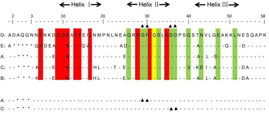
To eliminate the VH3 binding activity, the amino
acids at positions 29 and 30 of SpA A domain and the amino acids at positions 36 and 37 of
SpA C domain, which both interact with VH3 of
[image:5.612.94.523.73.256.2]Igs, were randomly mutated (Figure 1) [22]. A combinatorial phage library displaying the randomly-rearranged mutated A and C domains Figure 1. Alignment of the amino acid sequences of the five SpA domains and the mutation sites of SpA A domain
of SpA (library 1) was constructed and subject-ed to in vitro molecular evolution with the bait hIgG. As a control, the combinatorial phage library displaying randomly-rearranged A and C domains of SpA without any mutation (library 2) was also constructed and subjected to in vitro
molecular evolution as well. The library 1 had 5.3*106 members, and the titre of the phage
library was 1.8*1012 TU/ml (Table 1). The library
2 had 3.6*106 members, and the titre of the
phage library was 1.6*1012 TU/ml (Table 1).
The capacity of the two established libraries
satisfied the needs of the subsequent in vitro
molecular evolution.
To check the randomness of nucleotides in the mutation sites in A and C domain of SpA from library 1, eighty clones from the original library 1 were randomly selected and primers pCANTAB5S-1 and pCANTAB5S-6 was adopted
for the PCR amplification of the inserted frag -ments of phages [22]. The results are as fol-lows: there were three phage clones displaying three domains, twenty phage clones displaying two domains, forty-six phage clones displaying one domain and eleven phage clones with no inserted fragment. Among them, then twenty phage clones displaying two domains were cho-sen for sequencing analysis. The rearrange-ments of A and C domain of SpA in the twenty phage clones were as follows: three A-A com- binations, two AR-A combinations (R refers to
reverse complementary sequence), two A-AR,
two C-CR combinations, two CR-CR
combina-tions, two A-C combinacombina-tions, two A-CR
combina-tions, two AR-C combinations, one CR-C
combi-nation, one C-AR combination and one CR-AR
combination. Hence, the twenty phage clones included 22 A domains and 18 C domains. In
the A domain, the sequence analyses indi- cating that the A:T:C:G ratio in the NNS muta-tions of the amino acids at both posimuta-tions 29
and 30 was 11:10:11:12 for the first base N,
10:13:11:10 for the second base N, and the C:G ratio was 21:23 in the third base S. In the C domain, the A:T:C:G ratio in the NNS mutations of the amino acids at both positions 36 and 37
was 9:8:9:10 for the first base N, 7:8:11:10 for
the second base N, and the C:G ratio for the NNS mutation was 19:17 in the third base S. In summary, the variety and randomness of the
library 1 satisfied the needs of the subsequent
in vitro molecular evolution.
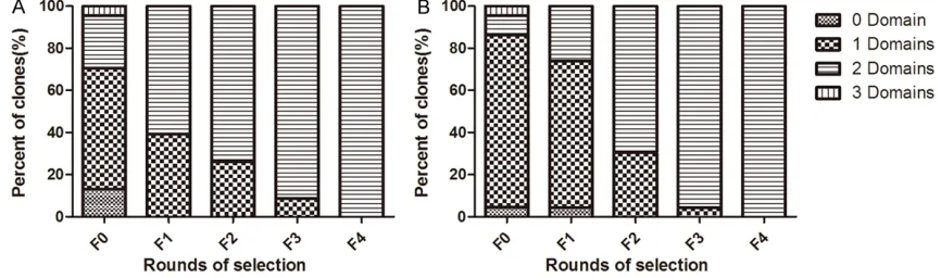
As observed in the previous studies [18, 22], the distribution of the inserted fragment sizes showed remarkable change throughout the in vitro evolution of both libraries in this study (Figure 2), which indicated an effective evolu-tion. As a result, the ratio of phage clones dis-playing two domains was less than 30% in the original library, and increased to 100% during four rounds of selection of both libraries.
Analysis of IgG binding activity of phage clones in the post-selection populations
[image:6.612.89.522.75.203.2]of AL29I30-AV29K30, and all ten phage clones from the library 2 also showed the same NEIBM com-bination of A-A.
The Ig binding properties of NEIBM AL29I30 -AV29K30
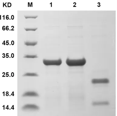
AL29I30-AV29K30 and A-A were expressed and
puri-fied as fusion proteins using pET-32a (+)
ex-pression vector to compare their Ig binding activities (Figure 4). ELISA analysis showed that AL29I30-AV29K30 and A-A exhibited comparable binding activities with hIgG. In contrast to the remarkable binding activities of A-A with hIgM and hIgA, AL29I30-AV29K30 presented no hIgM and
hIgA binding activities (Figure 5). Consistent with these results, the SPR data (Table 2) also demonstrated that AL29I30-AV29K30 showed hIgG binding potential which was comparable to that of A-A but its hIgM and hIgA binding potential was diminished.
Improved hIgG binding potential and anti-HIV detection effect of HRP-AL29I30-AV29K30
The conjugates of HRP-AL29I30-AV29K30 and HRP-A-A were produced, and their binding activities with hIgG, hIgM, and hIgA were compared. To our surprise, HRP-AL29I30-AV29K30 exhibited signif-icantly enhanced binding activities with hIgG compared with those of HRP-A-A (Figure 6). Then, we compared the detection effects of the HRP-AL29I30-AV29K30 and HRP-A-A in a panel
comprising of forty anti-HIV-positive human serum samples from HIV patients and forty anti-HIV-negative serum samples from healthy
blood donors, respectively. As shown in Figure 7, both HRP-AL29I30-AV29K30 and HRP-A-A showed the same detection effects for negative serum samples, whereas the HRP-AL29I30-AV29K30-based
assay presented significantly improved detec -tion effects for positive serum samples com-pared with HRP-A-A (P < 0.001).
AL29I30-AV29K30 affinity chromatography recov-ered pure IgG from human serum
To investigate whether AL29I30-AV29K30 has an
[image:7.612.93.516.71.223.2]application advantage in IgG purification, the purification efficiency of affinity chromatogra -phy columns made from AL29I30-AV29K30 or SpA was compared. As shown in Figure 8, the AL29I30 -AV29K30 affinity column recovered the compara -Figure 3. Detection of IgG binding activities of the phages clones in post-selection population through phage ELISA. (A): Library 1; (B): Library 2.
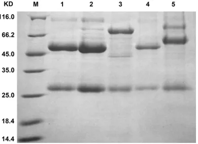
[image:7.612.97.290.275.467.2]ble amount of IgG antibodies from human
serum as that recovered by SpA affinity chro -matography. Additionally, no IgM and IgA anti-bodies were recovered by the AL29I30-AV29K30
affinity column, whereas they were recovered by SpA affinity chromatography. The data indi -cated that AL29I30-AV29K30 had the obvious
advan-tage in IgG purification.
Discussion
SpA, which is a natural Igs binding protein, especially for IgG, has fundamental applica-tions in IgG antibody diagnostic detection,
anti-body purification, immunoprecipitation assays
and immunoadsorption therapy. Crystal studies have shown that each SpA binding domain has two different binding interfaces: one is the Fc binding interface, which is located in helix I and helix II and interacts with one of Fc sites in two
IgG heavy chains; the other one is the VH3 Fab
binding interface, which is located in helix II and
helix III and interacts with the VH3 chain in one
Fab of IgG, IgM and IgA [10, 11]. Meanwhile,
SpA contains a tandem repeat of five highly
homologous Ig-binding domains and thus pres-ents a number of binding avidity models by
using some of the five Fc binding interfaces and the five VH3 binding interfaces. Among
these potential binding models, the two-site Fc binding mode (Fc-Fc, two Fc binding interfaces from two Ig-binding domains of SpA simultane-ously bind with two Fc binding sites on one IgG molecule) is the key binding model for IgG
and is utilized for the purification and specific detection of IgG. The VH3 binding interfaces may produce a low affinity for Fab of IgG, IgM
and IgA, and may thus complicate the
applica-tions that are specific for IgG. For example,
when a natural SpA chromatography was used
for the purification of recombinant IgG cons-isting of VH3, besides the Fc-Fc binding model,
it provided a number of other binding models
for a single IgG molecule, such as Fc, VH3-Fc-Fc, VH3-Fc-Fc-VH3, VH3-VH3 and
VH3-VH3-Fc. Namely, these models could recover IgG with different conformations along with that recovered by the Fc-Fc binding model, to gener-ate heterologous IgG conformations, and thus
to complicate IgG purification. Based on this consideration, the residues involved in VH3
binding, at positions 29 and 30 of SpA A domain and at positions 36 and 37 of the C domain, were chosen to randomly mutate to diminish
the VH3 binding potential and preserve the Fc
binding potential.
[image:8.612.95.522.71.181.2]In theory, proper combinations of the two SpA binding domains are necessary to produce the Fc-Fc binding avidity, and all four types of com-binations, A-A, A-C, C-C and C-A, may produce Figure 5. Binding activities of AL29I30-AV29K30 to hIgG, hIgM and hIgA compared to A-A according to ELISA analysis. The
plates were coated with purified AL29I30-AV29K30 and A-A, and 1:2 serial dilutions of biotin-labeled hIgG, hIgM and hIgA were incubated in the wells. The reactive complexes were detected using horseradish peroxidase (HRP)-conjugated streptavidin.
Table 2. Surface plasmon resonance analysis of the interactions between AL29I30-AV29K30 or A-A and IgG, IgM or IgA
Analyte Ligand Constant AL29I30-AV29K30 A-A IgG ka (M-1s-1) 1.00*105 1.53*105
kd (s-1) 2.86*10-4 3.61*10-4
KA (M-1) 3.50*108 4.24*108
IgM ka (M-1s-1) 6.56*10-3 1.71*103
kd (s-1) 5.91*10-5 1.99*10-5
KA(M-1) 1.11*102 8.59*107
IgA ka (M-1s-1) 10.8 1.08*104
kd (s-1) 1.51 2.34*10-4
[image:8.612.89.288.286.433.2]Fc-Fc binding avidity. To our surprise, in our study, only the A-A combinations (AL29I30-AV29K30 and A-A) were selected by in vitro molecular evolution of two phage libraries, which indicat-ed that the A-A combinations possess some advantages in generating the perfect Fc-Fc binding avidity compared to other combina-tions. Interestingly, only the mutated A-A combi-nation, AL29I30-AV29K30, was selected from the phage library displaying the randomly-rear-ranged mutated A and C domain, which should contain the A-A combination, indicating that the
mutations at positions 29 and 30 of AL29I30 -AV29K30 should favor Fc binding. In the present study, our strategy of in vitro molecular evolu-tion only guaranteed the selecevolu-tion of phage clones with the strongest Fc binding potential but did not guarantee the selection of phage
clones with eliminated VH3 binding potential. Hence, the elimination of VH3 binding
poten-tial of AL29I30-AV29K30 is not the result of in vitro
molecular evolution but is an incidental conse-quence of the mutations that favor IgG bind-
ing. A fine-resolution map of the sequence
[image:9.612.97.522.71.194.2] [image:9.612.93.282.253.437.2]function landscape of computational designed IgG binding protein FcB6 revealed that substi-tution of any of the core residues involving Fc binding are usually depleted and that most of the substitutions of any of the other residues are allowed or not allowed, part of them are depleted, and only few are favored [25]. In this Figure 6. Detection of the binding activities of HRP-AL29I30-AV29K30 and HRP-A-A with hIgG, hIgM and hIgA. The plates were coated with hIgG, hIgM and hIgA. 1:2 serial dilutions of AL29I30-AV29K30 and HRP-A-A were incubated in each well. Binding was detected by the addition of TMB.
Figure 7. Comparison of the detection effects of HRP-AL29I30-AV29K30 and HRP-A-A in anti-HIV ELISA. The strips were coated with HIV core antigen recom -binant protein. One hundred-microliter of 10-fold
dilutions of the forty anti-HIV-positive human serum
samples and forty anti-HIV-negative human serum
samples were separately added. One hundred-micro-liter of 1000-fold dilutions of HRP-AL29I30-AV29K30 and HRP-A-A were added, and the plates were incubated. The strips were developed upon the addition of TMB and detected at 450 nm on an ELISA Reader.
Figure 8. Comparison of IgG purification effects of
human serum with AL29I30-AV29K30 and SpA affinity
chromatography through SDS-PAGE analysis. M: pro-tein molecular weight marker; lane 1: human serum
[image:9.612.329.524.255.397.2]study, considering the randomly mutated resi-dues at positions 29 and 30 are adjacent to the residues at positions 28, 31, and 32, which are involved in Fc binding, the substitutions at posi-tions 29 or 30 are likely not allowed, or deplet-ed, and few are favored. The combined substi-tutions at both positions should have less chance to generate the mutants that favor Fc binding. However, on the other hand, the simul-taneous substitutions at positions 29 and 30 could possibly compensate for their individual negative effects, and increased the chance to
generate the mutants which benefit Fc binding.
In this study, only one mutant, AL29I30-AV29K30, which has combined substitutions at positions 29 and 30 both in A domain, was obligatorily
selected and favor Fc binding. This finding
should be helpful for designing mutations of targeted amino acids of functional domains or proteins to achieve successful protein engi-neering via in vitro molecular evolution.
Unexpectedly, HRP-labeled AL29I30-AV29K30 sho- wed obviously improved IgG binding activity compared to HRP-labeled A-A (Figure 6), where-as the binding where-assays clearly demonstrated that AL29I30-AV29K30 and A-A showed comparable IgG binding activities (Figure 5). Our explana-tion for these results is as follows. The SpA binding domains adopt different residues to
interact with Fc and VH3, respectively. The resi -dues involved in Fc binding are primarily locat-ed in helix I, and less involves in helix II [10, 11],
whereas the residues involved in VH3 binding
are located in helix II and helix III [5]. The substi-tutions of residues at positions 29 and 30 in AL29I30-AV29K30, which are involved in VH3 binding
and located in the second and third turns of helix II, respectively, possibly induce conforma-tion adjustment, and the neighboring residues at positions 28, 31 and 32 in helix II, which are involved in Fc binding, could be affected. Interestingly, the residue at position 29 in wild type A domain is G, which is conservative in
all five domains of SpA and has a strong pro
-pensity for breaking the α-helical structure. In
contrast, all the substituted amino acids in AL29I30-AV29K30, V, K, L and I, have a strong or medium propensity for forming α-helical struc -ture. These substitutions may contribute to the
stability of helix II and therefore benefit the Fc
binding of neighboring residues at positions 28, 31 and 32 in helix II. The reinforced helix II in AL29I30-AV29K30 could also confer more
resis-tance to the HRP labelling than wild-type SpA and may thus contribute to the improved IgG binding. This result revealed that in vitro mo- lecular evolution may have more substantial effects on protein property than those that we designed only according to their binding
prop-erty. This finding, together with our previous finding [22], might have a significant impact on
protein engineering via in vitro molecular evolu-tion to improve binding activity and applicaevolu-tion potential.
The NEIBM, AL29I30-AV29K30, with preserved IgG binding potential and diminished IgM and IgA binding potential, demonstrated some applica-tion advantages. In contrast to natural SpA, the AL29I30-AV29K30 affinity column recovered pure IgG
without the contamination of IgM and IgA from
human serum. This provides a novel affinity
chromatographic medium with a simple IgG
binding mode which could favor the purification
of IgG antibodies. How AL29I30-AV29K30 acts in the
purification of recombinant IgG drug production
remains an interesting question. With enhanced IgG binding potential, the HRP-AL29I30-AV29K30 -based ELISA exhibited a much better detection
effect for anti-HIV core antigen than
HRP-A-A-based ELISA, which implied the former’s
appli-cation advantage in the detection of specific
IgG antibodies, for the diagnosis of infections by pathogenic organisms. Moreover, AL29I30 -AV29K30 may contribute to the improvement of
detection of specific IgM antibody responses
against various pathogens by absorbing IgG from serum without any loss of IgM, which is usually absorbed by natural SpA, and eliminat-ing competitive antigen bindeliminat-ing between IgG and IgM.
In this study, a new NEIBM, AL29I30-AV29K30, with preserved IgG binding potential and diminished IgM and IgA binding potential was obtained through in vitro phage-based molecular evolu-tion, and it showed substantial application
advantages in IgG purification and detection.
This study demonstrates a successful example of functional protein engineering via in vitro
molecular evolution and provides a useful ap- proach to remold the Ig binding property of SpA for application purposes.
Acknowledgements
numbers 30872405, 30972632), Chinese Na-
tional Key Special Project for the Prevention
and Control of Major Infectious Diseases (grant number 2009ZX10004-105), Chinese National
Key Special Project for Major New Drug
Dis-covery (grant number 2011ZX09506-001) and the National 863 Project (grant number 2014- AA021403).
Disclosure of conflict of interest
None.
Address correspondence to: Dr. Wei Pan, Depart- ment of Medical Microbiology and Parasitology, School of Basic Medicine, The Second Military Me- dical University, No. 800 Xiangyin Road, Shanghai City, 200433, China. Tel: +8602181870989; E-mail: [email protected]
References
[1] Goward CR, Scawen MD, Murphy JP and Atkin-son T. Molecular evolution of bacterial cell-sur-face proteins. Trends Biochem Sci 1993; 18: 136-140.
[2] Kumar A, Tassopoulos AM, Li Q and Yu FS.
Staphylococcus aureus protein A induced
in-flammatory response in human corneal epithe -lial cells. Biochem Biophys Res Commun 2007; 354: 955-961.
[3] Housden NG, Harrison S, Roberts SE, Becking-ham JA, Graille M, Stura E and Gore MG. Immu-noglobulin-binding domains: protein L from pe- ptostreptococcus magnus. Biochem Soc Trans 2003; 31: 716-718.
[4] Nomellini JF, Duncan G, Dorocicz IR and Smit J. S-layer-mediated display of the immunoglobu-lin G-binding domain of streptococcal protein G on the surface of caulobacter crescentus: de-velopment of an immunoactive reagent. Appl Environ Microbiol 2007; 73: 3245-3253. [5] Graille M, Stura EA, Corper AL, Sutton BJ,
Taussig MJ, Charbonnier JB and Silverman GJ. Crystal structure of a Staphylococcus aureus protein A domain complexed with the fab frag-ment of a human IgM antibody: structural ba-sis for recognition of B-cell receptors and supe-rantigen activity. Proc Natl Acad Sci U S A 2000; 97: 5399-5404.
[6] Sjodahl J. Repetitive sequences in protein A from Staphylococcus aureus. Arrangement of
five regions within the protein, four being highly
homologous and Fc-binding. Eur J Biochem 1977; 73: 343-351.
[7] Vidal MA and Conde FP. Alternative mecha -nism of protein A-immunoglobulin interaction
the VH-associated reactivity of a monoclonal
human IgM. J Immunol 1985; 135: 1232-1238.
[8] Sasso EH, Silverman GJ and Mannik M. Hu-man IgM molecules that bind staphylococcal
protein A contain VHIII H chains. J Immunol
1989; 142: 2778-2783.
[9] Sasso EH, Silverman GJ and Mannik M. Hu-man IgA and IgG F(ab’)2 that bind to
staphylo-coccal protein A belong to the VHIII subgroup. J
Immunol 1991; 147: 1877-1883.
[10] Deisenhofer J. Crystallographic refinement
and atomic models of a human Fc fragment and its complex with fragment B of protein A from staphylococcus aureus at 2.9- and 2.8-A resolution. Biochemistry 1981; 20: 2361-2370.
[11] Gouda H, Shiraishi M, Takahashi H, Kato K, To -rigoe H, Arata Y and Shimada I. NMR study of the interaction between the B domain of staph-ylococcal protein A and the Fc portion of im-munoglobulin G. Biochemistry 1998; 37: 129-136.
[12] Hober S, Nord K and Linhult M. Protein A chro
-matography for antibody purification. J Chro -matogr B Analyt Technol Biomed Life Sci 2007; 848: 40-47.
[13] Bhullar SS, Kashyap RS, Chandak NH, Purohit
HJ, Taori GM and Daginawala HF. Protein A-based ELISA: its evaluation in the diagnosis of
herpes simplex encephalitis. Viral Immunol
2011; 24: 341-346.
[14] Poullin P, Announ N, Mugnier B, Guis S, Roudi-er J and Lefevre P. Protein A-immunoadsorp-tion (Prosorba column) in the treatment of rheumatoid arthritis. Joint Bone Spine 2005; 72: 101-103.
[15] Dickson C. Protein techniques: immunoprecipi-tation, in vitro kinase assays, and Western blotting. Methods Mol Biol 2008; 461: 735-744.
[16] Kelley B. Industrialization of mAb production
technology: the bioprocessing industry at a crossroads. MAbs 2009; 1: 443-452.
[17] Das RC, Morrow KJ Jr. Antibody therapeutics:
product development, market trends, and stra-tegic issues. D&MD Publications, Westbor-ough, MA 2004.
[18] Yang H, Cao J, Li LQ, Zhou X, Chen QL, Liao WT, Wen ZM, Jiang SH, Xu R, Jia JA, Pan X, Qi ZT and Pan W. Evolutional selection of a combina-torial phage library displaying randomly-rear-ranged various single domains of immunoglob-ulin (Ig)-binding proteins (IBPs) with four kinds of Ig molecules. BMC Microbiol 2008; 8: 137. [19] Jiang SH, Wang JF, Xu R, Liu YJ, Wang XN, Cao
J, Zhao P, Shen YJ, Yang T, Yang H, Jia JA, Chen QL and Pan W. Alternate arrangement of PpL B3 domain and SpA D domain creates
regions of fab. DNA Cell Biol 2008; 27: 423-431.
[20] Cao J, Chen Q, Zhang H, Qi P, Liu C, Yang X, Wang N, Qian B, Wang J, Jiang S, Yang H, Sun S and Pan W. Novel evolved immunoglobulin (Ig)-binding molecules enhance the detection of IgM against hepatitis C virus. PLoS One 2011; 6: e18477.
[21] Qi P, Ding YY, He T, Yang T, Chen Q, Feng J, Wang J, Cao M, Li X, Peng H, Zhu H, Cao J and Pan W. In vitro molecular evolution yields an NEIBM with a potential novel IgG binding prop-erty. Sci Rep 2014; 4: 6908.
[22] He T, Ding YY, Feng JJ, Chen QL, Zhu HM, Peng H, Rui B, Li XY, Cao MM and Pan W. In vitro molecular evolution of AL NEIBMs improved immunoglobulin (Ig) binding and antibody de-tection. J Biotechnol 2014; 184: 118-127.
[23] Chen Q, Li L, Liao W, Zhang H, Wang J, Sheng B, Zhang H, Huang X, Ding Y, Zhang T, Cao J, Wu H and Pan W. Characterization of tat anti-body responses in Chinese individuals infected
with HIV-1. PLoS One 2013; 8: e60825.
[24] Ding Y, Chen X, Qian B, Wu G, He T, Feng J, Gao C, Wang L, Wang J, Li X, Cao M, Peng H, Zhao C and Pan W. Characterization of the antibody
response against EV71 capsid proteins in
Chinese individuals by NEIBM-ELISA. Sci Rep 2015; 5: 10636.
Table S1. Primers for the amplification of DNA fragments encoding the mutants of A and C domains
of SpA
Name Description Sequence (5’-3’)
UA1-AX-1 Forward amplifying primer TCGTCAGACGCCGTACCTGCTCTAGAaGCTGACAACAATTTCAAC
DA1-A1-1 Reverse amplifying primer ATCTTTTAAGCTTTGGATSNNSNNbATTGCGTTGTTCTTCGTT
UA2-A1-2 Forward amplifying primer ATCCAAAGCTTAAAAGATGACCCAAGTCAAAGT
DA2-AX-2 Reverse amplifying primer TCGTCAGACGCCGTACCTGCTCTAGAaTTTCGGTGCTTGAGATTC
UC1-CX-1 Forward amplifying primer TCGTCAGACGCCGTACCTGCTCTAGAaGCTGACAACAAATTCAAC
DC1-C1-1 Reverse amplifying primer TTCTTTGCTCACTGAAGGSNNSNNbTTTAAGGCTTTGGATGAA
UC2-C1-2 Forward amplifying primer CCTTCAGTGAGCAAAGAAATTTTAGCAGAAGCT
DC2-CX-2 Reverse amplifying primer TCCTCAGACGCCGTACCTGCTCTAGAaTTTTGGTGCTTGAGCATC Note: aThe restriction sites are underlined, Xba I cutting site is “TCTAGA”; bNucleotide sequences of randomly mutational
[image:13.612.87.521.273.342.2]pep-tide are in bold (N = A/T/C/G, S = G/C).
Table S2. Primers for the amplification of DNA fragments encoding the A and C domains of SpA
Name Description Sequence (5’-3’)
UA1-AX-1 Forward amplifying primer TCGTCAGACGCCGTACCTGCTCTAGAaGCTGACAACAATTTCAAC
DA2-AX-2 Reverse amplifying primer TCGTCAGACGCCGTACCTGCTCTAGAaTTTCGGTGCTTGAGATTC
UC1-CX-1 Forward amplifying primer TCGTCAGACGCCGTACCTGCTCTAGAaGCTGACAACAAATTCAAC
DC2-CX-2 Reverse amplifying primer TCCTCAGACGCCGTACCTGCTCTAGAaTTTTGGTGCTTGAGCATC Note: aThe restriction sites are underlined, Xba I cutting site is “TCTAGA”.
Table S3. Primers for the DNA sequences amplification of AL29I30-AV29K30 and A-A
Name Description Sequence (5’-3’)
U-AA-BamH Forward amplifying primer CGCTCGGGATCC*GCCCAGCCGGCCTCT
D-AA-Sal Reverse amplifying primer GTGGGCGTCGAC*CTAAGGCCTGAGCTCTCT
[image:13.612.89.516.385.427.2]