Original Article
Anti-tumor effects of
Gecko
ethanol extract on
human cervical cancer SiHa cells
Wen-Jing Ge, Ling Liu, Rui-Fang Li, Leng-Xin Duan, Hong-Chao Liu, Jian-Gang Wang
Department of Pharmacology, Medical College, Henan University of Science and Technology, Anhui Road 31, Luoyang 471000, Henan Province, China
Received March 5, 2016; Accepted August 22, 2016; Epub October 15, 2016; Published October 30, 2016
Abstract: Gecko has been applied as an anti-tumor agent in traditional Chinese medicine for hundreds of years. Gecko ethanol extract (GEE) which was extracted from the powder of whole Gecko, has shown the obvious antitumor activity. It is still unclear the effect of GEE on human cervical cancer cells. In this study we investigated the anti-tumor effect of GEE on human cervical cancer SiHa cells and its potential molecular mechanism. The growth inhibi-tory effect of GEE on SiHa cells were assessed by 3-(4,5-dimethylthiazol-2yl)-2,5-diphenyltetrazolium bromide (MTT)
assay and colony forming assay in vitro. The results showed that GEE significantly inhibited proliferation of SiHa cells
in a dose- and time- dependent manners. The typical morphological changes of apoptosis were observed in SiHa cells, such as chromatin condensation, nuclear fragmentation and reduction of cell volume. The effects of GEE on proteins expression of SiHa cells were detected by Western blot assay. The protein expression levels of Bcl-2 and VEGF were down-regulated, whereas Bax, cytochrome c (Cyt c), caspase-9 and caspase-3 protein were up-regulated after treatment of GEE in vitro. In a word, these results suggested that GEE play an antineoplastic role on SiHa cells, which may be concerned with the effect of anti-proliferation, anti-angiogenesis and apoptosis-induction.
Keywords: Gecko ethanol extract, human cervical cancer, anti-tumor, apoptosis
Introduction
Cervical cancer is one of the most menace to women health, and the incidence and mortality of the cancer keep increasing all over the world [1-3]. Current treatment for cervical cancer includes surgery, radiation therapy, chemother-apy, and concurrent chemoradiation therapy [4]. However, it is important to note that lots of conventional chemotherapeutic drug cause serious cytotoxic, numerous side effects some-times, and multiple drug resistance [5]. There- fore, identification of novel antitumor agents from natural products with better effectiveness is an alternative choice for management of cer-vical cancer. Traditional Chinese medicine is more widely used to treat malignant tumors in Chinese clinic due to its high activity and low toxicity [6, 7]. Therefore, many research groups are actively investigating the role of different traditional Chinese medicine in anticancer pharmacological effect [8].
Gekko swinhonis Guenther, commonly known as Gecko, has been applied in traditional
Chinese medicine for hundreds of years [9, 10]. As “Compendium of Materia Medica” recorded, Gecko could cure “Blood plot into a ruffian, Pandora wind scrofula”. Recently, Gecko were reported to show its strong antineoplastic activ-ity on different cancer, such as liver cancer, colorectal cancer, bladder cancer and esopha-geal cancer [11-14]. Jin Long capsule, fresh gecko as the main ingredients, was used to treat malignant tumors for several years. Recent studies have shown that Jin Long capsule could improve the clinical effect, boost living quality and reduce the adverse reaction of chemotherapy [15, 16]. Although the anti- tumor activity of Gecko is explicitly confirmed, the investigation of its mechanism is still superficial.
antitu-mor effect of GEE on HepG2 cells in vitro and in a mouse xenograft model of ascites H22 tumors [18, 19]. Although the antitumor effect of Gecko has been reported, there is still limit-ed information about its effect in human cervi-cal cancer. Therefore, we emphaticervi-cally studied that the anti-cancer activity of GEE on human cervical cancer and its underlying mechanism of action in this paper.
Materials and methods
Preparation of Gecko ethanol extract (GEE)
The protocol used to obtain GEE from Gecko
was based on the previously method [17-19]. Whole-dried Gecko japonicus were purchased from Anhui Bozhou Yonggang Co. Ltd (Bozhou, China). In brief, dry powder of Gecko (400 g) was dissolved in 400 mL double distilled water, and then the mixture was put into a lapping machine to grind into homogenate four hours continuously. Following centrifugation at 5000 rpm for 5 min, the precipitation was collected and extracted by soaking in 400 mL 55% etha-nol solution for 4 h at 4°C. The supernatant solution was further concentrated using a rota-ry evaporator to remove the ethanol, and finally lyophilized in a freeze-dryer to collect golden extration powder GEE. Then the lyophilized of GEE were deposited at the -80°C refrigerator, which was used in the subsequent experi- ments.
Cell line and cultures
The human cervical cancer cell line SiHa was kindly provided by the First Affiliated Hospital of Henan University of Science and Technology. Dulbecco’s Modified Eagle Medium (DMEM) medium was obtained from Gibco (Grand Island, NY, USA), and fetal bovine serum (FBS) was purchased from Hangzhou Sijiqing Bio- logical Engineering Materials Co. Ltd. (Hang- zhou, China). The cells were cultured in DMEM medium supplemented with 10% fetal bovine serum, and incubated at 37ºC in a humiditive atmosphere with 5% CO2. Cells in the exponen-tial growth phase were collected for the subse-quent experiments.
MTT assay
The inhibition of cell growth was measured by MTT assay [20]. SiHa cells in logarithmic growth
were dispensed into 96-well plates at a den- sity of 2.5×10⁴ cells/mL with 200 μLand
incu-bated 24 h to allow the cells to attach. The cells were exposed to different doses of GEE (0.1, 0.15, 0.2, 0.25, 0.3, 0.4 and 0.5 mg/mL) for 24 h, 48 h, and 72 h, respectively, while cells cul-tured without GEE served as a control group and cells treated with 0.01 mg/mL 5-Fu served as a positive control group. After incubation for specified time at 37°C in a humidified incuba-tor, 20 μL MTT (5 mg/mL) was added to each well and additional incubated for 4 h. After 4 h, medium was removed and replaced by 200 μL dimethyl sulfoxide (DMSO) in each well to solu-bilize the formazan product, and the plate was placed on a plate shaker for 10 min at room temperature. Finally, the absorbance (A) was recorded on an ELX800 Universal Microplate Reader (Bio-Tek Instruments) at the wavelength of 490 nm. The inhibition rate (IR) was calcu-lated using the following formula: IR% = [1-Adrug/ Acontrol]×100%.
Plate clone formation assay
The multiplication capacity of SiHa cells were observed by plate clone formation assay. SiHa cells in the logarithmic growth phase were sus-pended and transferred into six-well plate at 5000 cells per well. After 24 h, cells were cul-tured with GEE (0, 0.04, 0.06 and 0.1 mg/mL) and 0.01 mg/mL 5-Fu, respectively. Then the plates were maintained at 37°C in a humidified incubator with 5% CO2 for 10 d, until the cell clones could be observed directly. At the end of the incubation period, the cells were stained with crystal violet for 20 min. The number of colonies was counted with an inverted micro-scope. Each experiment was repeated three times. The colony forming ability (CFA) was cal-culated using the following formula: CFA% = [Colony counts in experiment/Colony counts in control group] ×100%.
Cell morphological observation
Hoechst 33258 staining
Hoechst 33258 staining was used to observe morphologic changes of cell nuclei in vitro. SiHa cells, which were in exponential growth, were seeded in a six-well plate at a dose of 2.5×10⁴/
well for 24 h. Afterward, cells were exposed to GEE (0, 0.1, 0.2 and 0.3 mg/mL) and 0.01 mg/mL 5-Fu for an additional 48 h. Following treatment with GEE, cells were washed twice with PBS and fixed in 4% paraformaldehyde for 15 min at 4°C. After washing twice with PBS, cells were Hoechst 33258 staining solution for 10 min at room temperature in the dark.
Afterwards, cells were washed twice again before observed under the fluorescence micro-scope. All experiments were performed in triplicate.
Annexin V-FITC/PI (propidium iodide) double staining
Apoptosis was determined by Annexin V-FITC staining and PI labeling. To quantify apoptosis, prepared cells were washed twice with cold PBS and then resuspended in 500 μl binding buffer at a concentration of 1×10⁶ cells/ml.
Five microliters annexin-V-FITC and 5 µl PI were then added to these cells, which the were kept in the dark at RT (25°C) for 10 min. Data acquisition and analysis were performed in a FACScalibur flow cytometer (Becton Dickinson) and calculated by CellQuest software (BD Biosciences, Franklin Lakes, NJ).
Western bloting
SiHa cells were seeded into four 25 cm2 culture
flasks at a density of 5×10⁵ cells/mL and
[image:3.612.90.286.72.228.2]incu-bated for 24 h. Varied doses of GEE (0, 0.1, 0.2 and 0.3 mg/mL) and 0.01 mg/mL 5-Fu were added to each well and the cells were incubat-ed for 48 h. Afterward, the cells were centri-fuged and washed twice with pre-cooled PBS, and lysed in RIPA lysis buffer with protease inhibitors for 30 min on ice. Following centrifu-gation at 14,000 rpm for 20 min, the superna-tants were removed and total protein dose was measured using the BCA protein assay kit. BCA protein quantification kit was purchased from Solarbio Science and Technology Co. Ltd (Bei- jing, China), the antibodies used for Western blotting were purchased from Proteintech Group, Inc (Wuhan, China). Equal amounts of protein were separated in 10% sodium dodecyl sulfate polyacrylamide gel electrophoresis and then transferred to polyvinylidene difluoride (PVDF) membranes at 200 mA for 4 h. The membranes were blocked in 5% skim milk at room temperature for 1 h and then incubated overnight with primary antibody at 4°C. Sub- sequently, the membrane was further incubat-ed with horseradish peroxidase-conjugatincubat-ed secondary antibody for 1 h, and then washed with PBST 3 times. Finally, the protein bands were visualized using the DAB chromogenic reagent, and the intensity ratios of the bands compared with control bands.
Figure 1. The anti-proliferative effect of GEE on SiHa cells. The growth inhibitory effect of GEE on SiHa cells were assessed by MTT assay in vitro. Values are mean ± SD (n = 5).
[image:3.612.91.286.307.430.2]Statistical analysis
The results were expressed as the mean ± standard deviation (SD). Statistical analysis was performed using SPSS 16.0 software. One-way ANOVA was applied to analyze the data by SPSS 16.0 system to determine differ-ences between groups. P-values less than 0.05 (P<0.05) was considered statistically significant.
Results
Effects of GEE on the proliferation of SiHa cells
SiHa cells were treated with increasing doses of GEE for 24, 48, 72 h, respectively, and cell viability was assessed by the MTT assay. The MTT assay data indicated that exposure to GEE at 0.1~0.5 mg/mL significantly inhibited the viability and proliferation of SiHa cells, and these effects occurred in a dose- and time-dependent manner (Figure 1). Additionally, our data demonstrated that incubation with 5-Fu (0.01 mg/mL) for 24, 48, 72 h resulted in a marked decrease in cell viability, and the inhibi-tion ratio were 33.43%, 43.47% and 44.77%, respectively. After treatment of GEE for 24, 48,
72 h, the 50% inhibitory dose (IC50) values were 0.318 mg/mL, 0.239 mg/mL, 0.219 mg/ mL, respectively.
Effects of GEE on the colony forming ability of SiHa cells
To evaluate the effect of GEE on the clone abil-ity of SiHa cells, plate clone formation assay was employed. As shown in Figure 2, the colony forming ability of GEE cells was decreased com-pared with the control group. The colony forma-tion assay further confirmed that GEE may inhibit the proliferation of SiHa cells.
Effects of GEE on the morphologic change of SiHa cells
[image:4.612.84.524.72.171.2]Following treatment of GEE for 48 h, the mor-phological changes observed in SiHa cells via inverted microscope. As shown in Figure 3, cells of the control group were adherent, spin-dle-shaped, and tightly packed. Compared with the control group, cells treated with varying doses of GEE (0.1, 0.2, 0.3 mg/mL) were mark-edly shrunken, cell adhesion reduced, and the cell membrane were partially broken. These changes were more severe or more evident with increasing doses of GEE.
Figure 3. The effects of GEE on the morphologic change of SiHa cells (100×). Following treatment for 48 h, the morphological changes observed in SiHa cells via inverted microscope. Abbreviations as above. Representative pictures of the morphologic change in SiHa cells.
[image:4.612.89.524.230.329.2]Effects of GEE on the apoptosis of SiHa cells
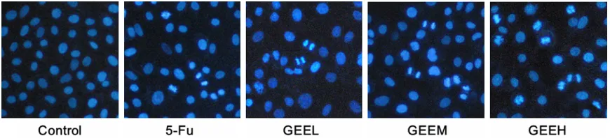
The nuclei morphological changes were obser- ved in SiHa cells via Hoechst 33258 staining
[image:6.612.90.287.75.249.2]assay are shown in Figure 4. As indicated in
Figure 4, the nuclei of SiHa cells in control group were similarly sized, regularly shaped, and evenly stained. However, cells treated with GEE showed conspicuous morphological changes, such as chromatin condensation, nuclear fragmentation and reduction of cell vol-ume. Especially in 0.3 mg/mL GEE group, cells revealed serious nuclear condensation, frag-mentation, and apoptotic bodies, all of which are characteristics of apoptosis.
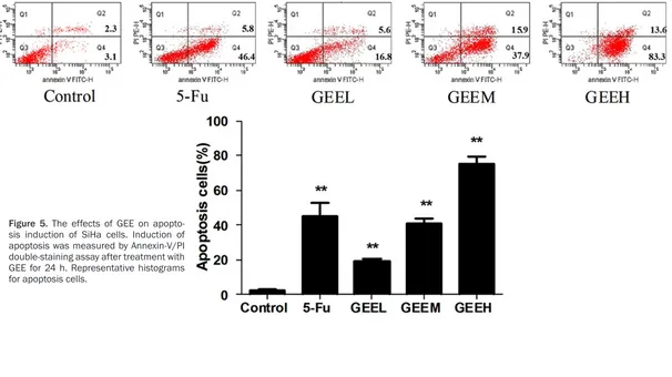
GEE-induced apoptosis in SiHa cells was fur-ther determined using an Annexin V-FITC+/PI
-staining assay. As shown in Figure 5, compared to the control group, GEE dramatically triggered apoptosis in SiHa cells in a dose-dependent manner. The percentage of apoptotic cells in the control group was 3.6%. After treatment with GEE for 24 h, the percentages of apopto- tic cells were 16.8%, 37.9% and 83.3%, respectively.
Effects of GEE on the expression of VEGF in SiHa cells
Obviously, angiogenesis have been suggested a prerequisite progress for tumor growth, meanwhile VEGF play an important role in angiogenesis. Therefore, Western blot analysis was employed to detect the protein expression of VEGF. As shown in Figure 6, compared to the control group, VEGF protein expression de- creased in the GEE-treated groups. This indi-cated that there was decrease in the VEGF/β -actin ratio.
Effects of GEE on the expression of apoptotic-related proteins
[image:6.612.89.288.372.544.2]Western blot analysis was applied to observe the expression of apoptotic-related proteins and investigate the mechanism responsible for the apoptosis induced by GEE in Siha cells. As shown in Figure 7, western blotting analysis demonstrated that Bcl-2 protein expression was down-regulated while Bax protein expres-sion was increased. Compared with control group, both of them are statistically significant (P<0.05). As shown in Figure 8, Cyt c, cas-pase-3 and caspase-9 were significantly enhanced in GEE groups. These data together suggested that intrinsic mitochondrial pathway may be involved in GEE-induced apoptosis.
Figure 6. The effect of GEE on the expression of VEGF in SiHa cells. The expression of VEGF in SiHa cells was examined via Western blot analysis. A. Rep-resentative Western blot band photographs of VEGF. B. Ratio of the protein expression of VEGF to β-actin.
Abbreviations as above. Changes were significantly
different compared with control group (*P<0.05, **P<0.01). β-actin was used as the internal loading
control.
Figure 7. The effect of GEE on the expression of
Bcl-2, Bax and β-actin in SiHa cells. Western blot analysis
was employed to detect the protein expression of Bcl-2, Bax and β-actin. A. Representative Western blot band photographs. B. Ratio of the protein expression of Bax to Bcl-2 in SiHa cells. Abbreviations as above.
Changes were significantly different compared with
control group (*P<0.05, **P<0.01). β-actin was used
Discussion
In recent years, many traditional Chinese medi-cine have been gradually discovered to be potential sources of antitumor drugs for its role in killing tumor cells more actively and less toxi-cally [8]. Because of the good therapeutic effects in cancer, the active ingredients of
Gecko were searched by several labs [7, 9, 14]. A number of studies have demonstrated that
Gecko could inhibit proliferation and also could induce apoptosis in liver cancer, colorectal can-cer, bladder cancer and esophageal cancer [11-14]. In addition, Gecko has also been indicated as a suppressant in angiogenesis and cell motility of liver cancer [7, 9, 21, 22]. All these findings provide a certain rationale for thera-peutic properties of Gecko on carcinoma. However, the potential molecular mechanisms are still elusive and require further validation. Therefore, we aimed to elucidate the inhibitory proliferation effect of GEE on human cervical cancer SiHa cells in this study. In the present study, we demonstrated that GEE could
[image:7.612.93.522.74.359.2]signifi-cantly inhibit the proliferation of SiHa cells in a dose- and time- dependent manners via MTT assay and plate clone formation assay. Moreover, Hoechst staining revealed that GEE could induce the apoptosis of SiHa cells in vitro. Angiogenesis, the growth of new blood vessels from the pre-existing ones, is essential for the development and progression of malignant tumors. It supplies nutrients and oxygen for cell proliferation, penetrating the whole growth of tumor [23, 24]. VEGF is regarded as the impor-tant regulatory protein of the angiogenic pro-cess. It can promote proliferation of endothelial cells, angiogenesis and increase the permea-bility of blood vessels [25, 26]. Several studies reveal that blocking of VEGF function could inhibit angiogenesis, and then suppress tumor growth and metastasis [27, 28]. In the current study, the expression of VEGF was decreased following the treatment with GEE. This finding indicates that GEE may have an effect on angio-genesis. Moreover, suppressed expression of VEGF can induce release of Cyt c and activation
Figure 8. The effect of GEE on the expression of apoptosis-related proteins in SiHa cells. Western blot analysis was employed to detect the protein expression of Cyt c, caspase-3, caspase-9 and β-actin. A. Representative Western
blot band photographs. B. Ratio of the protein expression of Cyt c to β-actin in SiHa cells. C. Ratio of the protein ex
-pression of caspase-9 to β-actin in SiHa cells. D. Ratio of the protein ex-pression of caspase-3 to β-actin in SiHa cells. Abbreviations as above. Changes were significantly different compared with control group (*P<0.05, **P<0.01).
of caspase-3, which ultimately results in apop-tosis [29].
Apoptosis is essential for maintaining the phys-iologic balance between cell death and cell growth. When disrupted, an imbalance between life and death of cells can lead to tumor initia-tion, progression and metastasis [30, 31]. Currently, inducing apoptosis plays a major role in cancer treatment, serving as the main effec-tor function of anti-cancer therapies [32]. Consequently, we aimed to elucidate that whether apoptosis is involved in the antineo-plastic effect of GEE on human cervical cancer SiHa cells in this study. Mainly two apoptotic pathways are known as the extrinsic(death receptor-mediated) and the intrinsic (mitochon-drial-mediated) pathway [33, 34]. In the intrin-sic pathway, mitochondria play a pivotal role in mediating apoptosis [35]. The mitochondrial pathway is regulated by the Bcl-2 family pro-teins which includes the anti-apoptotic propro-teins (Bcl-2, etc.) and the proapoptotic proteins (Bax and Bak) [31, 36, 37]. Our data showed that after GEE treatment, the expression of Bcl-2 in SiHa cells was significantly decreased in a dose-dependent manner. On the contrary, the activity of Bax was decreased when the amounts of GEE were increased. Accumulation of proapoptotic proteins on the mitochondrial outer membrane results in the increase of mitochondrial membrane permeability, and causing the release of Cytochrome C (Cyt c) into the cytoplasm [38, 39]. Release of Cyt c from the intermembrane spaces of the mito-chondria into the cytosol is a key event in apop-tosis [40]. In this study, we demonstrated that the expression of Cyt c was down-regulated. Cyt c binds to the cytosolic protein Apaf-1 to facilitate the formation of apoptosomes, which can then recruit and activate the inactive pro-caspase-9 [41]. Only the propro-caspase-9 bound to the apoptosome is able to efficiently cleave and activate downstream executioner caspas-es such as caspase-3, and trigger a cascade of events leading to apoptosis [42-44]. To clarify whether the mitochondrial pathway is involved in GEE-induced apoptosis, we examined the activation of some apoptosis-associated pro-teins by Western blotting. As shown, both cas-pase-9 and caspase-3 were up-regulated in cells following GEE treatment. These results suggested that the GEE-induced apoptosis might be through the intrinsic mitochondrial
pathway. In brief, these results suggested that GEE induced the apoptosis of human cervical cancer SiHa cells through promoting the release of Cyt c, then activation of caspase family, which may be concerned with the endog-enous mitochondrial pathway.
In summary, these results demonstrated that GEE play a antineoplastic role on SiHa cells, which may be concerned with the effect of anti-proliferation, anti-angiogenesis and apoptosis-induction. These effects may owe to the increase of Cyt c, caspase-3, caspase-9 and the decrease of VEGF, Bcl-2 proteins. Although GEE could inhibit the proliferation of SiHa cell, the antitumor mechanism is quite complicated, which is need further investigations.
Disclosure of conflict of interest
None.
Acknowledgements
This study was supported by Medical Science and Technology Research Project of Henan Province, China, No. 102102310063. We thank Mr. Xi Shou-min and Dr Li Rui-fang for their excellent technical supports and Dr Wang Jian-gang and Liu Ling for their proofreading the article.
Address correspondence to: Dr. Jian-Gang Wang, Department of Pharmacology, Medical College, Henan University of Science and Technology, Anhui Road 31, Luoyang 471000, Henan Province, China. Tel: +86-0379-64820862; E-mail: ylwjg@163.com
References
[1] Arbyn M, Castellsague X, de Sanjose S, Bruni L, Saraiya M, Bray F and Ferlay J. Worldwide bur-den of cervical cancer in 2008. Ann Oncol 2011; 22: 2675-2686.
[2] Nahvijou A, Hadji M, Marnani AB, Tourang F, Bayat N, Weiderpass E, Daroudi R, Sari AA and Zendehdel K. A systematic review of economic aspects of cervical cancer screening strate-gies worldwide: discrepancy between econom-ic analysis and poleconom-icymaking. Asian Pac J Cancer Prev 2014; 15: 8229-8237.
telomer-ase activity in human cervical cancer cells (SiHa). BMC Complement Altern Med 2015; 15: 23.
[4] Kim HS, Yoon G, Ryu JY, Cho YJ, Choi JJ, Lee YY, Kim TJ, Choi CH, Song SY, Kim BG, Bae DS and Lee JW. Sphingosine kinase 1 is a reliable prognostic factor and a novel therapeutic tar-get for uterine cervical cancer. Oncotartar-get 2015; 6: 26746-26756.
[5] Promraksa B, Daduang J, Khampitak T, Tavichakorntrakool R, Koraneekit A, Palasap A, Tangrassameeprasert R and Boonsiri P. Anticancer Potential of Cratoxylum formosum
Subsp. Pruniflorum (Kurz.) Gogel Extracts
Against Cervical Cancer Cell Lines. Asian Pac J Cancer Prev 2015; 16: 6117-6121.
[6] Liu S, Sun Y and Louie W. Symptom distress and its association with traditional Chinese medicine use in Chinese American women with cancer. Oncol Nurs Forum 2015; 42: E24-32.
[7] Tang Z, Huang SQ, Liu JT, Jiang GX and Wang CM. Anti-angiogenic activity of gecko aqueous extracts and its macromolecular components in CAM and HUVE-12 cells. Asian Pac J Cancer Prev 2015; 16: 2081-2086.
[8] Sun Y. The role of Chinese medicine in clinical oncology. Chin J Integr Med 2014; 20: 3-10. [9] Wu XZ, Chen D and Han XQ. Anti-migration
ef-fects of Gekko sulfated glycopeptide on hu-man hepatoma SMMC-7721 cells. Molecules 2011; 16: 4958-4970.
[10] Jeong AJ, Chung CN, Kim HJ, Bae KS, Choi S, Jun WJ, Shim SI, Kang TH, Leem SH and Chung JW. Gecko Proteins Exert Anti-Tumor Effect against Cervical Cancer Cells Via PI3-Kinase/ Akt Pathway. Korean J Physiol Pharmacol 2012; 16: 361-365.
[11] Liu F, Wang JG, Wang SY, Li Y, Wu YP and Xi SM. Antitumor effect and mechanism of Ge- cko on human esophageal carcinoma cell lines in vitro and xenografted sarcoma 180 in Kunming mice. World J Gastroenterol 2008; 14: 3990-3996.
[12] Song Y, Wang JG, Li RF, Li Y, Cui ZC, Duan LX and Lu F. Gecko crude peptides induce apopto-sis in human liver carcinoma cells in vitro and exert antitumor activity in a mouse ascites H22 xenograft model. J Biomed Biotechnol 2012; 2012: 743573.
[13] Amiri A, Namavari M, Rashidi M, Fahmidehkar MA and Seghatoleslam A. Inhibitory effects of Cyrtopodion scabrum extract on growth of hu-man breast and colorectal cancer cells. Asian Pac J Cancer Prev 2015; 16: 565-570. [14] Kim GY, Park SY, Jo A, Kim M, Leem SH, Jun WJ,
Shim SI, Lee SC and Chung JW. Gecko proteins induce the apoptosis of bladder cancer 5637 cells by inhibiting Akt and activating the
intrin-sic caspase cascade. BMB Rep 2015; 48: 531-536.
[15] Yang ZH, Wu M, Liu R and Liu HM. Clinical ob-servationof Jin Long capsule combined che-motherapy on late cancer of colon. Modern Medical Journal 2013; 41: 908-910.
[16] Bai JW and Wu WM. Efficacy analysis of Jin
Long Capsule (JLC) in neoadjuvant chemother-apy of breast cancer. Chin J Clin Oncol 2014; 41: 246-249.
[17] Cui CC, Wang JG, Duan LX, Qian X, Wang CE and Xu XL. Apoptosis-Inducing Activities of Gekko Ethanol Extract on Human Laryngeal Carcinoma Hep2 Cells. Nat Prod Res Dev 2013; 25: 551-554.
[18] Cui CC, Wang JG, Li RF, Xu XL, Zhao HS and Duan LX. Study on the anti-cancer effects of gekko ethanol extract in mice subrenal cap-sule xenograft model of h22 hepatocellular carcinoma. Lishizhen Med Mater Med Res 2013; 24: 1142-1144.
[19] XU XL, Wang JG, Li RF, Li SP and Duan LX. Inhibitory effect of Gecko peptides mixture on growth of human esophageal squamous carci-noma cell line EC109 cells. Chin J Clin Pharmacol 2013; 29: 602-604.
[20] Al-Sheddi ES, Farshori NN, Al-Oqail MM, Musarrat J, Al-Khedhairy AA and Siddiqui MA. Cytotoxicity of Nigella sativa seed oil and ex-tract against human lung cancer cell line. Asian Pac J Cancer Prev 2014; 15: 983-987. [21] Chen D, Yao WJ, Zhang XL, Han XQ, Qu XY, Ka
WB, Sun DG, Wu XZ and Wen ZY. Effects of Gekko sulfated polysaccharide-protein com-plex on human hepatoma SMMC-7721 cells: inhibition of proliferation and migration. J Ethnopharmacol 2010; 127: 702-708.
[22] Zhang SX, Zhu C, Ba Y, Chen D, Zhou XL, Cao R, Wang LP, Ren Y and Wu XZ. Gekko-sulfated gly-copeptide inhibits tumor angiogenesis by
tar-geting basic fibroblast growth factor. J Biol
Chem 2012; 287: 13206-13215.
[23] Yadav L, Puri N, Rastogi V, Satpute P and Sharma V. Tumour Angiogenesis and Angio- genic Inhibitors: A Review. J Clin Diagn Res 2015; 9: XE01-XE05.
[24] Detmar M. Tumor angiogenesis. J Investig Dermatol Symp Proc 2000; 5: 20-23.
[25] Ferrara N. Role of vascular endothelial growth factor in regulation of physiological angiogen-esis. Am J Physiol Cell Physiol 2001; 280: C1358-1366.
[26] Lee SH, Jeong D, Han YS and Baek MJ. Pivotal role of vascular endothelial growth factor path-way in tumor angiogenesis. Ann Surg Treat Res 2015; 89: 1-8.
PI3K/AKT signaling pathway. Int J Clin Exp Med 2015; 8: 12411-12417.
[28] Zhao P, Li Q, Shi Z, Li C, Wang L, Liu X, Jiang C, Qian X, You Y, Liu N, Liu LZ, Ding L and Jiang BH. GSK-3beta regulates tumor growth and an-giogenesis in human glioma cells. Oncotarget 2015; 6: 31901-31915.
[29] Frost P, Berlanger E, Mysore V, Hoang B, Shi Y, Gera J and Lichtenstein A. Mammalian target of rapamycin inhibitors induce tumor cell apoptosis in vivo primarily by inhibiting VEGF expression and angiogenesis. J Oncol 2013; 2013: 897025.
[30] Koff JL, Ramachandiran S and Bernal-Mizrachi L. A time to kill: targeting apoptosis in cancer. Int J Mol Sci 2015; 16: 2942-2955.
[31] Lopez J and Tait SW. Mitochondrial apoptosis: killing cancer using the enemy within. Br J Cancer 2015; 112: 957-962.
[32] Labi V and Erlacher M. How cell death shapes cancer. Cell Death Dis 2015; 6: e1675. [33] Kim JH, Choi YW, Park C, Jin CY, Lee YJ, Park da
J, Kim SG, Kim GY, Choi IW, Hwang WD, Jeong YK, Kim SK and Choi YH. Apoptosis induction of human leukemia U937 cells by gomisin N, a dibenzocyclooctadiene lignan, isolated from Schizandra chinensis Baill. Food Chem Toxicol 2010; 48: 807-813.
[34] Nguyen VT, Lee JS, Qian ZJ, Li YX, Kim KN, Heo SJ, Jeon YJ, Park WS, Choi IW, Je JY and Jung WK. Gliotoxin isolated from marine fungus Aspergillus sp. induces apoptosis of human cervical cancer and chondrosarcoma cells. Mar Drugs 2014; 12: 69-87.
[35] Elmore S. Apoptosis: a review of programmed cell death. Toxicol Pathol 2007; 35: 495-516. [36] Mignotte B and Vayssiere JL. Mitochondria and
apoptosis. Eur J Biochem 1998; 252: 1-15.
[37] Gross A, McDonnell JM and Korsmeyer SJ. BCL-2 family members and the mitochondria in apoptosis. Genes Dev 1999; 13: 1899-1911. [38] Wang X. The expanding role of mitochondria in
apoptosis. Genes Dev 2001; 15: 2922-2933. [39] Seervi M, Joseph J, Sobhan PK, Bhavya BC and
Santhoshkumar TR. Essential requirement of cytochrome c release for caspase activation by
procaspase-activating compound defined by
cellular models. Cell Death Dis 2011; 2: e207. [40] Ju HK, Lee HW, Chung KS, Choi JH, Cho JG,
Baek NI, Chung HG and Lee KT. Standardized
flavonoid-rich fraction of Artemisia princeps
Pampanini cv. Sajabal induces apoptosis via mitochondrial pathway in human cervical can-cer HeLa cells. J Ethnopharmacol 2012; 141: 460-468.
[41] Garrido C, Galluzzi L, Brunet M, Puig PE, Didelot C and Kroemer G. Mechanisms of cyto-chrome c release from mitochondria. Cell Death Differ 2006; 13: 1423-1433.
[42] Porter AG and Janicke RU. Emerging roles of caspase-3 in apoptosis. Cell Death Differ 1999; 6: 99-104.
[43] Wall DM and McCormick BA. Bacterial secret-ed effectors and caspase-3 interactions. Cell Microbiol 2014; 16: 1746-1756.