Original Article
Long non-coding RNA H19 promotes colorectal cancer
metastasis via negative modulation of miR-148b
Buning Tian, Xiaorong Li, Ni Gong, Gui Hu, Jianyu Zhou
Departments of General Surgery, The Third Xiangya Hospital of Central South University, Changsha, P. R. China Received November 30, 2015; Accepted February 29, 2016; Epub June 15, 2016; Published June 30, 2016
Abstract: Cancer cell invasion and metastasis are the main reason for high mortality (approximately 30%) of colorec-tal cancer (CRC) patients. Thus, it will be helpful for CRC treatment to understand the potential factors promoting CRC cell metastasis. Long coding RNAs (lncRNAs) are newly discovered RNAs which make up 80% of non-coding RNAs, which have been linked in every stage of cell life, including cell proliferation, differentiation, apoptosis and motor. Although H19 has a vital role in cancer, little is known about the mechanism through which H19 exerts its oncogenic activity, and the interaction between H19 and miRNAs remains largely unknown. In the present study, we demonstrated that H19 was upregulated in CRC tissues. H19 upregulated the miRNA-148b target gene chole-cystokinin-2 receptor (CCK2R) by competitively ‘sponging’ miRNA-148b and then promoted the proliferation and invasiveness of CRC cells. Our findings suggest that lncRNA H19 acts as a competing endogenous RNA for miR-148b, and subsequent regulates CCK2R expression. Thus, H19/miR-148b/CCK2R axis may serve as a potential target for CRC therapy.
Keywords: Long non-coding RNAs, microRNA, colorectal cancer, metastasis, H19
Introduction
The number of deaths caused by colorectal cancer (CRC) is ranked the third in the world [1]. Cancer cell invasion and metastasis are the main reasons for high mortality (approximately 30%) of CRC patients [2]. Thus, it will be helpful for CRC treatment to understand the potential factors promoting CRC cell metastasis.
Non-coding RNAs, including microRNA and long non-coding RNAs (lncRNAs), have been report-ed to be involvreport-ed in CRC progress [3]. Although microRNAs have been well studied in CRC, lncRNAs, which are newly discovered RNAs making up 80% of non-coding RNAs, are need-ed to be deeply illustratneed-ed [4]. lncRNAs have been linked in every stage of cell life, including cell proliferation, differentiation, apoptosis and motor [5]. And LncRNAs have been identified as oncogene or tumor suppressor, or a predictor of prognosis [6, 7]. High lncRNA-GHET1 levels are correlated with tumor size, tumor invasion and poor survival in gastric cancer, which pro-motes gastric carcinoma cell proliferation via increasing c-Myc mRNA stability and
expres-sion [8]. The expression of IncRNA-CCAT1 in colorectal tumor tissue is significantly higher than that in normal para-carcinoma tissue, and significantly correlated with local infiltration depth, tumor staging, and vascular invasion [9]. Both of miRNAs and lncRNAs have been well characterized that they can regulate protein-coding genes. Recently, emerging evidence suggests that miRNAs and lncRNAs also can form a well-orchestrated regulatory interaction network: miRNAs and lncRNAs are able to inter-act with each other, imposing an additional level of posttranscriptional regulation [10]. For example, lncRNA GAS5 can bind miRNA-222 and form a regulatory interaction [11]. It is reported that stable expression of lncRNA H19 significantly promotes EMT progression and accelerates colorectal tumor growth in vivo and
in vitro [12].
Interaction of H19 and miR-148b in CRC
we demonstrated that H19 was upregulated in CRC tissues. H19 upregulated the miRNA-148b target gene cholecystokinin-2 receptor (CCK2R) by competitively ‘sponging’ miRNA-148b and then promoted the proliferation and invasive-ness of CRC cells. H19 may function as a part of the ‘competitive endogenous RNA (ceRNA)’ network.
Materials and methods
Tissue samples
A total of 15 colorectal carcinoma tissues and the paired adjacent tissues were collected from the Third Xiangya Hospital of Central South University. Informed consents have been signed by all subjects. All samples were collect-ed and indentificollect-ed by histopathological evalua-tion, and stored at -80°C until used.
Cell culture
The human colorectal cancer cell lines, Lovo, HCT-116 and Caco-2, and the human colonic epithelial cells, HCoEpiC, were obtained from American Type Culture Collection (ATCC, Manassas, VA, USA). All the cells were cultured in RPMI1640 medium (Invitrogen Life Tech- nologies, Carlsbad, CA, USA), supplemented with 10% (v/v) fetal bovine serum (FBS) (Invitrogen Life Technologies) at 37°C in a humidified 5% CO2 incubator.
Transfection
The wild type H19 (Wt-H19) and mutated H19 (Mut-H19) expressed vector were designed and constructed in Fulengen (Guangzhou, China). And the empty pcDNA3.1 plasmid was used as a negative control. To overexpress the Wt-H19 or the Mut-H19, Lovo and HCT-116 cells were transfected with empty vector, Wt-H19 or Mut-H19 for 48 h by using Lipofectamine 2000 (Life Technologies, Carlsbad, CA). The expression of H19 was detected by real time PCR, and the transfected cells were used for further analysis.
To knock down the expression of H19, Lovo and HCT-116 cells were transfected with scramble sequences or H19-siRNA for 48 h by using Lipofectamine 2000 (Life Technologies). The siRNA sequences of H19 were designed and purchased from Ribobio Co. Ltd. (cat no. Q000283120-1-B, Guangzhou, China).
Ectopic expression of miR-148b was achiev- ed by transfecting Lovo and HCT-116 cells with miR-148b mimics or inhibitors (cat no. miR10000759-1-5 and miR20000759-1-5, RiboBio, Guangzhou, China) through using Lipofectamine 2000 (Life Technologies) accord-ing to the manufacturer’s instructions.
Real time quantitative PCR (qPCR) analysis
Trizol reagent (Invitrogen, USA) was used to extract total RNA from the indicated cells and tissues according to the manufacturer’s instruc-tions. One-Step Real-Time RT-PCR Master Mixes (Thermo Fisher Scientific, Grand Island, NY, USA) was used for real time PCR to detect the expression of H19 and CCK2R. The primers of H19, CCK2R and β-actin are as follows: H19, forward, CGCTTTTGAACCAGCAGGG, reverse, TTCCCGAGGCTTTGGTGTG; CCK2R, forward, ATCTGTCCAGCCACGAATCA, reverse, ATTAGCA- CCTCCATCCAGCA. β-actin, forward, AGGGGCC- GGACTCGTCATACT, reverse, GGCGGCACCACC- ATGTACCCT. The expression of H19 and CCK2R was normalized by β-actin. The MiScript Reverse Transcription kit (Qiagen, Hilden, Germany) was used to reversely transcribe RNA into cDNA, and the MiScript SYBR-Green PCR Kit (Qiagen, Hilden, Germany) was used for real-time PCR to detect the expression of miR-148b. Specific primer sets for miRNA-148b (cat no. HmiRQP0205) and U6 (cat no. HmiRQP9001) were purchased from Fulengen (Guangzhou, China). U6 expression was used as an endogenous control. All the qPCR data were processed using 2-ΔΔCT method.
Dual luciferase reporter assay
Wild type (WT) and mutant (MUT) forms of the 3’UTR of H19 were inserted downstream of the dual luciferase reporter vector. For the lucif-erase assay, 5×10⁴ HCT-116 cells were
cul-tured in 96-well plates to reach approximately 80% confluence. The fibroblasts were co-trans-fected with 50 nM 148b mimics or miR-148b inhibitors and 30 ng of the WT/MUT 3’UTR of H19 dual luciferase reporter vector using Lipofectamine 2000 (Life Technologies, Carlsbad, CA). After 48 h of transfection, a Dual-Luciferase-Reporter Assay System (Pro- mega, Madison, WI) was used to detect lucifer-ase activity using a GloMax®-Multi+Lumino-
CCK-8 cell proliferation assay
Cell growth was measured by CCK-8 assay. 1000/well Lovo or HCT-116 cells transfected with Wt-H19 or H19-siRNA were seeded in each 96-well plate for 12 h, and further incubated for 0h, 24 h, 48 h and 72 h, respectively. 1 hour before the ending of incubation, 10 μl CCK-8 reagents (Dojindo, Japan) were added to each well. OD value at 570 nm in each well was determined by an enzyme immunoassay analyzer.
Transwell assay
The indicated cells were starved for 24 h, and then resuspended in serum-free medium and added to the upper chamber. The lower cham-ber was filled with medium containing 10% FBS. Following 48 hours culture, cells attached to the bottom were fixed and stained with crys-tal violet for 45 min and dried in air. The optical density (OD) at 570 nm of crystal violet dis-solved by 10% acetic acid was detected by an enzyme immunoassay analyzer (Synergy™ Mx; BioTek, Winooski, VT, USA).
Statistical analysis
Statistical analyses were performed using GraphPad Prism 5 software (Graphpad Soft- ware, Inc., La Jolla, CA, USA) and the data are presented as the mean ± standard deviation (SD). An unpaired two-tailed Student’s t-test or one way analysis of variance (ANOVA) with
Bonferroni t post-test was used to analyze the data depending on conditions. P<0.05 was considered to indicate a statistically significant difference.
Results
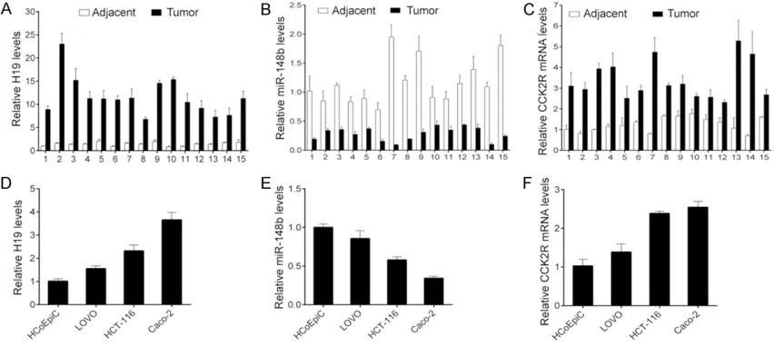
H19 and CCK2R are significantly upregulated, but miR-148b is reduced in CRC cancer tis-sues and cell lines
To investigate the role of H19 in CRC, we firstly analyzed the expression of H19 in CRC cancer tissues. We found that H19 expression was sig-nificantly increased in the CRC samples com-pared with the adjacent tissues (Figure 1A). Furthermore, we found that H19 is a putative target of miR-148b through the informatics tool, start base (http://www.lncrnablog.com/ tag/starbase-v2-0/). Thus, we detected the expression of miR-148b and its target CCK2R in the cancer tissues and their matched adja-cent tissues by using real time qPCR. All of the cancer tissues had lower miR-148b levels and higher CCK2R levels than their matched adja-cent tissues (Figure 1B, 1C). In addition, the similar results were observed in the CRC cell lines (Figure 1D-F). These results indicate that H19 and miR-148b/CCK2R signaling may con-tribute to CRC tumor development.
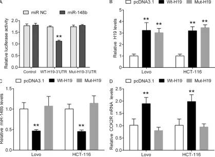
Wild type H19 regulates miR-148b/CCK2R signaling
[image:3.612.93.519.75.266.2]Interaction of H19 and miR-148b in CRC
and found that miR-148b significantly repressed the luciferase activity of wt-H19 3’UTR, but did not alter the luciferase activity of Mut-H19 3’UTR, indicating that miR-148b directly bound to 3’UTR of H19 (Figure 2A). Furthermore, we investigated whether H19 could regulate the expression of miR-148b. The wild type and mutated type H19 expressed vector were trans-fected into Lovo and HCT-116 cells (Figure 2B). Then, the expression of miR-148b was ana-lyzed. We found that miR-148b expression was significantly decreased by wild type H19, and subsequently resulting in upregulation of CCK2R. However, the mutated type of H19 could not change the expression of miR-148b and CCK2R. In contrast, we knocked down the expression of H19 by using siRNA transfection, and found that knockdown of H19 was able to upregulate the expression of miR-148b, and subsequent reduction of CCK2R (Figure 3A-C). In the other hand, we further investigated
whether miR-148b could regulate H19 expres-sion because of their interaction. We knocked down or overexpressed miR-148b in Lovo and HCT-116 cells (Figure 3D), and found that miR-148b negatively regulated the expression of CCK2R, but not alter the expression of H19. These findings suggest that H19 regulates miR-148b/CCK2R signaling through its interaction with miR-148b.
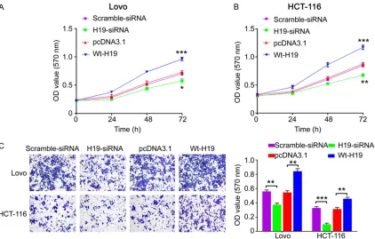
Knockdown of H19 inhibits cell proliferation and invasion in CRC cells
[image:4.612.97.516.77.383.2]Transwell assay showed that the invasive abili-ty was significantly decreased in H19 siRNA group compared with the negative control
[image:5.612.96.523.77.263.2]group, while the invasive ability was enhanced in Wt-H19 group than the pcDNA3.1 control group (Figure 4C). These results indicate that Figure 3. H19 regulated miR-148b/CCK2R signaling. Real-time PCR analysis for H19 (A), miR-148b (B) and CCK2R (C) in Lovo and HCT-116 cells transfected with scramble sequences or H19 siRNA sequences. Real-time PCR analy-sis for 148b (D), H19 (E) and CCK2R (F) in Lovo and HCT-116 cells transfected with 148b mimic, or miR-148b inhibitor or their negative control. Data are presented as mean ± SD. Experiments are independently repeated three times. *P<0.05, **P<0.01, ***P<0.001. ns, no significance.
[image:5.612.96.518.346.615.2]Interaction of H19 and miR-148b in CRC
knockdown of H19 is able to repress CRC cell growth and invasion.
Discussion
Non-coding RNAs play a vital role in carcinogen-esis, cancer development and cancer metasta-sis [13]. The studies on non-coding RNA provide new insights into the biology of cancers. Over the past decade, miRNAs have been well dem-onstrated in almost all the cancers [14, 15]. It would facilitate the development of lncRNA-directed therapy that understanding the pre-cise molecular mechanism by which lncRNAs function. Emerging evidence also shows that lncRNAs contribute to the development of can-cers in every stage, including CRC [16]. However, the role of lncRNAs in the carcinogenesis of CRC remains largely unknown. In this study, we provide evidence that H19 exhibits oncogenic activities partly through modulation of miRNA-148b and then its target CCK2R.
Recent evidence suggests that there is an interaction network involving competitive endogenous RNA (ceRNA), where lncRNAs could regulate miRNA by binding and removing them from miRNAs’ binding sites on protein coding messengers [17, 18]. It was reported that lncRNA HULC was highly upregulated in liver cancer, and HULC exhibited an inhibitory effects on the expression and activity of miRNA-372 [19]. And lncRNA MALAT1 acted as an oncogene in bladder cancer, its expression was suppressed by miR-125b [20]. There also was an interaction on loc285194 and miR-211 in colon cancer [21]. The interaction between lncRNA-miRNA in CRC cancer remains to be explored. Kallen et al. demonstrated that H19 could interact with let-7 and modulate let-7 availability, and inhibits muscle differentiation through antagonizing let-7 [22].
The imprinted oncofetal lncRNA H19 is expressed in the embryo, down-regulated at birth and then reappears in tumors [23]. Higher expression of H19 was positively correlated with advanced tumor-node-metastasis stage and tumor size in non-small-cell lung cancer [24]. Recent studies suggest that lncRNAs may exert functions through targeting miRNAs. H19 enhanced the aggressive phenotype of breast cancer cells through targeting miR-675 and then decreasing both c-Cbl and Cbl-b
expres-sion [25]. Tsang WP et al. demonstrated that there was an inverse relationship between the expressions of RB and H19/miR-675 in human CRC tissues and colon cancer cell lines. These findings revealed that H19-derived miR-675 regulated the CRC development through down-regulation of its target RB and thus may serve as a potential target for CRC therapy [26]. Liang WC found that H19 was characterized as a competing endogenous RNA (ceRNA) for miR-138 and miR-200a and acted as a novel regula-tor of epithelial to mesenchymal transition (EMT) in CRC [27]. And H19 was highly expressed in mesenchymal-like cancer cells and primary CRC cancer tissues [27, 28]. Herein, we found that H19 interacted with miR-148b in CRC cancer cells. There were signifi-cant changes in the expression of miR-148b in response to wild type H19 knockdown or over-expression. And ectopic expression of H19 also induced the upregulation of CCK2R, a target of miR-148b. However, miR-148b was unable to regulate the expression of H19 in CRC cancer cells. MiR-148b have been identified as a tumor suppressor in CRC, overexpression of miR-148b can inhibit cell proliferation and arrest cell cycle progression in CRC cell lines and decrease tumor growth in vivo. Furthermore, p53 expression is associated with miR-148b expression, and p53 directly activates the tran-scription of miR-148b by binding to its promoter [29]. The expression of miR-148b is significant-ly downregulated in human colorectal cancer tissues and three cell lines compared with non-tumor adjacent tissues, which is associated with tumor size in colorectal cancer patients. Moreover, overexpression of miR-148b in HCT-116 cells can inhibit cell proliferation in vitro
and suppress tumorigenicity in vivo by targeting cholecystokinin-2 receptor gene (CCK2R) [30]. The present study revealed that H19 was shown to promote cell proliferation and inva-sion in CRC cell lines. These findings suggest that H19 may promote tumor development through ‘sponging’ miRNA-148b, and then reg-ulating CCK2R. However, the precise down-stream mechanism requires further investiga-tion in the future.
Disclosure of conflict of interest
None.
Address correspondence to: Dr. Jianyu Zhou, De- partments of General Surgery, The Third Xiangya Hospital of Central South University, 87 Tongzipo Road, Changsha 410013, Hunan, P. R. China. Tel: +86-738-8527888; Fax: +86-738-8215760; E-mail: [email protected]
References
[1] Rui Q, Xu Z, Yang P, He Z. Long noncoding RNA expression patterns in lymph node metastasis in colorectal cancer by microarray. Biomed Pharmacother 2015; 75: 12-18.
[2] Ji Q, Zhang L, Liu X, Zhou L, Wang W, Han Z, Sui H, Tang Y, Wang Y, Liu N, Ren J, Hou F, Li Q. Long non-coding RNA MALAT1 promotes tu-mour growth and metastasis in colorectal can-cer through binding to SFPQ and releasing on-cogene PTBP2 from SFPQ/PTBP2 complex. Br J Cancer 2014; 111: 736-748.
[3] Wang J, Song YX, Ma B, Wang JJ, Sun JX, Chen XW, Zhao JH, Yang YC, Wang ZN. Regulatory Roles of Non-Coding RNAs in Colorectal Can-cer. Int J Mol Sci 2015; 16: 19886-19919. [4] Guo Q, Zhao Y, Chen J, Hu J, Wang S, Zhang D,
Sun Y. BRAF-activated long non-coding RNA contributes to colorectal cancer migration by inducing epithelial-mesenchymal transition. Oncol Lett 2014; 8: 869-875.
[5] Angrand PO, Vennin C, Le Bourhis X, Adriaens-sens E. The role of long non-coding RNAs in genome formatting and expression. Front Gen-et 2015; 6: 165.
[6] Zhao J, Liu Y, Huang G, Cui P, Zhang W, Zhang Y. Long non-coding RNAs in gastric cancer: ver-satile mechanisms and potential for clinical translation. Am J Cancer Res 2015; 5: 907-927.
[7] Han D, Wang M, Ma N, Xu Y, Jiang Y, Gao X. Long noncoding RNAs: novel players in colorec-tal cancer. Cancer Lett 2015; 361: 13-21. [8] Yang F, Xue X, Zheng L, Bi J, Zhou Y, Zhi K, Gu
Y, Fang G. Long non-coding RNA GHET1 pro-motes gastric carcinoma cell proliferation by increasing c-Myc mRNA stability. FEBS J 2014; 281: 802-813.
[9] Ye Z, Zhou M, Tian B, Wu B, Li J. Expression of lncRNA-CCAT1, E-cadherin and N-cadherin in colorectal cancer and its clinical significance. Int J Clin Exp Med 2015; 8: 3707-3715. [10] Wang J, Lei ZJ, Guo Y, Wang T, Qin ZY, Xiao HL,
Fan LL, Chen DF, Bian XW, Liu J, Wang B. miRNA-regulated delivery of lincRNA-p21 suppresses beta-catenin signaling and tumori-genicity of colorectal cancer stem cells. Onco-target 2015; 6: 37852-70.
[11] Zhao X, Wang P, Liu J, Zheng J, Liu Y, Chen J, Xue Y. Gas5 Exerts Tumor-suppressive Func-tions in Human Glioma Cells by Targeting miR-222. Mol Ther 2015; 23: 1899-911.
[12] Liang WC, Fu WM, Wong CW, Wang Y, Wang WM, Hu GX, Zhang L, Xiao LJ, Wan DC, Zhang JF, Waye MM. The lncRNA H19 promotes epi-thelial to mesenchymal transition by function-ing as miRNA sponges in colorectal cancer. Oncotarget 2015; 6: 22513-22525.
[13] Ye LC, Ren L, Qiu JJ, Zhu DX, Chen T, Chang WJ, Lv SX, Xu J. Aberrant expression of long non-coding RNAs in colorectal cancer with liver me-tastasis. Tumour Biol 2015; 36: 8747-54. [14] Muhammad S, Kaur K, Huang R, Zhang Q,
Kaur P, Yazdani HO, Bilal MU, Zheng J, Zheng L, Wang XS. MicroRNAs in colorectal cancer: role in metastasis and clinical perspectives. World J Gastroenterol 2014; 20: 17011-17019. [15] Zhou M, Liu Z, Zhao Y, Ding Y, Liu H, Xi Y, Xiong
W, Li G, Lu J, Fodstad O, Riker AI, Tan M. Mi-croRNA-125b confers the resistance of breast cancer cells to paclitaxel through suppression of pro-apoptotic Bcl-2 antagonist killer 1 (Bak1) expression. J Biol Chem 2010; 285: 21496-21507.
[16] Shi J, Li X, Zhang F, Zhang C, Guan Q, Cao X, Zhu W, Zhang X, Cheng Y, Ou K, Chen Q, Hu S. Circulating lncRNAs associated with occur-rence of colorectal cancer progression. Am J Cancer Res 2015; 5: 2258-2265.
[17] Guo G, Kang Q, Zhu X, Chen Q, Wang X, Chen Y, Ouyang J, Zhang L, Tan H, Chen R, Huang S, Chen JL. A long noncoding RNA critically regu-lates Bcr-Abl-mediated cellular transformation by acting as a competitive endogenous RNA. Oncogene 2015; 34: 1768-1779.
[18] Liu XH, Sun M, Nie FQ, Ge YB, Zhang EB, Yin DD, Kong R, Xia R, Lu KH, Li JH, De W, Wang KM, Wang ZX. Lnc RNA HOTAIR functions as a competing endogenous RNA to regulate HER2 expression by sponging miR-331-3p in gastric cancer. Mol Cancer 2014; 13: 92.
[19] Wang J, Liu X, Wu H, Ni P, Gu Z, Qiao Y, Chen N, Sun F, Fan Q. CREB up-regulates long non-cod-ing RNA, HULC expression through interaction with microRNA-372 in liver cancer. Nucleic Ac-ids Res 2010; 38: 5366-5383.
[20] Han Y, Liu Y, Zhang H, Wang T, Diao R, Jiang Z, Gui Y, Cai Z. Hsa-miR-125b suppresses blad-der cancer development by down-regulating oncogene SIRT7 and oncogenic long non-cod-ing RNA MALAT1. FEBS Lett 2013; 587: 3875-3882.
[21] Liu Q, Huang J, Zhou N, Zhang Z, Zhang A, Lu Z, Wu F, Mo YY. LncRNA loc285194 is a p53-reg-ulated tumor suppressor. Nucleic Acids Res 2013; 41: 4976-4987.
Interaction of H19 and miR-148b in CRC
Gregory RI, Ding Y, Huang Y. The imprinted H19 lncRNA antagonizes let-7 microRNAs. Mol Cell 2013; 52: 101-112.
[23] Raveh E, Matouk IJ, Gilon M, Hochberg A. The H19 Long non-coding RNA in cancer initiation, progression and metastasis - a proposed unify-ing theory. Mol Cancer 2015; 14: 184. [24] Zhang E, Li W, Yin D, De W, Sun S, Han L.
c-Myc-regulated long non-coding RNA H19 indi-cates a poor prognosis and affects cell prolif-eration in non-small-cell lung cancer. Tumour Biol 2015; [Epub ahead of print].
[25] Vennin C, Spruyt N, Dahmani F, Julien S, Ber-tucci F, Finetti P, Chassat T, Bourette RP, Le Bourhis X, Adriaenssens E. H19 non coding RNA-derived miR-675 enhances tumorigene-sis and metastatumorigene-sis of breast cancer cells by downregulating c-Cbl and Cbl-b. Oncotarget 2015; 6: 29209-29223.
[26] Tsang WP, Ng EK, Ng SS, Jin H, Yu J, Sung JJ, Kwok TT. Oncofetal H19-derived miR-675 regu-lates tumor suppressor RB in human colorec-tal cancer. Carcinogenesis 2010; 31: 350-358.
[27] Liang WC, Fu WM, Wong CW, Wang Y, Wang WM, Hu GX, Zhang L, Xiao LJ, Wan DC, Zhang JF, Waye MM. The lncRNA H19 promotes epi-thelial to mesenchymal transition by function-ing as miRNA sponges in colorectal cancer. Oncotarget 2015; 6: 22513-22525.
[28] Tsang WP, Ng EK, Ng SS, Jin H, Yu J, Sung JJ, Kwok TT. Oncofetal H19-derived miR-675 regu-lates tumor suppressor RB in human colorec-tal cancer. Carcinogenesis 2010; 31: 350-358. [29] Wang G, Cao X, Lai S, Luo X, Feng Y, Wu J, Ning
Q, Xia X, Wang J, Gong J, Hu J. Altered p53 reg-ulation of miR-148b and p55PIK contributes to tumor progression in colorectal cancer. Onco-gene 2015; 34: 912-921.