Original Article
Multiple linear regression model
for predicting functional recovery
in obstructive uropathy due to stones
Yuchao Lu, Henglong Hu, Baolong Qin, Deng He, Jiaqiao Zhang, Yufeng Wang, Qing Wang, Shaogang Wang, Jihong Liu
Department of Urology, Tongji Hospital, Tongji Medical College, Huazhong University of Science and Technology, Wuhan, Hubei, China
Received December 17, 2015; Accepted May 5, 2016; Epub June 15, 2016; Published June 30, 2016
Abstract: This study aimed to develop a mathematical model for predicting recovery potential of an obstructed kidney with poor function by a noninvasive method. We analyzed a cohort study of 93 patients who suffered from
unilateral renal or ureteral stones with hydronephrosis and renography showing an ipsilateral glomerular filtration
rate (GFR) <15 ml/min. We recorded clinical parameters, including age, sex, body mass index, residual renal volume (RRV), unilateral GFR, and split renal function (SRF) before percutaneous nephrostomy (PCN). After 38.16±5.87 days of drainage. Renal function was re-evaluated by renography. Almost all of the kidneys showed changes in GFR and SRF. Univariate and multivariate analysis showed that the patient’s age, RRV, pre-GFR, and pre-SRF were
sig-nificant factors associated with recoverability of renal function. A multiple linear regression model was developed to
predict the recovery potential of the obstructed kidney.
Keywords: Urolithiasis, ureteral obstruction, renal function, linear models, percutaneous nephrostomy
Introduction
Urolithiasis can result in hydronephrosis and impairment of renal function, eventually lead-ing to atrophy of the kidneys [1]. Management of poorly functioning kidneys due to obstruc- tion by calculi still remains debatable. Some urologists prefer nephrectomy, especially when split renal function (SRF) is <10%. While others favor renal salvage by drainage and obse- rvation because predicting the potential func-tional recovery of an obstructed kidney after relief from obstruction is difficult and inaccu-rate [2-4]. Several methods or indices have been used to assess renal function, including serum creatinine levels, glomerular filtration rate (GFR), excretory urography, computed to- mography (CT), and magnetic resonance imag-ing [5]. Currently, radionuclide renographyisone of the most popular methods to evaluate over-all and split renal function, but it is useless for prediction of functional recoverability [3]. Percutaneous nephrostomy (PCN), first descri- bed by Goodwin et al. [6], has been performed
to gain temporary relief of obstruction of the kidneys worldwide. Some researchers have used PCN as an effective means of determining recoverability of renal function in ureteropelvic junction obstruction (UPJO) [7, 8], the finding shows improved function in a large proportion of kidneys in both children and adults. However, there are limited data on the outcome of PCN in obstructed kidneys with urinary stones. If renal functional recoverability following PCN could be predicted, this would be helpful for choosing a treatment method.
This study aimed to develop a mathematical model for predicting the recovery potential of an obstructed kidney with poor renal function following PCN.
Materials and methods
Patients
Technology. Between March 2013 and February 2015, all patients who suffered from unilateral renal or ureteral stones with severe hydrone-phrosis were evaluated preoperatively with uri-nalysis, urine culture, a complete blood count, serum biochemistry, coagulation tests, ab- dominal ultrasonography and three-dimension-al CT (3D-CT) image reconstruction software (GE workstation 4.6, Fairfield, CT) was used to calculate the residual renal volume (RRV). Tech- netium-99m diethylenetriamine pentaacetic acid (99mTc DTPA) was used for diuretic renogra-phy to assess the GFR and SRF. We applied the criteria introduced by Lee et al. [9] to categorize the degree of hydronephrosis as severe, mod-erate, or mild. Patients who had ipsilateral GFR <15 ml/min and contralateral GFR >40 ml/min were included in this study. Exclusion criteria included infection, UPJO, and compression of the ureter. Finally, 93 patients were included in this study.
Surgical procedures
With the patient in the prone position under local or general anesthesia, PCN was per-formed in the affected side. The dilated collect-ing system was punctured by a 4F puncture needle under ultrasound guidance. A guide wire was then inserted and fixed. The puncture
nee-dle was withdrawn and dilation of the percuta-neous tracts were performed serially over the guidewire with a fascial dilator to 14F. At the end of the procedure, a 12F nephrostomy tube was left in place.
Outcomes evaluation
Drainage was maintained for approximately 1 month. Changes in hydronephrosis and renal function were re-evaluated with abdominal ultrasound and radionuclide renography.
Statistical analysis
All of the data were analyzed using the SPSS version 17.0 statistical software package (SPSS Inc., Chicago, IL). Analysis of variance of repeat-ed measurement design was performrepeat-ed to evaluate the between-group differences of con-tinuous variables. Levene’s tests were comput-ed to examine the homogeneity of variance. Correlations of changes in GFR with various variables were examined by Pearson correla-tions. A multiple linear regression model using the stepwise method was performed to predict the relationships between renal functional recoverability and the selected variables. All p values were two-tailed and P<0.05 was consid-ered as statistically significant.
Results
The patients’ demographics are shown in Table 1. The mean age and body mass index were 47.80±13.72 years (range, 18-78 years) and 23.41±3.58, respectively. The sex distribution was slightly skewed toward women (58.06% vs. 41.94%). The left to right side radio was 48:45. Before PCN, the mean GFR (pre-GFR) was 8.14±3.55 ml/min and SRF (pre-SRF) was 12.61%±5.42%. PCN tubes were success- fully placed in all patients and none of the patients had bleeding or urinary tract infection. The mean volume of urine that was drained from the nephrostomy tubes was 1542.38± 796.43 ml, which indicated the volume of hydronephrosis.
[image:2.612.91.289.84.297.2]After a mean of 38.16±5.87 days of PCN drainage, almost all kidneys showed changes in GFR and SRF on radionuclide renography. In 39 male patients, the mean GFR and SRF of affected kidneys before PCN were 8.10±3.97 ml/min and 14.14%±8.05%, and these values increased to 12.87±6.24 ml/min and 19.83%±
Table 1. Demographics of the patients
Characteristic† (n=93) Sex
Male 39 (41.94)
Female 54 (58.06)
Age, year 47.80±13.72
BMI 23.41±3.58
Affected side
Left 48 (51.61)
Right 45 (48.39)
Pre-GFR‡, ml/min 8.14±3.55
Pre-SRF‡, % 12.61±5.42
RRV§, ml 42.15±17.44
Hydronephrosis, ml 1542.38±796.43 PCN duration, day 38.16±5.87 Post-GFR¶, ml/min 13.90±7.14
Post-SRF¶, % 19.25±8.74
†Data are shown as mean ± SD or n (%). ‡
Pre-GFR/pre-SRF, ipsilateral GFR/SRF before PCN drainage. §RRV,
residual renal volume. ¶Post-GFR/post-SRF, ipsilateral
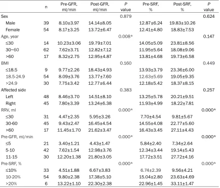
10.26%, respectively, after drainage (Table 2). Similar findings were observed in women, but there were no differences between the two groups (P>0.05). The side of the affected kid-ney and body mass index also had no effect on renal functional recoverability (i.e., changes in GFR and SRF).
When comparing changes in GFR among the different age groups (≤30, 30-60, >60 years), we found that younger adults received better results than elderly patients (P=0.008). How- ever, no significant difference in changes in SRF was observed among the age groups. RRV was divided into three categories (≤30, 30-60, >60 ml). The pre-GFR and pre-SRF were also categorized according to pre-PCN renography into three groups (≤5, 5-10, 10-15 ml/min; ≤10%, 10-20%, >20%, respectively). As pre-sented in Table 2, statistical analysis showed distinct differences in RRV, GRF, and
pre-SRF groups, regardless of changes in GFR or SRF (all P<0.05).
There was a significant negative correlation between renal GFR recoverability and the pa- tient’s age (r=-0.34, P=0.001, Figure 1A). RRV (r=0.89, P<0.001), pre-GFR (r=0.89, P<0.001), and pre-SRF (r=0.72, P<0.001) maintained po- sitive associations with restoration of renal function (Figure 1B-D).
After stepwise multiple linear regression analy-sis, only the patient’s age (X1), RRV (X2), pre-GFR (X3), and pre-SRF(X4) remained in the regression equation (Table 3). The multiple regression equation with a high adjusted R2 value of 0.975 is shown below:
[image:3.612.90.524.84.454.2]ΔGFR=-0.354X1+0.623X2+0.995X3-0.336X4 Using standardized coefficients, we found that pre-GFR was the first factor affecting renal
Table 2. Comparison of changes in GFR and SRF among different subgroups of variables
n Pre-GFR, ml/min Post-GFR, ml/min valueP Pre-SRF, % Post-SRF, % valueP
Sex 0.879 0.624
Male 39 8.10±3.97 14.14±8.05 12.87±6.24 19.83±10.26
Female 54 8.17±3.25 13.72±6.47 12.41±4.80 18.83±7.53
Age, year 0.008* 0.147
≤30 14 10.23±3.06 19.79±7.01 14.05±5.09 23.81±8.56
30~60 62 7.62±3.71 12.82±7.12 11.95±5.64 18.08±9.06
>60 17 8.32±2.75 12.95±4.87 13.81±4.68 19.73±6.58
BMI 0.160 0.449
≤18.5 9 9.77±2.26 18.43±4.93 13.93±3.79 23.36±6.00
18.5-24.9 54 8.09±3.76 13.77±7.60 12.63±5.69 19.05±9.35
>24.9 30 7.75±3.42 12.77±6.44 12.18±5.42 18.37±8.15
Affected side 0.383 0.257
Left 48 8.46±3.70 14.51±8.10 13.25±5.78 20.21±9.51
Right 45 7.80±3.39 13.24±6.38 11.93±4.99 18.22±7.81
RRV, ml 0.000* 0.000*
≤30 31 4.47±2.35 5.95±3.26 7.70±4.54 9.81±5.67
30-60 45 9.43±2.47 16.45±4.54 14.55±4.08 22.77±5.60
>60 17 11.45±1.70 21.62±3.47 16.43±3.45 27.11±4.43
Pre-GFR, ml/min 0.000* 0.000*
≤5 21 3.40±1.21 4.43±1.47 5.84±2.40 7.34±2.64
5-10 42 7.62±1.54 12.98±3.76 12.34±3.44 19.14±5.43
11-15 30 12.20±1.38 21.80±3.05 17.72±3.51 27.72±4.16
Pre-SRF, % 0.000* 0.000*
≤10% 33 4.51±1.88 6.67±3.83 6.74±2.39 9.56±4.21
10-20% 54 9.80±2.38 17.38±5.10 15.04±2.80 23.63±4.69
>20% 6 13.22±1.10 22.30±2.38 22.96±1.45 33.11±1.47
functional improvement in our study, followed by RRV, pre-SRF, and the patient’s age.
Discussion
Upper urinary tract stones are a major con- tributory factor to worsening renal function. These patients can have severe hydronephro-sis and dramatically decreased renal function [10]. Treatment of a patient with severe renal functional impairment resulting from obstru- ction of stones can be challenging and irre- solute. This is because doctors find it difficult to decide on whether they should perform ne- phrectomy directly or provide temporary relief
[image:4.612.94.520.75.363.2]helpful for making relevant clinical decisions [3]. Common renal functional tests, such as blood urea nitrogen, serum creatinine, and the creatinine clearance rate, are of little use in a normal contralateral kidney [11, 12]. Reno- graphy is widely used to assess SRF, but it is also helplessness for accurate prediction of recovery [2, 7]. Moreover, reduced kidney func-tion due to obstrucfunc-tion may decrease the sensi-tivity and specificity of renography in assessing GFR. During obstruction, elevated intrapelvic pressure reduces the effective glomerular filtration pressure. Additionally, because of an obstructed urinary tract, 99mTc DTPA may be trapped in the collecting system and this could
Figure 1. Correlations between renal GFR recoverability and different variables. There was a significant negative cor -relation between renal GFR recoverability and the patient’s age (A). RRV, pre-GFR, and pre-SRF maintained positive associations with restoration of renal function (B-D).
Table 3. Multiple linear regression analysis of the factors af-fecting recoverability of GFR
Variables Unstandardized coefficients Standardized coefficients valuet valueP Adjusted R2
Age -0.049 -0.354 -9.716 0.000* 0.975
RRV 0.094 0.623 9.166 0.000*
Pre-GFR 0.771 0.995 9.595 0.000* Pre-SRF -0.168 -0.336 -3.489 0.001*
*p<0.05 was considered statistically significant.
from obstruction and secondary surgery, owing to the fact that functional recovery cannot be well predicted.
[image:4.612.91.356.454.530.2]affect radiologists in identifying the region of interest.
Goodwin et al.[6] first described using PCN for relief of an obstructed kidney and for assess-ment of renal function to perform reconstruc-tive surgery in optimal conditions. Some researchers have reported the use of PCN as an effective means to determine the recover-ability of renal function in children or adults with UPJO [7, 12]. They showed improved func-tion in a large proporfunc-tion of kidneys, even with an SRF <10%. However, there are limited stud-ies on the outcome of PCN in obstructed kid-neys with urinary stones. Obstruction from cal-culi is different from UPJO, which usually com-prises partial obstruction and has a longer duration in adults.
We evaluated renal functional recovery follow-ing PCN in 93 patients with severe hydrone-phrosis due to stone obstruction. The GFR of the affected kidney was less than 15 ml/min. The majority of the patients obtained a certain extent of renal functional recovery after drain-age for approximately 1 month. We found that four factors were significantly associated with the improvement of renal function: patient’s age, pre-GFR, pre-SRF, and RRV. Patients with larger residual renal tissue before PCN appeared to be more likely to obtain improve-ment in kidney function. Mibu et al. [13] report-ed that renal volumeis closely relatreport-ed to renal function. In addition, better GFR and SRF of an affected kidney are also beneficial for function-al recovery. A young age showed a markedly protective effect on recoverability of GFR, but there was no significant difference in a change in SRF among the three groups (≤30, 30-60, >60 years). A reasonable explanation for this finding is that young patients often have a high-er contralathigh-eral GFR than eldhigh-erly patients. Therefore, on the same level of improvement in SRF, younger patients always have better GFR recoverability than older patients. Our study showed a similar conclusion by univariate analysis.
Multiple linear regression analysis showed that the patient’s age, RRV, pre-GFR, and pre-SRF remained significant as independent risk fac-tors for renal functional recovery. Automated curve fitting yielded quality of fit statistics that uniformly converged to coefficient of deter- mination values (R2=0.975), indicating a good
quality of curve fit to the datasets. The vari-ables adopted in the mathematical model need to be assessed before the surgical procedure. Our findings suggest that urologists can use these noninvasive test results to predict the recoverability of an obstructive kidney accord-ing to the regression equation.
Examples of using this equation in clinical practice are as follows: A 39-year-old patient suffers from severe hydronephrosis due to stone obstruction. The residual renal volume calculated based on 3D-CT is 40 ml, and renog-raphy shows that the individual GFRs of two kidneys are 10 ml/min and 60 ml/min. The following equation can be adopted: ΔGFR=-0.354 × 39+0.623 × 40+0.995 × 10-0.336 × [10/(10+60) × 100]=16.26 ml/min. However, for a 70-year-old patient, whose RRV is 40 ml, and the GFRs of two kidneys are 10 ml/min and 45 ml/min, the predictive ΔGFR would be 3.98 ml/min.
Hussain and colleagues [12] predicted renal functional recovery in obstructive renal failure due to stones. They found that a pre-operative DTPA scan was correlated with a post-operative decrease in serum creatinine levels. Moreover, a urine pH of 6 or less, post-PCN diuresis, and natriuresis were good prognostic indicators. Additionally, they found that PCN was the most reliable method of predicting future recovery of renal function after relief of obstruction, with 97.8% accuracy. However, in our study, some invasive test results, including drainage urine volume, pH, natriuresis, and urine specific grav-ity, were excluded. These factors were excluded because we wanted to develop a mathematical model for predicting recovery potential using simple clinical data only by a noninvasive meth-od, which might be helpful in making decisions and saving time for waiting and observation. Moreover, in contrast to other reports [14, 15], routine biochemistry measurements, such as serum creatinine, urea, and potassium levels, were not chosen as variables in our research because their collinearity with GFR on renogra-phy might have reduced the efficacy of the regression equation. To the best of our knowl-edge, no studies to date have directly predicted the recovery potential of an obstructed kidney by a noninvasive method.
observation time (38.16±5.87 days) was not long. Patients, especially those who had litho-tripsy performed after PCN, should be carefully evaluated in the postoperative follow-up peri-od. Additionally, we excluded kidneys with infec-tion. Further prospective studies with these patients are necessary in future. We anticipate that other researchers will test our model and add other factors from their own clinical practice.
In conclusion, our study shows that patients with larger residual renal tissue, and better GFR and SRF of the affected kidney before PCN are more likely to obtain improvement in kidney function. Young age shows a markedly protec-tive effect on renal functional recoverability. We have developed a mathematical model for pre-dicting the recovery potential of an obstructed kidney using noninvasive parameters. This will be helpful for surgeons in making decisions regarding the management of hydronephrosis due to urinary calculi with poor renal function.
Acknowledgements
We wish to thank Dr. Shuping Sang (School of Medicine, Yunnan University, Kunming, P. R. China) for her excellent statistical support.
Disclosure of conflict of interest
None.
Address correspondence to: Shaogang Wang, De- partment of Urology, Tongji Hospital, Tongji Medi- cal College, Huazhong University of Science and Technology, No. 1095, Jiefang Avenue, Wuhan, Hu- bei, China. Tel: 83663460; Fax: +86-27-83663460; E-mail: [email protected]; sgwang- [email protected]
References
[1] Iravani O, Tay EW, Bay BH, Ng YK. Unilateral ureteric stone associated with gross hydrone-phrosis and kidney shrinkage: a cadaveric re-port. Anat Cell Biol 2014; 47: 267-70.
[2] Bansal R, Ansari MS, Srivastava A, Kapoor R. Long-term results of pyeloplasty in poorly func-tioning kidneys in the pediatric age group. J Pediatr Urol 2012; 8: 25-8.
[3] Zhang S, Zhang Q, Ji C, Zhao X, Liu G, Zhang S, Li X, Lian H, Zhang G, Guo H. Improved split renal function after percutaneous nephrosto-my in young adults with severe hydronephrosis due to ureteropelvic junction obstruction. J Urol 2015; 193: 191-5.
[4] Ransley PG, Dhillon HK, Gordon I, Duffy PG, Dillon MJ, Barratt TM. The postnatal manage-ment of hydronephrosis diagnosed by prenatal ultrasound. J Urol 1990; 144: 584-7; discus-sion 593-4.
[5] O'Reilly PH. Role of modern radiological inves-tigations in obstructive uropathy. Br Med J (Clin Res Ed) 1982; 284: 1847-51.
[6] Goodwin WE, Casey WC, Woolf W. Percutaneous trocar (needle) nephrostomy in hydronephro-sis. J Am Med Assoc 1955; 157: 891-4. [7] Wagner M, Mayr J, Hacker FM. Improvement of
renal split function in hydronephrosis with less than 10 % function. Eur J Pediatr Surg 2008; 18: 156-9.
[8] Gupta DK, Chandrasekharam VV, Srinivas M, Bajpai M. Percutaneous nephrostomy in chil-dren with ureteropelvic junction obstruction and poor renal function. Urology 2001; 57: 547-50.
[9] Lee YH, Tsai JY, Jiaan BP, Wu T, Yu CC. Prospective randomized trial comparing shock wave lithotripsy and ureteroscopic lithotripsy for management of large upper third ureteral stones. Urology 2006; 67: 480-4; discussion 484.
[10] Huang TY, Lin JP, Huang CN. Dyspnea as an unexpected presentation of huge hydrone-phrosis. Iran J Kidney Dis 2014; 8: 25. [11] Wimpissinger F, Springer C, Kurtaran A, Stackl
W, Turk C. Functional aspects of silent ureteral stones investigated with MAG-3 renal scintigra-phy. BMC Urol 2014; 14: 3.
[12] Hussain M, Ali B, Ahmed S, Zafar N, Naqvi SA, Rizvi SA. Prediction of renal function recovery in obstructive renal failure due to stones. J Pak Med Assoc 1997; 47: 159-61.
[13] Mibu H, Tanaka N, Hosokawa Y, Kumamoto H, Margami N, Hirao Y, Fujimoto K. Estimated functional renal parenchymal volume predicts the split renal function following renal surgery. World J Urol 2015; 33: 1571-7.
[14] Teenan RP, Ramsay A, Deane RF. Percutaneous nephrostomy in the management of malignant ureteric obstruction. Br J Urol 1989; 64: 238-40.