1,4-Bis(5-methyl-1
H
-1,2,4-triazol-3-yl)-benzene tetrahydrate
Ai-Xin Zhu,a* Xiu-Li Chen,bZhen Li,aYuan-Chao Duaand Hong-Can Wanga
aFaculty of Chemistry and Chemical Engineering, Yunnan Normal University, Kunming 650092, People’s Republic of China, andbDepartment of Chemistry, Zhengzhou Normal University, Zhengzhou 450044, People’s Republic of China Correspondence e-mail: zaxchem@126.com
Received 9 April 2011; accepted 21 April 2011
Key indicators: single-crystal X-ray study;T= 293 K; mean(C–C) = 0.003 A˚; Rfactor = 0.052;wRfactor = 0.148; data-to-parameter ratio = 15.1.
In the title compound, C12H12N64H2O, the two triazole rings adopt acisconfiguration with a crystallographic twofold axis passing through the central benzene group. The benzene and triazole rings are almost coplanar with a dihedral angle of 5.5 (1). In the crystal, water molecules are joined together by
OW—H OW hydrogen bonds to form a one-dimensional zigzag chain. These water chains are further connected to the organic molecule, forming a three-dimensional network by intermolecular OW—H N and N—H OW hydrogen bonds. Moreover,–stacking interactions between triazole rings [centroid–centroid distances = 3.667 (1)–3.731 (1) A˚ ] are observed. One of the water molecules shows one of the H atoms to be disordered over two positions.
Related literature
For applications of 1,2,4-triazole and its derivatives in coor-dination chemistry, see: Zhang et al. (2005); Ouelletteet al. (2006); Zhu et al. (2009). For the structures of ruthenium complexes with pyridine-2-yl-1,2,4-triazole-based ligands, see: Passanitiet al.(2002). For the previous synthesis of the title compound, see: Bahc¸eciet al.(2005).
Experimental
Crystal data
C12H12N64H2O
Mr= 312.34
Monoclinic,C2=c
b= 13.937 (2) A˚
c= 9.0648 (14) A˚
= 100.893 (3)
MoKradiation
= 0.10 mm1
0.350.280.08 mm
Data collection
Bruker APEX CCD diffractometer Absorption correction: multi-scan
(SADABS; Sheldrick, 1996)
Tmin= 0.966,Tmax= 0.992
4670 measured reflections 1542 independent reflections 1286 reflections withI> 2(I)
Rint= 0.021
Refinement
R[F2> 2(F2)] = 0.052
wR(F2) = 0.148
S= 1.04 1542 reflections
102 parameters
H-atom parameters constrained
max= 0.27 e A˚
3
min=0.25 e A˚3
Table 1
Hydrogen-bond geometry (A˚ ,).
D—H A D—H H A D A D—H A
N1—H1D O1W 0.86 1.88 2.736 (2) 173 O1W—H1WA N2i 0.85 2.08 2.926 (2) 172 O1W—H1WB O2Wii
0.85 1.96 2.801 (2) 170 O2W—H2WA N3iii
0.85 1.95 2.800 (2) 173 O2W—H2WB O2Wiii 0.85 1.93 2.754 (3) 164 O2W—H2WC O2Wiv
0.85 1.92 2.774 (3) 178
Symmetry codes: (i)x;yþ1;z1 2; (ii)xþ
1 2;yþ
1 2;z
1
2; (iii)xþ1;y;z; (iv) xþ1;y;zþ1
2.
Data collection:SMART(Bruker, 2004); cell refinement:SAINT
(Bruker, 2004); data reduction:SAINT; program(s) used to solve structure:SHELXS97(Sheldrick, 2008); program(s) used to refine structure: SHELXL97 (Sheldrick, 2008); molecular graphics:
DIAMOND(Brandenburg, 1999); software used to prepare material for publication:SHELXTL(Sheldrick, 2008).
The authors thank the Youth Foundation (grant No. 10QZ02) of Yunnan Normal University, the Science Founda-tion of the EducaFounda-tion Department (grant No. 2010Y004) as well as the Science and Technology Department (grant No. 2010ZC070) of Yunnan Province for supporting this work.
Supplementary data and figures for this paper are available from the IUCr electronic archives (Reference: IM2280).
References
Bahc¸eci, S., Yu¨ksek, H. & Serdar, M. (2005).Indian J. Chem. Sect. B,44, 568– 572.
Brandenburg, K. (1999).DIAMOND. Crystal Impact GbR, Bonn, Germany. Bruker (2004).SMARTandSAINT. Bruker AXS Inc., Madison, Wisconsin,
USA.
Ouellette, W., Prosvirin, A. V., Chieffo, V., Dunbar, K. R., Hudson, B. & Zubieta, J. (2006).Inorg. Chem.45, 9346–9366.
Passaniti, P., Browne, W. R., Lynch, F. C., Hughes, D., Nieuwenhuyzen, M., James, P., Maestri, M. & Vos, J. G. (2002).J. Chem. Soc. Dalton Trans.pp. 1740–1746.
Sheldrick, G. M. (1996).SADABS. University of Go¨ttingen, Germany. Sheldrick, G. M. (2008).Acta Cryst.A64, 112–122.
Zhang, J.-P., Lin, Y.-Y., Huang, X.-C. & Chen, X.-M. (2005).J. Am. Chem. Soc.
127, 5495–5506.
Zhu, A.-X., Lin, J.-B., Zhang, J.-P. & Chen, X.-M. (2009).Inorg. Chem.48, 3882–3889.
Structure Reports
Online
supporting information
Acta Cryst. (2011). E67, o1255 [doi:10.1107/S1600536811015133]
1,4-Bis(5-methyl-1
H
-1,2,4-triazol-3-yl)benzene tetrahydrate
Ai-Xin Zhu, Xiu-Li Chen, Zhen Li, Yuan-Chao Du and Hong-Can Wang
S1. Comment
In the past few years, 1,2,4-triazole and its derivatives have attracted increasing attention as N-heterocyclic aromatic
ligands, since they are effective bridging ligands combining the coordination modes of both imidazoles and pyrazoles. In
addition, metal-triazolate frameworks have demonstrated high thermal and chemical stabilities, and interesting
luminescent, magnetic and gas-adsorption properties (Zhang et al. 2005; Ouellette et al. 2006; Zhu et al. 2009). However,
there are rare crystal structure reports of 1,2,4-triazole derivatives attached to aromatic groups (Passaniti et al. 2002).
Although the synthesis of the title compound 1,4-bis(5-methyl-1H-1,2,4-triazol-3-yl)benzene has been reported by
Bahçeci et al. (2005), no crystallographic study has been reported on this ligand and related metal coordination
compounds. We reported herein another synthetic method and the crystal structure of the title compound.
The asymmetric unit of the title compound contains one-half organic molecule, which adopts a cis-configuration with a
crystallographic mirror plane passing through the central benzene group, and two water molecules (Fig. 1). The bond
lengths and angles are within normal ranges in accordance with the corresponding values reported (Passaniti et al. 2002).
The benzene and the triazole rings are almost coplanar, with a dihedral angle of 5.4 (1)°. In the crystal structure, water
molecules are joined together by OW—H···OW hydrogen bonds to form a one-dimensional zig-zag water chain (Fig. 2,
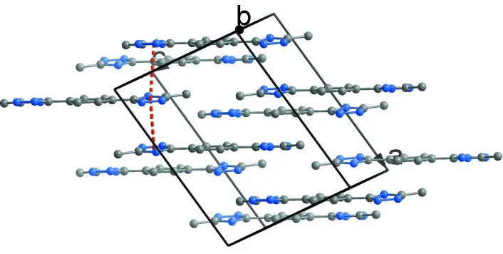
Table 1). These water chains are further connected to the organic molecule producing a three-dimensional network (Fig.
2) by intermolecular OW—H···N and N—H···OW hydrogen bonds (Table 1). Moreover, π-π stacking interactions
between triazole rings (centroid-centroid distance = 3.665 (1)–3.732 (1) Å) are observed (Fig. 3).
S2. Experimental
The ligand 1,4-bis(5-methyl-1H-1,2,4-triazol-3-yl)benzene was synthesized according to a literature method (Bahçeci et
al. 2005). Yellow, plate-like single crystals of the title compound are obtained from a solution of
1,4-bis(5-methyl-1H-1,2,4-triazol-3-yl)benzene (24 mg, 0.1 mmol) in methanol (1 ml) and water (5 ml) if the solution is placed in a
Teflon-lined stainless steel vessel (15 ml), heated at 453 K for 24 h and then cooled to room temperature at a rate of 5 K
h-1.
S3. Refinement
All H atoms were placed in idealized positions (O—H = 0.85 Å, N—H = 0.86 Å and C—H = 0.95 Å) and refined as
riding atoms with Uiso(H) = 1.2Ueq(C, N) and Uiso(H) = 1.5Ueq(O). One hydrogen atom from O2W is disordered over two
Figure 1
Molecular structure of the title compound, non-H atoms are depicted as 30% probability displacement ellipsoids.
Figure 2
Packing diagram of the title compound showing the hydrogen bonding interactions as dashed lines. H atoms not involved
[image:3.610.129.482.309.559.2]Figure 3
π-π Stacking interactions between triazole rings, H and O atoms are omitted for clarity.
5-methyl-3-[4-(5-methyl-1H-1,2,4-triazol-3-yl)phenyl]-1H- 1,2,4-triazole tetrahydrate
Crystal data
C12H12N6·4H2O
Mr = 312.34 Monoclinic, C2/c
Hall symbol: -C 2yc
a = 12.7343 (19) Å
b = 13.937 (2) Å
c = 9.0648 (14) Å
β = 100.893 (3)°
V = 1579.8 (4) Å3
Z = 4
F(000) = 664
Dx = 1.313 Mg m−3
Mo Kα radiation, λ = 0.71073 Å
µ = 0.10 mm−1
T = 293 K Plate, yellow
0.35 × 0.28 × 0.08 mm
Data collection
Bruker APEX CCD diffractometer
Radiation source: fine-focus sealed tube Graphite monochromator
ω scans
Absorption correction: multi-scan (SADABS; Sheldrick, 1996)
Tmin = 0.966, Tmax = 0.992
4670 measured reflections 1542 independent reflections 1286 reflections with I > 2σ(I)
Rint = 0.021
θmax = 26.0°, θmin = 2.2°
h = −15→15
k = −15→17
l = −11→11
Refinement
Refinement on F2
Least-squares matrix: full
R[F2 > 2σ(F2)] = 0.052
wR(F2) = 0.148
S = 1.04 1542 reflections 102 parameters 0 restraints
Primary atom site location: structure-invariant direct methods
Secondary atom site location: difference Fourier map
Hydrogen site location: inferred from neighbouring sites
H-atom parameters constrained
w = 1/[σ2(F
o2) + (0.0816P)2 + 0.7602P]
where P = (Fo2 + 2Fc2)/3
(Δ/σ)max < 0.001
Δρmax = 0.27 e Å−3
Geometry. All e.s.d.'s (except the e.s.d. in the dihedral angle between two l.s. planes) are estimated using the full covariance matrix. The cell e.s.d.'s are taken into account individually in the estimation of e.s.d.'s in distances, angles and torsion angles; correlations between e.s.d.'s in cell parameters are only used when they are defined by crystal symmetry. An approximate (isotropic) treatment of cell e.s.d.'s is used for estimating e.s.d.'s involving l.s. planes.
Refinement. Refinement of F2 against ALL reflections. The weighted R-factor wR and goodness of fit S are based on F2,
conventional R-factors R are based on F, with F set to zero for negative F2. The threshold expression of F2 > σ(F2) is used
only for calculating R-factors(gt) etc. and is not relevant to the choice of reflections for refinement. R-factors based on F2
are statistically about twice as large as those based on F, and R- factors based on ALL data will be even larger. One hydrogen atom from O2W is disordered over two positions in a 0.52 (3):0.48 (3) ratio, which is freely refined with command ′PART′.
Fractional atomic coordinates and isotropic or equivalent isotropic displacement parameters (Å2)
x y z Uiso*/Ueq Occ. (<1)
N1 0.66106 (13) 0.34969 (11) −0.19565 (17) 0.0531 (5)
H1D 0.6874 0.3847 −0.2578 0.064*
N2 0.62354 (14) 0.38395 (11) −0.07499 (17) 0.0522 (5)
N3 0.60877 (12) 0.22386 (10) −0.09140 (16) 0.0454 (4)
C1 0.68428 (18) 0.19667 (15) −0.3248 (2) 0.0590 (6)
H1A 0.7292 0.1449 −0.2805 0.088*
H1B 0.7230 0.2361 −0.3830 0.088*
H1C 0.6218 0.1711 −0.3887 0.088*
C2 0.65166 (14) 0.25529 (13) −0.20444 (18) 0.0448 (4)
C3 0.59264 (14) 0.30486 (12) −0.01538 (19) 0.0425 (4)
C4 0.54472 (14) 0.30544 (12) 0.12072 (19) 0.0421 (4)
C5 0.52248 (17) 0.22014 (13) 0.1863 (2) 0.0510 (5)
H5A 0.5379 0.1622 0.1443 0.061*
C6 0.52170 (16) 0.39108 (13) 0.1860 (2) 0.0510 (5)
H6A 0.5358 0.4491 0.1429 0.061*
O1W 0.73472 (14) 0.45314 (11) −0.41166 (19) 0.0757 (5)
H1WA 0.6969 0.4969 −0.4609 0.091*
H1WB 0.8001 0.4691 −0.4017 0.091*
O2W 0.44076 (12) −0.02791 (9) 0.10493 (16) 0.0602 (4)
H2WA 0.4240 −0.0868 0.0928 0.072*
H2WB 0.4790 −0.0006 0.0500 0.072* 0.52 (3)
H2WC 0.4784 −0.0277 0.1930 0.072* 0.48 (3)
Atomic displacement parameters (Å2)
U11 U22 U33 U12 U13 U23
N1 0.0657 (11) 0.0516 (10) 0.0484 (8) −0.0032 (8) 0.0274 (8) 0.0074 (7)
N2 0.0662 (11) 0.0437 (9) 0.0523 (9) −0.0025 (7) 0.0257 (8) 0.0035 (7)
N3 0.0540 (9) 0.0428 (8) 0.0425 (8) −0.0035 (7) 0.0166 (7) −0.0015 (6)
C1 0.0649 (13) 0.0672 (13) 0.0495 (10) −0.0045 (10) 0.0232 (9) −0.0060 (9)
C2 0.0467 (10) 0.0488 (10) 0.0401 (9) −0.0022 (8) 0.0115 (7) 0.0016 (7)
C3 0.0452 (10) 0.0437 (9) 0.0397 (9) −0.0003 (7) 0.0108 (7) 0.0012 (7)
C6 0.0603 (12) 0.0380 (10) 0.0604 (11) −0.0004 (8) 0.0260 (9) 0.0049 (8)
O1W 0.0804 (11) 0.0679 (10) 0.0871 (11) 0.0058 (8) 0.0373 (9) 0.0277 (8)
O2W 0.0809 (11) 0.0447 (7) 0.0611 (9) −0.0064 (7) 0.0289 (8) −0.0014 (6)
Geometric parameters (Å, º)
N1—C2 1.322 (3) C4—C5 1.382 (2)
N1—N2 1.360 (2) C4—C6 1.388 (2)
N1—H1D 0.8600 C5—C5i 1.383 (4)
N2—C3 1.320 (2) C5—H5A 0.9300
N3—C2 1.324 (2) C6—C6i 1.376 (4)
N3—C3 1.358 (2) C6—H6A 0.9300
C1—C2 1.484 (3) O1W—H1WA 0.8500
C1—H1A 0.9600 O1W—H1WB 0.8500
C1—H1B 0.9600 O2W—H2WA 0.8500
C1—H1C 0.9600 O2W—H2WB 0.8501
C3—C4 1.476 (2) O2W—H2WC 0.8499
C2—N1—N2 110.87 (14) N2—C3—C4 122.67 (15)
C2—N1—H1D 124.6 N3—C3—C4 123.69 (15)
N2—N1—H1D 124.6 C5—C4—C6 118.63 (17)
C3—N2—N1 102.31 (15) C5—C4—C3 120.35 (15)
C2—N3—C3 104.00 (15) C6—C4—C3 121.01 (15)
C2—C1—H1A 109.5 C4—C5—C5i 120.67 (10)
C2—C1—H1B 109.5 C4—C5—H5A 119.7
H1A—C1—H1B 109.5 C5i—C5—H5A 119.7
C2—C1—H1C 109.5 C6i—C6—C4 120.69 (10)
H1A—C1—H1C 109.5 C6i—C6—H6A 119.7
H1B—C1—H1C 109.5 C4—C6—H6A 119.7
N1—C2—N3 109.18 (15) H1WA—O1W—H1WB 108.3
N1—C2—C1 123.88 (16) H2WA—O2W—H2WB 121.0
N3—C2—C1 126.93 (17) H2WA—O2W—H2WC 102.0
N2—C3—N3 113.64 (16) H2WB—O2W—H2WC 105.4
Symmetry code: (i) −x+1, y, −z+1/2.
Hydrogen-bond geometry (Å, º)
D—H···A D—H H···A D···A D—H···A
N1—H1D···O1W 0.86 1.88 2.736 (2) 173
O1W—H1WA···N2ii 0.85 2.08 2.926 (2) 172
O1W—H1WB···O2Wiii 0.85 1.96 2.801 (2) 170
O2W—H2WA···N3iv 0.85 1.95 2.800 (2) 173
O2W—H2WB···O2Wiv 0.85 1.93 2.754 (3) 164
O2W—H2WC···O2Wi 0.85 1.92 2.774 (3) 178