O R I G I N A L A R T I C L E
Prenatal exposure to maternal smoking and offspring
DNA methylation across the lifecourse:
fi
ndings from
the Avon Longitudinal Study of Parents and Children
(ALSPAC)
Rebecca C. Richmond
1
, Andrew J. Simpkin
1
, Geoff Woodward
1
, Tom R. Gaunt
1
,
Oliver Lyttleton
1
, Wendy L. McArdle
1
, Susan M. Ring
1
, Andrew D.A.C. Smith
1
,
Nicholas J. Timpson
1
, Kate Tilling
1
, George Davey Smith
1,
†
and Caroline L. Relton
1,2,
†
,
*
1
MRC Integrative Epidemiology Unit (IEU), School of Social and Community Medicine, University of Bristol, Bristol
BS8 2BN, UK and
2Institute of Genetic Medicine, Newcastle University, Newcastle upon Tyne NE1 3BZ, UK
*To whom correspondence should be addressed at: Oakfield House, Oakfield Grove, Bristol BS8 2BN, UK. Tel: +44 1173314028; Fax: +44 1173310123;
Email: caroline.relton@bristol.ac.uk
Abstract
Maternal smoking during pregnancy has been found to influence newborn DNA methylation in genes involved in fundamental developmental processes. It is pertinent to understand the degree to which the offspring methylome is sensitive to the intensity and duration of prenatal smoking. An investigation of the persistence of offspring methylation associated with maternal smoking and the relative roles of the intrauterine and postnatal environment is also warranted. In the Avon Longitudinal Study of Parents and Children, we investigated associations between prenatal exposure to maternal smoking and offspring DNA methylation at multiple time points in approximately 800 mother–offspring pairs. In cord blood, methylation at 15 CpG sites in seven gene regions (AHRR,MYO1G,GFI1,CYP1A1,CNTNAP2,KLF13andATP9A) was associated with maternal smoking, and a dose-dependent response was observed in relation to smoking duration and intensity. Longitudinal analysis of blood DNA methylation in serial samples at birth, age 7 and 17 years demonstrated that some CpG sites showed reversibility of methylation (GFI1,KLF13andATP9A), whereas others showed persistently perturbed patterns (AHRR,MYO1G,CYP1A1andCNTNAP2). Of those showing persistence, we explored the effect of postnatal smoke exposure and found that the major contribution to altered methylation was attributed to a critical window ofin uteroexposure. A comparison of paternal and maternal smoking and offspring methylation showed consistently stronger maternal associations, providing further evidence for causal intrauterine mechanisms. Thesefindings emphasize the sensitivity of the methylome to maternal smoking during early development and the long-term impact of such exposure.
†These authors contributed equally to the work.
Received:September 22, 2014.Revised and Accepted:December 22, 2014
© The Author 2014. Published by Oxford University Press.
This is an Open Access article distributed under the terms of the Creative Commons Attribution License (http://creativecommons.org/licenses/by/4.0/), which permits unrestricted reuse, distribution, and reproduction in any medium, provided the original work is properly cited.
Human Molecular Genetics, 2015, Vol. 24, No. 8 2201–2217
doi: 10.1093/hmg/ddu739
Advance Access Publication Date: 30 December 2014 Original Article
2201
at University of the West of England on December 2, 2015
http://hmg.oxfordjournals.org/
Introduction
Despite the known health risks to both mothers and newborns, maternal smoking during pregnancy remains a significant public health problem in high-income countries and recent reports sug-gest that∼12% of mothers in England are still smoking at the time of delivery (1). Exposure of the fetus to maternal smokingin utero
has been associated with adverse perinatal outcomes, including low birth weight (2–4), elevated blood pressure (5,6), obesity (7,8) and behavioural difficulties in childhood (9,10). It has been pro-posed that epigenetic modifications such as DNA methylation may mediate the adverse developmental consequences asso-ciated with smoking during pregnancy (11).
Cigarette smoke is an established environmental associate of DNA methylation (12–17) and maternal smoking in pregnancy has recently been found to be associated with levels of DNA methylation in large-scale epigenome wide association studies (EWAS) of cord blood (18) and infant whole blood shortly after delivery (19). Of particular importance is the observation that maternal smoking during pregnancy is associated with changes in methylation in genes involved in fundamental developmental processes (18,19).
The associations found between maternal cotinine levels, an objective biomarker of smoking and DNA methylation in new-borns imply a dose-dependent effect of maternal smoking in pregnancy (18). The sensitivity of the offspring methylome to the intensity (19) and duration (20) of smoking during pregnancy has been further explored. Of potential relevance is the impact of maternal smoking in early pregnancy, when women may not be aware that they are pregnant. During the early phases of embryo-genesis, the products of tobacco smoke may induce soma-wide modification of DNA methylation in the exposed offspring, which may be then be maintained into postnatal life (21,22). Con-versely, a recent study of DNA methylation in newborns found no difference in methylation between the offspring of mothers who never smoked and those who smoked early in pregnancy (23). It has also been shown that the effect ofin uteroexposure on new-born methylation is stronger when the mother smoked past 18 weeks than when she quit earlier in pregnancy (20). Thesefi nd-ings warrant further investigation in an independent study.
Associations between own smoking and methylation at later time points have been found (15,24), with one study of former smokers showing that methylation in a key gene region asso-ciated with smoking (AHRR) approaches the levels of never smo-kers within thefirst few years of quitting, but never completely returns to normal levels (15). Two recent studies have also inves-tigated prospective associations between maternal smoking in pregnancy and peripheral blood methylation in offspring when they were children (25) and adolescents (26). A high degree of similarity was found with smoking-associated DNA methylation in newborns (18), implying a lasting effect of maternal smoking in pregnancy on offspring DNA methylation profiles. However, a more comprehensive longitudinal assessment of intrauterine exposure and methylation levels in the same offspring assessed at multiple time points is required.
The relative roles of the intrauterine and postnatal environ-ment in the persistence of DNA methylation changes associated with maternal smoking are yet to be determined. Previous stud-ies have shown that associations between prenatal exposure to maternal cigarette smoking and offspring methylation during adolescence are not attenuated with adjustment for postnatal smoking of the parents or the offspring themselves (25,26). However, the method of adjusting for a potential mediator in standard regression models to estimate the direct effect of an
exposure may produce spurious conclusions (27,28). Alternative methods are therefore required to test the hypothesis that mater-nal smoking in pregnancy is the‘critical period’for influencing offspring methylation profiles in childhood and adolescence (29). Finally, given that some of the signals found for prenatal smoke exposure have also been identified in a study of personal smoking of adolescents (30), any apparent persistent effect of maternal smoking on offspring methylation profile at later ages may be explained by smoking of the adolescents themselves.
Epigenetic markers, in contrast to germ-line genetic variation (31), are phenotypic and are therefore subject to the same poten-tial problems of confounding which afflict observational epi-demiology (32,33). Hence, there is a need to apply a range of tools for strengthening causal inference in epigenetic epidemi-ology (34,35). One such method for inferring a causal intrauterine effect involves the use of paternal exposures as negative controls for maternal exposures thought to have an intrauterine influence on offspring outcomes (34,36–40). Paternal smoking may show associations with offspring methylation similar to those of maternal smoking in pregnancy if the associations are con-founded either by shared familial factors or by parental geno-types. However, if there is an intrauterine influence of maternal smoking, then only maternal exposure would be expected to show an independent association with the outcome.
We use the Accessible Resource for Integrated Epigenomic Studies (ARIES), a large collection of genome-wide DNA methyla-tion data from multiple time points in mothers and offspring from the Avon Longitudinal Study of Parents and Children (ALSPAC) (41,42) to (1) replicatefindings of a recently reported EWAS for maternal cotinine (18) by investigating associations between self-reported maternal smoking in pregnancy and off-spring cord blood methylation using the Illumina Infinium®
HumanMethylation450 (HM450) BeadChip; (2) explore the dose-dependent effect of maternal smoking by investigating associa-tions between the duration and intensity of maternal smoking and offspring cord blood methylation at key CpG sites; (3) exam-ine the persistence of DNA methylation changes at key CpG sites by investigating longitudinal associations at multiple time points, from birth to 17 years; (4) investigate the relative roles of the intrauterine and postnatal environment in the persistence of DNA methylation modifications; (5) assess potential causality in associations between maternal smoking during pregnancy and offspring DNA methylation at multiple time points, using pater-nal smoking as a negative control.
Results
Baseline characteristics
Compared with offspring in the core ALSPAC sample who are not part of the ARIES project, those in ARIES were more likely to be singletons, had a higher birth weight on average, had a longer gestation and had mothers who were: older at time of delivery, more highly educated, from a higher social class, more likely to drink alcohol in pregnancy and less likely to smoke in pregnancy (Table1).
Of the 1018 mother–offspring pairs in the ARIES project, 916 offspring had cord blood methylation data, which successfully passed quality control (QC). Seven hundred and ninety had data on both sustained smoking in pregnancy and cord blood DNA methylation. Of these, 699 were classified as non-smokers and 91 were classified as sustained smokers during pregnancy. Com-pared with the non-smokers, sustained smokers were more likely to be younger at time of delivery, less well educated, from a lower
2202 | Human Molecular Genetics, 2015, Vol. 24, No. 8
at University of the West of England on December 2, 2015
http://hmg.oxfordjournals.org/
social class, less likely to drink in pregnancy and more likely to have partners who also smoked in pregnancy (Table2).
EWAS for maternal smoking in pregnancy and cord blood methylation
In an unadjusted analysis of the associations between maternal smoking in pregnancy and cord blood epigenome-wide methyla-tion levels, 15 CpG sites fell below the Bonferroni threshold for significance of 1.07 × 10⁻⁷and 28 CpG sites fell below the false
dis-covery rate (FDR) cut-off of 0.05 (Fig.1and Table3). Of the CpG sites falling below the Bonferroni threshold, these were located in seven gene regions and most have been previously identified in EWAS for maternal smoking, with the top hit inAHRR(cg05575921) being consistently replicated (18,19). The effects of smoking on methyla-tion levels were direcmethyla-tionally consistent with previous studies (23,24) for all of these sites, with hypomethylation of sites at
AHRR,GFI1andCNTNAP2and hypermethylation ofMYO1Gand
CYP1A1in the offspring of smokers compared with non-smokers. Of the CpG sites which fell below the FDR but not the Bonferroni threshold,five of these CpG sites were also located in theAHRR,
GFI1,CYP1A1andMYO1Ggene regions. Other gene regions har-bouring CpG sites associated with maternal smoking at Bonferroni significance wereKLF13andATP9Aand at FDR significance were
GNG12, ENSG00000225718, CTNNA2, NOTCH1, ALS2CL, CHI3L1,
ZNF710andSPATS2. Sites atATP9A,GNG12andENSG00000225718
have previously identified in other EWAS for maternal smoking (18,19), but the other sites appear to be novel.
The sample size was reduced to 744 participants once all cov-ariates were included in the adjusted model. Results were slightly attenuated in the model adjusting for a number of potential con-founding factors and 12 probes no longer reached the FDR cut-off for epigenome-wide significance (Table3). This reduction in the number of CpG sites reaching epigenome-wide significance with adjustment for confounders is likely due to a loss of power with a reduced sample size, because the magnitude and direction of methylation difference at all the sites were similar.
We next investigated whether any of the CpG sites that reached epigenome-wide significance in our main analysis were identified as being either single nucleotide polymorphism (SNP)-con-founded or cross-hybridizing based on a comprehensive assess-ment reported by Naeemet al.(43). Five CpG sites identified in the original analysis wereflagged by this study as sites to exclude as SNPs are known to overlap the probe region (Table3).
Evidence for a difference in four of the six estimated cell proportions was found between non-smokers and sustained smokers (Supplementary Material, Table S1). To establish the
effect of correcting for cell type, we added the predicted cell-type components as covariates in the main model. Results were largely unaltered with this adjustment (Supplementary Material, Table S2).
We also explored whether there were any sex-specific asso-ciations by stratifying the analysis based on sex of the offspring (Supplementary Material, Table S3). This analysis involved 388 boys and 402 girls. In boys, three CpG sites reached the FDR threshold for epigenome-wide significance, located inAHRR,
MYO1GandCYP1A1. In girls, three CpG sites reached the FDR threshold for epigenome-wide significance, located inAHRR,
MYO1GandGFI1. These same sites were among the top hits in the combined analysis. There was some evidence for an inter-action by sex atAHRR (cg05575921), where the methylation change associated with sustained smoking was larger in girls than in boys and atCYP1A1(cg05549655) where the methylation change was larger in boys than in girls. However, there was lim-ited evidence for a difference in effect size between boys and girls at the other CpG sites in these same gene regions, providing no strong evidence for sex-specific associations.
Given that most of the CpG sites falling below the Bonferroni threshold were located within common genomic regions, we used coMET (44), a web-based plotting tool, to visualize the genomic regions of interest from our EWAS (Supplementary Material, Figs S1–S7). There was some evidence for localized clustering around the top CpG site (that with the smallestP-value in the EWAS) in
AHRR,MYO1G,GFI1andCYP1A1, although there was little evidence for strong co-methylation within the gene regions indicating independence in methylation levels at each CpG site. However, we decided to only take forward the CpG site with the smaller
P-value in each gene region to focus our downstream analyses.
Dose-dependence of cord blood methylation on maternal smoking
To investigate dose-dependent effects of maternal smoking on cord blood methylation in the offspring, we ran an exploratory analysis for the top CpG sites in each of the seven gene regions identified in the main combined analysis: AHRR (cg05575921), MYO1G
(cg22132788),GFI1(cg09935388),CYP1A1(cg05549655),CNTNAP2
[image:3.595.60.541.73.210.2](cg25949550), KLF13 (cg26146569) and ATP9A (cg07339236). We found that cord blood methylation differences between the off-spring of mothers who smoked in pregnancy compared with that of non-smokers were more extreme with both increased duration (number of trimesters; Fig.2) and intensity (average number of ci-garettes per day) of smoking in pregnancy (Fig.3), though this trend was more pronounced at some sites than others, e.g.AHRR Table 1.Baseline characteristics for individuals in ARIES compared with those in the ALSPAC core cohort who were not part of ARIES
Individuals in ARIES (N= 1018)a Individuals not in ARIES (N= 14 062)a P-value for difference
Sex (% males) 48.8 51.7 0.056
Multiple births (% singletons) 99.6 97.1 <0.001
Birth weight (g) 3487.4 (488.1) 3377.1 (577.9) <0.001
Gestational age (weeks) 39.6 (1.5) 39.3 (2.1) <0.001
Maternal age (years) 30.0 (4.4) 28.3 (5.0) <0.001
Maternal parity (% parous) 53.6 55.5 0.26
Maternal education (% university degree) 20.5 12.2 <0.001
Household social class (% non-manual) 88 79.8 <0.001
Maternal BMI (kg/m2) 22.8 (3.7) 22.9 (3.8) 0.18
Alcohol during pregnancy? (% yes) 66.7 63.1 0.024
Mother smoked during pregnancy? (% yes) 14.3 30.2 <0.001
aNvaries according to completeness of data on baseline characteristics.
Human Molecular Genetics, 2015, Vol. 24, No. 8 | 2203
at University of the West of England on December 2, 2015
http://hmg.oxfordjournals.org/
(cg05575921) (P= 2.7 × 10−42) versusATP9A(cg07339236) (P= 9.9 × 10⁻³) for the duration of smoking in pregnancy.
Longitudinal analysis of maternal smoking in pregnancy and offspring methylation
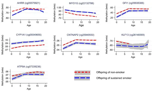
Longitudinal analyses were performed to investigate whether the effect of smoking on offspring methylation at birth was transient or persisted into later life. Methylation data were available for offspring in ARIES at age 7 [mean age when blood samples were taken 7.5 (SD 0.1)] and at age 17 [mean age 17.1 (SD 1.0)]. We inves-tigated changes in methylation levels for the CpG sites that were found to be associated with maternal smoking in cord blood using multilevel modelling (Fig.4 and Supplementary
Material, Table S4). For the seven CpG sites, there were changes in methylation found during childhood, while the magnitude of change was quite small during adolescence. At CYP1A1
(cg05549655) andCNTNAP2(cg25949550), while there was some evidence for change in methylation among the offspring of the smokers and non-smokers over time, the difference in methyla-tion between groups persisted. Evidence for differing rates of change in methylation level between the offspring of smokers and non-smokers was found at AHRR (cg05575921), MYO1G
(cg22132788),GFI1(cg09935388) andKLF13(cg26146569) between birth and age 7 (P-value for difference in methylation change 0.01–1 × 10⁻¹⁶).
AtMYO1G(cg22132788), methylation level for the offspring of smokers deviated more from the level of the offspring of non-smokers over time, whereas atGFI1 (cg09935388) andKLF13
(cg26146569) there was some recovery of methylation towards the level of those not exposed to prenatal maternal smoke. At
AHRR (cg05575921) during childhood, methylation increased at a faster rate in the offspring of smokers with evidence for a
‘catchup’ in methylation among the offspring of smokers [a 2.04% (95% CI 1.72, 2.36%) average yearly increase in methyla-tion for the offspring of sustained smokers compared with a 1.28% (95% CI 0.97, 1.59%) increase in methylation for the off-spring of non-smokers, between birth and age 7]. However, dur-ing adolescence, levels of AHRR (cg05575921) methylation decreased among both the smoker and non-smoker offspring, with methylation in the smoker offspring decreasing at a faster rate [a 0.33% (95% CI 0.26, 0.40%) average yearly decrease in methylation for the offspring of sustained smokers compared with a 0.17% (95% CI 0.12, 0.22%) decrease in methylation for the offspring of non-smokers, between age 7 and 17], leading again to a difference in methylation levels. A similar trend was found forATP9A(cg07339236), although this was not as robust.
For the CpG sites which showed a persistent difference in methylation between the offspring of smokers and non-smokers [MYO1G (cg22132788), CYP1A1 (cg05549655) and CNTNAP2
(cg25949550); Fig.4], we sought to determine whether the asso-ciations with maternal smoking were explained by a direct‘ crit-ical period’effect of smoking in pregnancy or via an indirect pathway involving postnatal smoke exposure. Identifying these underlying mechanisms of association is hampered by the high correlation (0.87) between sustained smoking in pregnancy and maternal smoking at 8 weeks postnatally. To disentangle the effect of smoking in pregnancy on offspring methylation versus smoking postnatally, we implemented a structured approach to model the effects of the binary maternal smoking exposure at three time points (in pregnancy and postnatally at 8 weeks and 61 months) on offspring methylation at age 7 (Table4). The hy-pothesis for anin uterocritical period is supported by data at all three CpG sites, with this model not being substantially different from the saturated model (P≥0.06). However, a model for effect modification of the intrauterine exposure was also supported by the data (P≥0.06) and forCYP1A1andCNTNAP2a critical per-iod at 8 weeks postnatally could not be ruled out (P≥0.18). As the
in uterocritical period model is nested within the effect modifi ca-tion model, we further performed a direct ANOVA test to investi-gate whether effect modification provided a betterfit of the data than thein uterocritical period model. This was found to be the case forCYP1A1(P= 0.009), but not forMYO1GorCNTNAP2(P≥
0.22), where thein uterocritical period was found to be the best model (Table4).
[image:4.595.56.286.94.546.2]For the CpG sites which showed evidence of methylation difference between the offspring of smokers and non-smokers at age 17 [AHRR (cg05575921), MYO1G (cg22132788), CYP1A1 Table 2.Differences in potential confounding factors between
individuals in ARIES whose mothers did not smoke in pregnancy compared with sustained smokers
Non-smoker (N= 699)a
Sustained (N= 91)a
P-value for difference
Sex
Male 49.4 47.3 0.71
Female 50.6 52.8
Maternal age (years) 30.5 (4.1) 27.9 (5.4) <0.001 Maternal age (categories)
<25 7.6 30.8 <0.001
25–30 39.2 37.4
>30 53.2 31.9
Parity
0 45.5 51.1 0.53
1 37.7 32.2
2 13.2 11.1
3+ 3.6 5.6
Maternal education
CSE/vocational 13.9 33.0 <0.001
O-level 32.7 40.9
A-level 30.4 18.2
Degree 23.1 8.0
Social class
I 22.9 4.9 <0.001
II 44.0 39.0
III (NM) 24.4 20.7
III (M) 5.3 22.0
IV or V 3.5 13.4
Maternal BMI 22.8 (3.7) 23.0 (3.6) 0.63 Maternal BMI (categories)
<18.5 3.3 3.5 0.59
18.5–25 79.1 76.5
25–30 13.1 14.1
30+ 4.6 5.9
Maternal weight 61.6 (10.4) 61.5 (10.5) 0.95 Alcohol
Non-drinker 34.2 40.9 0.040
Drank before 18 weeks of gestation
15.6 5.7
Still drinking at 18 weeks of gestation
50.2 53.4
Paternal smoking
Non-smoker 78.6 20.9 <0.001
Smoker 21.4 79.1
aNvaries according to completeness of data on baseline characteristics.
2204 | Human Molecular Genetics, 2015, Vol. 24, No. 8
at University of the West of England on December 2, 2015
http://hmg.oxfordjournals.org/
(cg05549655) andCNTNAP2(cg25949550); Fig.4], we followed up the association between methylation at these sites and own smoking among the adolescents (Supplementary Material, Table S4). The correlation between sustained smoking in preg-nancy and own smoking (of the adolescent) was found to be 0.16. For the majority of CpG sites for which the association with smoking in pregnancy was evident at age 17, there was directional consistency of the association between own smoking and methy-lation at this time point.However, the magnitude of the associ-ation with own smoking was smaller than for the sustained maternal prenatal smoking analysis at all CpG sites, with the ex-ception ofAHRR(cg05575921), where the effect size for sustained smoking in pregnancy was−3.6% (95% CI−4.6,−2.6%) compared with−3.4% (95% CI−4.4, 2.4%) for the own smoking analysis, sug-gesting that not all of the association between maternal smoking in pregnancy and age 17 methylation can be explained through the mediating role of the adolescent’s own smoking.
We next investigated the associations between sustained smoking in pregnancy and methylation at age 17, excluding those offspring who reported smoking themselves (Supplemen-tary Material, Table S5). The magnitude and direction of associ-ation of the CpG sites were comparable to those in the full analysis, providing more evidence that own smoking is not fully mediating the observed association between maternal smoking in pregnancy and offspring methylation at age 17. In addition, this was with the notable exception of CpG sites atAHRR, where the effect size was halved [from−3.6% (95% CI−4.6,−2.6%) to
−1.8% (95% CI 0.0,−3.6%)] when adolescents who reported smoking were excluded from the analysis. This provides some indication that personal smoking by adolescents and its correlation with ma-ternal smoking could be driving the apparent persistent methyla-tion difference in this gene region at age 17.
Assessing causality of intrauterine associations using paternal smoking as a negative control
Finally, parental comparisons of associations between smoking during pregnancy and methylation levels at the top CpG sites showed consistently larger effect estimates for prenatal maternal
smoking than for paternal smoking at all three time points (Fig.5). In addition, adjusting for paternal smoking in maternal associations made little difference to affect estimates while ad-justing for maternal smoking attenuated all paternal associa-tions. For example, in the analysis of methylation in cord blood, any smoking by mothers during pregnancy was associated with a 6.1% (95% CI−7.1,−5.1%) reduction in cord blood methylation at cg05575921 (AHRR), which was not substantially attenuated with adjustment for partner’s smoking (−5.6%; 95% CI −6.7,
−4.5%). Smoking by partners during pregnancy was associated with a 2.1 (−2.8,−1.3%) reduction in cord blood methylation at cg05575921 (AHRR), which was fully attenuated with adjustment for maternal smoking (−0.01%; 95% CI−0.01, 0.00%).
Discussion
In a large longitudinal birth cohort with genome-wide methyla-tion measured at three different time points in the offspring, wefirst identified 15 CpG sites that were differentially methy-lated in cord blood at birth. These sites are located in seven gene regions, six of which have been previously identified in other EWAS for exposure to maternal smokingin utero(18,19). The top hit in this analysis was located within theAHRR[aryl hydrocarbon receptor (AHR) repressor] gene. CpG sites located in this gene region have previously been shown to be differential-ly methylated in smokers in several studies (13–18,30). In particu-lar, the top hit in this analysis (cg05575921,P= 1.41 × 10⁻³⁰) was
identified in previous studies including an epigenome-wide as-sociation study for maternal smoking and both cord and neo-natal blood DNA methylation (18).
[image:5.595.59.538.57.283.2]At this site, an 8.1% (95% CI 6.9, 9.3%) reduction in cord blood methylation with sustained prenatal smoking exposure was identified, which is in line with the median methylation differ-ence of medium and high cotinine versus no exposure in a previ-ous EWAS (18), which was 5.4 and 9.9%, respectively. These associations were largely robust to adjustment for a number of genetic, environmental and cell-type specific confounding factors, supporting a causal effect of maternal smoking during pregnancy on offspring methylation at birth. However, for two
Figure 1.Manhattan and QQ plot for epigenome-wide association study of sustained smoking in pregnancy on cord blood DNA methylation. *Results are from the analysis
adjusted for batch only (N= 790).
Human Molecular Genetics, 2015, Vol. 24, No. 8 | 2205
at University of the West of England on December 2, 2015
http://hmg.oxfordjournals.org/
Table 3.Differential methylation in cord blood DNA for the offspring of mothers with sustained smoking in pregnancy compared with non-smokers
CpG site Chromosome Gene region Positionc Unadjusted model (N= 790†)a Adjusted model (N= 744‡)b
Effect size Standard error P-value FDR Effect size Standard error P-value FDR
cg05575921 5 AHRR 373 378 −0.081 0.006 1.41 × 10⁻³⁰ 6.59 × 10⁻²⁵ −0.075 0.007 7.64 × 10⁻¹⁸ 3.56 × 10⁻¹²
cg22132788 7 MYO1G 45 002 486 0.062 0.010 1.49 × 10⁻¹⁷ 3.47 × 10⁻¹² 0.054 0.012 1.72 × 10⁻¹⁰ 3.03 × 10⁻⁵
cg12803068 7 MYO1G 45 002 919 0.152 0.021 1.22 × 10⁻¹⁶ 1.90 × 10⁻¹¹ 0.126 0.025 4.47 × 10⁻⁹ 3.47 × 10⁻⁴
cg09935388 1 GFI1 92 947 588 −0.167 0.023 1.14 × 10⁻¹³ 1.19 × 10⁻⁸ −0.174 0.028 1.95 × 10⁻¹⁰ 3.03 × 10⁻⁵
cg14179389 1 GFI1 92 947 961 −0.078 0.012 1.28 × 10⁻¹³ 1.19 × 10⁻⁸ −0.070 0.015 2.49 × 10⁻⁸ 1.45 × 10⁻³
cg18146737 1 GFI1 92 946 700 −0.144 0.019 2.67 × 10⁻¹³ 2.07 × 10⁻⁸ −0.141 0.024 2.10 × 10⁻⁹ 1.96 × 10⁻⁴
cg05549655 15 CYP1A1 75 019 143 0.015 0.002 1.05 × 10⁻¹² 6.99 × 10⁻⁸ 0.016 0.002 4.73 × 10⁻¹⁰ 5.51 × 10⁻⁵
cg06338710 1 GFI1 92 946 187 −0.214 0.032 1.32 × 10⁻¹⁰ 7.69 × 10⁻⁶ −0.200 0.039 5.93 × 10⁻⁷ 0.02
cg12876356 1 GFI1 92 946 825 −0.179 0.029 1.60 × 10⁻⁹ 8.27 × 10⁻⁵ −0.206 0.035 1.36 × 10⁻⁸ 9.09 × 10⁻⁴
cg25949550 7 CNTNAP2 145 814 306 −0.006 0.001 3.06 × 10⁻⁹ 1.43 × 10⁻⁴ −0.006 0.001 8.08 × 10⁻⁶ 0.09
cg11902777 5 AHRR 3 68 843 −0.005 0.001 1.59 × 10⁻⁸ 6.74 × 10⁻⁴ −0.004 0.001 5.62 × 10⁻⁵ 0.17
cg12101586 15 CYP1A1 75 019 203 0.060 0.010 2.18 × 10⁻⁸ 8.49 × 10⁻⁴ 0.070 0.013 5.71 × 10⁻⁸ 2.96 × 10⁻³
cg18316974d
1 GFI1 92 947 035 −0.083 0.019 4.05 × 10⁻⁸ 1.45 × 10⁻³ −0.086 0.023 4.96 × 10⁻⁶ 0.08
cg26146569 15 KLF13 31 637 592 −0.072 0.013 4.61 × 10⁻⁸ 1.54 × 10⁻³ −0.083 0.016 2.97 × 10⁻⁷ 0.01
cg07339236d
20 ATP9A 50 312 490 −0.016 0.004 8.71 × 10⁻⁸ 2.71 × 10⁻³ −0.014 0.006 3.60 × 10⁻⁴ 0.24
cg09662411 1 GFI1 92 946 132 −0.127 0.024 1.26 × 10⁻⁷ 3.66 × 10⁻³ −0.147 0.029 6.60 × 10⁻⁷ 0.02
cg18092474 15 CYP1A1 75 019 302 0.077 0.016 2.17 × 10⁻⁷ 5.94 × 10⁻³ 0.057 0.019 5.12 × 10⁻⁴ 0.24
cg04180046 7 MYO1G 45 002 736 0.050 0.011 2.29 × 10⁻⁷ 5.94 × 10⁻³ 0.043 0.012 4.23 × 10⁻⁵ 0.16
cg25189904 1 GNG12 68 299 493 −0.055 0.011 4.32 × 10⁻⁷ 0.01 −0.052 0.013 1.20 × 10⁻⁴ 0.20
cg04598670 7 ENSG00000225718 68 697 651 −0.074 0.015 4.80 × 10⁻⁷ 0.01 −0.074 0.018 4.35 × 10⁻⁵ 0.16
cg27629977 2 CTNNA2 80 531 633 0.009 0.002 5.82 × 10⁻⁷ 0.01 0.012 0.003 4.07 × 10⁻⁷ 0.01
cg10835306 9 NOTCH1 139 396 760 −0.079 0.016 1.04 × 10⁻⁶ 0.02 −0.061 0.019 2.34 × 10⁻³ 0.30
cg00483459 3 ALS2CL 46 735 782 −0.049 0.010 1.32 × 10⁻⁶ 0.03 −0.056 0.011 2.19 × 10⁻⁶ 0.05
cg22549041d
15 CYP1A1 75 019 251 0.069 0.014 1.42 × 10⁻⁶ 0.03 0.089 0.017 2.60 × 10⁻⁷ 0.01
cg22937882d
5 AHRR 4 05 774 0.036 0.008 1.98 × 10⁻⁶ 0.04 0.036 0.011 3.13 × 10⁻⁴ 0.24
cg11196333 1 CHI3L1 203 154 370 −0.060 0.013 2.58 × 10⁻⁶ 0.05 −0.066 0.015 1.06 × 10⁻⁵ 0.11
cg00624799d
15 ZNF710 90 605 618 −0.029 0.006 2.79 × 10⁻⁶ 0.05 −0.037 0.007 4.19 × 10⁻⁷ 0.01
cg00560284 12 SPATS2 49 783 222 −0.016 0.003 2.84 × 10⁻⁶ 0.05 −0.013 0.004 1.79 × 10⁻³ 0.29
Effect size = difference in methylation level (beta) between offspring of sustained smokers and non-smokers.
aAdjusted for batch only:†N= 91 sustained smokers;N= 699 non-smokers (defined as those who did not smoke pre-pregnancy or in pregnancy).
bAdjusted for maternal age, maternal education, household social class, paternal smoking, maternal alcohol in pregnancy and batch:‡N= 76 sustained smokers;N= 668 non-smokers (defined as those who did not smoke pre-pregnancy
or in pregnancy).
cChromosomal position based on NCBI human reference genome assembly Build 37.3.
dNaeemflagged CpG site (43).
2206
|
Human
Molecular
Genetics,
2015,
V
ol.
24,
No.
8
Figure 2.Methylation level (beta) at key CpG sites associated with sustained smoking in pregnancy by the duration of smoking (number of trimesters).
Human
Molecular
Gene
tics,
2015,
V
ol.
24,
No.
8
|
2207
Figure 3.Methylation level (beta) at key CpG sites associated with sustained smoking in pregnancy by the intensity of smoking (number of cigarettes per day).
2208
|
Human
Molecular
Genetics,
2015,
V
ol.
24,
No.
8
of the CpG sites followed up in downstream analysis,CNTNAP2
(cg25949550) andATP9A(cg07339236), the FDRP-values were 0.09 and 0.24, respectively, in the adjusted model, but effect esti-mates were largely unchanged between the adjusted and un-adjusted model. In addition,ATP9A(cg07339236) wasflagged up in Table3as a low-quality probe based on a comprehensive as-sessment reported by Naeemet al.(43).
A regional analysis of EWAS hits provided some evidence for localized clustering around the top CpG site (that with the smallest
P-value in the EWAS) inAHRR,MYO1G,GFI1andCYP1A1, although there was little evidence for strong co-methylation within the gene regions indicating independence of methylation levels at each CpG site, supporting our use of single site analysis in the EWAS. We assessed the biological gradient of smoke exposure in pregnancy and identified a dose-dependent response of
methylation with both increased intensity and duration of smok-ing. In this analysis, we found that methylation in the offspring of mothers who smoked only in one trimester, namely thefirst, was largely comparable to that of unexposed offspring. Thesefi nd-ings are in line with previous studies, which showed no differ-ence in mean methylation at AHRR between mothers who never smoked and those who smoked early in their pregnancy (20,23), suggesting that sustained exposure to maternal smoking
[image:9.595.54.538.56.348.2]in uterois required to induce changes in methylation which are detectable in cord blood. In contrast to the view that early preg-nancy represents a critical window for environmentally induced epigenetic change, epigenetic reprogramming appears to occur throughout prenatal development and postnatally (45) and thesefindings imply a cumulative effect of smoke exposure throughout pregnancy on offspring methylation at birth.
Figure 4.Longitudinal trajectories of methylation at key CpG sites in the offspring of non-smokers and sustained smokers during pregnancy from birth to age 17.
Table 4.An exploration of critical period and other lifecourse effects that may underlie the persistence of associations between maternal smoke exposure in pregnancy and offspring methylation at age 7
Lifecourse hypothesis MYO1G (cg22132788) CYP1A1 (cg05549655) CNTNAP2 (cg25949550)
F-statistic P-value F-statistic P-value F-statistic P-value
Critical period in pregnancy 0.68 0.66 1.62 0.14 2.05 0.06
Critical period at 8 weeks postnatally 3.74 0.001 1.48 0.18 1.00 0.42
Critical period at 61 months postnatally 4.72 9.74 × 10⁻⁵ 5.07 4.02 × 10⁻⁵ 3.35 0.003
Accumulation of risk over time 3.89 7.77 × 10⁻⁴ 5.83 5.77 × 10⁻⁶ 4.19 3.68 × 10⁻⁴
Effect modification of critical period in pregnancy by postnatal exposure
0.82 0.51 0.08 0.98 2.32 0.06
Critical period in pregnancy nested within effect modification modela
0.40 0.67 4.8 0.009 1.50 0.22
Results of ANOVA test against a saturated model; a smallerF-statistic (and largerP-value) provides evidence of a betterfit of data to the hypothesized model. aResults of ANOVA test against effect modification model.
Human Molecular Genetics, 2015, Vol. 24, No. 8 | 2209
at University of the West of England on December 2, 2015
http://hmg.oxfordjournals.org/
[image:9.595.55.541.409.526.2]Figure 5.Parental comparisons of associations between any versus no smoking in pregnancy on offspring methylation at CpG sites most associated with sustained maternal smoking in pregnancy at all three time points.
2210
|
Human
Molecular
Genetics,
2015,
V
ol.
24,
No.
8
Nonetheless, knowledge of smoking intensity in thefirst tri-mester is important as a predictive marker of smoking later in pregnancy and hence of epigenetic change in the offspring. This has been confirmed in an analysis of 374 ARIES mothers–
child pairs for whom urinary cotinine was collected in thefirst trimester of pregnancy. An EWAS of maternal cotinine levels and cord blood methylation in this subsample was able to identify a signal atAHRR (cg05575921), which surpassed the Bonferroni threshold (P-value = 3.31 × 10⁻⁸; Supplementary Material, Fig. S8).
Whether pregnancy represents a critical period for determin-ing offsprdetermin-ing methylation patterns at later time points in child-hood in response to maternal smoking was also investigated. A longitudinal assessment of methylation marks associated with maternal smoke exposure in pregnancy found that whereas some CpG sites showed recovery of methylation to the level of offspring not exposed (GFI1, KLF13 and ATP9A), other sites showed persistently perturbed patterns (AHRR,MYO1G,CYP1A1
andCNTNAP2).
This prospective study design coupled with serial sampling at multiple time points provides powerful evidence of the persist-ence of DNA methylation changes inducedin utero. In addition, longitudinal modelling of the effects of exposure‘windows’ pro-vides evidence that prenatal exposure to smoking has persistent effects on later offspring DNA methylation, which outweighs the postnatal influence of maternal smoking or own smoking in adolescence at some CpG sites. Effect modification of prenatal exposure at later time points was also evident, indicating that postnatal exposure might have some further impact on the per-sistence of the methylation marks in those exposed to smoking
in utero. However, there was no strong evidence that the effect modification model was more consistent with the observed data than thein uterocritical period model at most sites. These observations are consistent with and significantly extend previ-ous analyses of long-term smoking-induced perturbations of DNA methylation (15,23,24,26). One exception to this is thefi nd-ing that own smoknd-ing by the offsprnd-ing at age 17 is strongly asso-ciated withAHRRmethylation at this time point which might therefore be underlying the apparently persistent effect of mater-nal smoking in pregnancy at this gene region, as shown in Figure4. This is perhaps because this site is most sensitive to smoking exposure and would therefore detect adolescent own smoking most readily.
In addition, the use of paternal smoking as a negative control demonstrates the biological effect of thisin uteroexposure at all time points considered. While methylation differences identified between maternal and paternal smoking at later time points might be attributed to the differential influences of these expo-sures postnatally, the similar trends identified in cord blood when no influence of the postnatal environment had been pre-sent provide further support for the causal effect of maternal smoking in pregnancy on offspring methylation. In addition, the low levels of tobacco exposure from partner smoking in non-smoking pregnant women in this cohort suggest that the use of partner’s smoking as a negative control for investigating intrauterine effects is valid (40). Mendelian randomization is an-other technique that may be used to bolster causal inference in this context (35,46,47). A SNP, rs1051730, located in the
CHRNA5-CHRNA3-CHRNB4nicotinic acetylcholine receptor gene cluster (chromosome 15q25), is robustly associated with smoking heaviness (48) and has also been associated with a reduced abil-ity of women to quit smoking in pregnancy (49). If an association was observed between maternal rs1051730 and offspring DNA methylation of mothers who smoked during pregnancy, this would provide further evidence of an intrauterine effect.
However, we were underpowered to investigate this formally within our sample of sustained smokers.
Strengths of our study include the application of the Illumina Infinium®HM450 BeadChip technology to assess genome-wide
methylation profiles at multiple time points from birth until late adolescence in a large, longitudinal cohort study. The wealth of phenotypic data in ALSPAC has aided the thorough assess-ment of potential confounding factors, a detailed analysis of the dose-dependence of methylation to smoke exposurein utero
and an investigation into the relative roles of intrauterine and postnatal smoking using questionnaire data on smoking habits taken from multiple time points in both parents and offspring. Longitudinal modelling (50,51) and robust statistical methods (34,39,52) have also been used to strengthen causal inference.
Limitations of the analysis include differential follow-up of smokers compared with non-smokers, where only 14.3% of mothers in the ARIES sample (selected based on blood sample availability up to 17 years postnatally) smoked in pregnancy com-pared with 30.2% in the wider cohort. Technical limitations relate to the HM450 BeadChip in that it covers only 1.7% of CpG sites across the genome. A more comprehensive appraisal may elicit additional relationships between the exposure in question and DNA methylation, or indeed locus-specific paternal effects which were not evident here (53–55). In addition, the use of ques-tionnaires to obtain data on parental smoking may result in under-reporting of smoking behaviour. However, to minimize the influence of maternal under-reporting, we excluded from the analyses individuals who had reported smoking before but not during pregnancy. In addition, a strong correlation between self-reported questionnaire data on smoking behaviour and plas-ma cotinine levels has been found (18,40) and maternal cotinine was found to be highly correlated with the sustained smoking variable (r= 0.76) in the subsample of ARIES participants with
first trimester maternal cotinine data available (N= 323). This analysis was limited to blood samples with mixed cell composition. Although no differences were found in the analysis with estimated cell-type correction, as has been shown previous-ly (18,19), it is unclear how effective the method used to correct for cell-type proportions is in these samples since the reference data sets are available only for adult peripheral blood (56). In add-ition, some of the DNA samples included in this analysis came from buffy coats rather than whole blood and there is no refer-ence cell-type correction available for buffy coat DNA. It should also be emphasized that the associations identified may be spe-cific to blood as an analysis of buccal epithelium and placenta did not identify the same smoking-associated methylation differ-ences in these tissues (23). This limitation of tissue specificity, as well as the lack of expression data currently available on these samples, limits the assessment of functional conse-quences of these methylation changes.
Given evidence for causal associations between maternal smoking in pregnancy and methylation changes in the offspring, it is important to consider whether these induced changes are also associated with the adverse perinatal and offspring out-comes associated with exposure to smokingin utero. Further work is required which may link smoking-responsive DNA methylation variation to health and development (57–59). In addition, whether DNA methylation is a true mediating mechan-ism of these associations or simply an exposure indicator may be explored by extending causal inference (35,47,60).
Transient environmental exposures during critical windows of development are known to affect the establishment of epigen-etic marks, which are evident at birth (57) and may persist until later life (58,59). Findings from this study highlight the sensitivity
Human Molecular Genetics, 2015, Vol. 24, No. 8 | 2211
at University of the West of England on December 2, 2015
http://hmg.oxfordjournals.org/
of the methylome to maternal smoking during fetal development and the long-term impact of such an exposure. Results strength-en causal inferstrength-ence in this area and thefinding that sustained smoking appears to be necessary to induce methylation changes has profound implications for antenatal care and the long-term effects on offspring health, directing potential intervention strat-egies at cessation of smoking early in pregnancy, such as at the
first antenatal appointment.
Thesefindings could also have very useful applications in epi-demiological studies. Given the magnitude and persistence of methylation change in relation to maternal exposurein utero, it is important to consider the potential confounding effect of maternal smoking in future EWAS studies attempting to identify sites associated with own smoking. The persistence of some (but not all) methylation marks at later time points presents the opportunity to use methylation signatures as an archive of historical exposure, particularly if methylation patterns can be robustly modelled over time and previous exposure inferred. A simplistic example would be to define prenatal smoking expos-ure, using DNA methylation signatures in children of women where no smoking history had been collected. In addition, the contrast between stable and reversible sites may be useful in dis-criminating betweenin uteroexposure and later life exposure.
Materials and Methods
Study design
We examined offspring DNA methylation in relation to self-reported maternal smoking during pregnancy in a subset of par-ticipants from ALSPAC using methylation data from the Illumina Infinium®HumanMethylation450 BeadChip assay (Illumina, Inc.,
CA, USA) (61).
Cohort and selection of participants
ALSPAC is a large, prospective cohort study based in the South West of England. A total of 14 541 pregnant women resident in Avon, UK, with expected dates of delivery 1 April 1991 to 31 December 1992 were recruited and detailed information has been collected on these women and their offspring at regular intervals (41,42). The study website contains details of all the data that are available through a fully searchable data dictionary (http://www. bris.ac.uk/alspac/researchers/data-access/data-dictionary/).
As part of the ARIES (http://www.ariesepigenomics.org.uk/) project, the Infinium HM450 BeadChip has been used to generate epigenetic data on 1018 mother–offspring pairs in the ALSPAC co-hort. The ARIES participants were selected based on availability of DNA samples at two time points for the mother (antenatal and at follow-up when the offspring was in adolescence) and at three time points for the offspring [neonatal, childhood (age 7) and adolescence (age 17)].
Written informed consent has been obtained for all ALSPAC participants. Ethical approval for the study was obtained from the ALSPAC Ethics and Law Committee and the Local Research Ethics Committees.
Laboratory methods, quality control and pre-processing
Cord blood and peripheral blood samples (whole blood, buffy coats or blood spots) were collected according to standard proce-dures. The DNA methylation wet laboratory and pre-processing analyses were performed at the University of Bristol as part of the ARIES project. Following extraction, DNA was bisulphite-converted using the Zymo EZ DNA MethylationTM kit (Zymo,
Irvine, CA, USA). Following conversion, genome-wide methyla-tion status of over 485 000 CpG sites was measured using the Infinium HM450 BeadChip according to the standard protocol. The arrays were scanned using an Illumina iScan and initial qual-ity review was assessed using GenomeStudio (version 2011.1).
The Infinium HM450 BeadChip assay detects the proportion of molecules methylated at each CpG site on the array. For the sam-ples, the methylation level at each CpG site was calculated as a beta value (β), which is the ratio of the methylated probe intensity and the overall intensity and ranges from 0 (no cytosine methy-lation) to 1 (complete cytosine methymethy-lation) (62,63). Methylation data were pre-processed using in R (version 3.0.1), with back-ground correction and subset quantile normalization performed using the pipeline described by Touleimat and Tost (64).
Samples from all time points in ARIES were distributed across slides using a semi-random approach (sampling criteria were in place to ensure that all time points were represented on each array) to minimize the possibility of confounding by batch effects. In addition, during the data generation process a wide range of batch variables were recorded in a purpose-built labora-tory information management system (LIMS). The main batch variable was found to be the bisulphite conversion (BCD) plate number. Samples were converted in batches of 48 samples and each batch identified by a plate number.
The LIMS also reported QC metrics from the standard control probes on the 450K BeadChip for each sample. Samples failing QC (average probeP-value of≥0.01) were repeated and if unsuc-cessful excluded from further analysis. As an additional QC step genotype probes were compared with SNP-chip data from the same individual to identify and remove any sample mismatches. For individuals with no genome-wide SNP data, samples wereflagged if there was a sex-mismatch based on X-chromosome methylation.
In addition to these QC steps, probes that contained <95% of signals detectable above background signal (detectionP-value of <0.01;N= 7938) were excluded from analysis. After excluding these probes, as well as control probes and probes on sex chro-mosomes, a total of 466 432 CpG sites were included in the main analysis for cord blood methylation. At age 7, 471 347 CpG sites were included and at age 17, 469 902 CpG sites were included in the main analysis, following the same exclusion criteria.
In uteroexposure variables
Information on mothers’smoking status during pregnancy was obtained in questionnaires administered at 18 and 32 weeks of gestation. Information was obtained about whether the mother smoked in each trimester of pregnancy and the number of cigar-ettes smoked on average per day. From these data, a dichotom-ous variable for sustained maternal smoking during pregnancy was derived. A mother was classified as a sustained smoker if she smoked in all three trimesters, smoked in thefirst and third trimester but not the second or smoked in the second and third trimesters but not thefirst. The reference group consisted of mothers who had reported not smoking in all three trimesters or before pregnancy. We excluded all individuals who smoked in one trimester only (i.e. not sustained) or who had missing infor-mation of smoking for two or more trimesters. Of those with missing information on one trimester, women were classified as a sustained smoker if they said they smoked in the other two trimesters.
For investigating the dose-dependent effects of maternal smoking in pregnancy on DNA methylation in cord blood, a vari-able was derived for the duration of smoking in pregnancy (0, 1, 2
2212 | Human Molecular Genetics, 2015, Vol. 24, No. 8
at University of the West of England on December 2, 2015
http://hmg.oxfordjournals.org/
or all three trimesters) as well as the intensity of smoking in preg-nancy (0, 1–4, 5–9, 10–14 and 15+ cigarettes/day).
Data on cotinine levels were available for a small subset of the ARIES mothers (n= 374). Cotinine levels (ng/ml) were assessed from a single urine sample taken during thefirst trimester of pregnancy. For most mothers, the samples were collected as part of routine clinical care but some samples were obtained specifically for ALSPAC. Urine samples were stored at−20°C and allowed to thaw at room temperature before use. Cotinine was measured using the Cozart Cotinine Enzyme Immunoassay (Concateno UK, Abingdon) urine kit. Where required, samples were diluted using cotinine-free serum (fetal calf serum). Absorb-ance was measured spectrophotometrically at a wavelength of 450 nm. Maternal cotinine levels were categorized into four groups: <70, 70–900, 900–3000 and >3000 ng/ml, which roughly correspond with self-reported non-smoking, 1–4 cigar-ettes per day, 5–14 cigarettes per day and >14 cigarettes per day, respectively (40).
Offspring methylation outcome
The main outcome measure in this analysis was DNA methyla-tion level at each of the CpG sites in cord blood samples. However, we also undertook an EWAS for maternal prenatal smoking in samples of peripheral blood when the children were age 7 and 17 years and followed up sites that reached genome-wide signifi -cance to investigate the persistence of methylation marks in the offspring over time.
Confounders
Variables considered as potential confounders in this analysis were maternal age, pre-pregnancy BMI, pre-pregnancy weight, parity, educational attainment, social class, alcohol intake and paternal smoking. Maternal age at delivery was derived from date of birth, which was recorded at that time. At enrolment, the mother was asked to record her height and pre-pregnancy weight, from which BMI was calculated. Mother’s parity was also recorded in a questionnaire completed during pregnancy. Based on questionnaire responses, highest educational qualifi ca-tion for the mother was collapsed into one of thefive categories from none/ Certificate of Secondary Education (CSE, the lower level of two national school exams that were taken when these women were in school at age 16) to university degree. In addition, the highest parental occupation was used to allocate the children to family social class groups using the 1991 British Office of Popu-lation Censuses and Surveys classification. Self-reported alcohol use was obtained in the questionnaire administered at 18 weeks of gestation and individuals were categorized based on whether they were non-drinkers, drank before 18 weeks of gestation or were still drinking alcohol at 18 weeks of gestation. Information on partners’smoking during pregnancy was obtained from self-reports at 18 weeks of gestation. Where self-reported data on partner smoking were not available (16.3% of partners), maternal reports were used. The bisulphite conversion batch for each sam-ple was also included in the analysis to adjust for batch effects.
Statistical analysis
Using offspring DNA samples taken from cord blood (at birth), we investigated methylation levels at 466 488 CpG sites across the genome. Methylationβvalues at each CpG site were transformed to obtainM-values [log2(β/(1−β)] for statistical analysis (62).
Multivariable linear regression was used to perform associ-ation tests between maternal cigarette smoking andM-values
at each CpG site as the outcome. The main exposure measure in our analysis was sustained smoking in pregnancy versus no smoking and the main outcome was cord blood DNA methylation level. Analyses were run with and without adjustment for a num-ber of potential confounders found to be associated with smok-ing status in pregnancy (Table2). DNA methylation sites were annotated based on data provided by Illumina (63).
Wefirst identified‘EWAS-significant’hits using a Bonferroni correction, where associations below a threshold of 1.07 × 10⁻⁷
were considered a likely true positive worthy of further examin-ation. However, this Bonferroni correction assumes independent tests and so, as correlation of DNA methylation within gene regions means that CpG sites may not be truly independent, a less conservative FDR procedure based on the Benjamini–
Hochberg method was also used to account for multiple testing (65). For this, CpG sites with FDR less than a 0.05 threshold were labelled as EWAS-significant.
It has been demonstrated that differences in methylation can arise as a result of variability of cell composition in whole blood (56). As smoking is known to influence cell composition (66), in order to ensure the results are not influenced by variation in cell-type fraction between samples, we estimated the fraction of CD8T-, CD4T-, NK- and B-cells, monocytes and granulocytes in the samples using theestimateCellCountsfunction in theminfi
Bioconductor package implemented in R (67). We investigated differences in estimated cell count by smoking status and ana-lyses were repeated adjusting for cell composition by including each blood cell fraction as a covariate in the multivariate linear regression.
Given the previous evidence for sex-specific DNA methylation differences in relation to prenatal smoke exposure (68), we under-took EWAS stratified by sex of the offspring and investigated whether there were any sex-specific CpG sites found to be asso-ciated with maternal smoke exposure.
We next used a web-based plotting tool, coMET (44), to inves-tigate the genomic regions of interest from our main EWAS analysis. This tool permits the visualization of methylation cor-relation between CpG sites, which was limited to a maximum of 75 CpG sites around to the top site of interest and within the gene region identified. In addition, the plots were annotated with functional genomic features based on the ENCODE project (geneENSEMBL, CGI, ChromHMM, DNAse, RegENSEMBL and SNPs).
Further analyses were performed to investigate whether the level of methylation differed depending on the duration and in-tensity of smoking to which the offspring were exposedin utero. For this, the untransformed methylationβvalues for the top CpG sites in each gene region reaching genome-wide significance were plotted against a variable for the duration of smoking in pregnancy as well as the intensity of smoking in pregnancy.
We also investigated whether the methylation alterations associated with prenatal exposure to maternal smoking per-sisted when the offspring were age 7 and 17 years. Longitudinal methylation data were extracted from each of probe which exceeded the Bonferroni threshold in cord blood. A multilevel model (50,51) including a random intercept and a linear regres-sion spline term to allow forflexibility was fitted to each of these CpG sites sequentially:
methij¼β0þu0iþβ1sustainediþβ2ageijþβ3ðageij7Þþ
þβ4sustainediageijþβ5sustainediðageij7Þþ
þconfoundersþεij
Human Molecular Genetics, 2015, Vol. 24, No. 8 | 2213
at University of the West of England on December 2, 2015
http://hmg.oxfordjournals.org/
εijeNð0;σ2εÞ
u0ieNð0;σ2uÞ
wherei= 1, . . . (770) indexes the offspring in the analyses,j= 1, 2, 3 indexes the measurement occasion anda+=aifa> 0 or 0
other-wise.β1gives the average difference between smoker and
non-smoker offspring;β2gives the average change in methylation
from birth to adolescence;β3tells us whether there is any change
in this trend (i.e.β2) from childhood to adolescence;β4tells us
whether there is a difference in methylation change between smoker and non-smoker offspring andβ5tells us whether
off-spring of smokers and non-smokers have adifferentchange to the trend (i.e.β2) of methylation change from birth to childhood.
From these we can calculate the change in methylation from 0 to 7 for children of non-smokers (β2) and smokers (β2+β4), and the
change from 7 to 17 for children of non-smokers (β2+β3) and
smo-kers (β2+β3+β4+β5). For each CpG site, we used a multilevel
model, adjusting for batch and thefirst 20 independent surrogate variable components (which account for heterogeneity between the cord blood and peripheral blood samples).
Strategies were then implemented to estimate the potential role of non-intrauterine mechanisms in the observed associa-tions at later time points. Wefirst considered the potential role of postnatal parental smoking in explaining the persistence of methylation differences at age 7 and both parental and own smoking at age 17 in the offspring of mothers who smoked com-pared with the offspring of mothers who did not smoke in preg-nancy. Additional information about mothers’smoking status postnatally was obtained in several questionnaires administered after birth, including 8 weeks postpartum and 61 months post-partum. In addition, information about own smoking status was obtained in questionnaires completed by the offspring when they were age 17.
For offspring methylation at age 7, we wished to disentangle a potential causal effect of maternal smoke exposurein utero(i.e. a
‘critical period’hypothesis) from other lifecourse effects, includ-ing the existence of postnatal critical periods of maternal smoke exposure, an accumulation of risk with exposure over time or effect modification ofin uteroexposure by postnatal exposure (52). We implemented a structured approach to model the effects of the binary maternal smoking exposure at three time points (in pregnancy and postnatally at 8 weeks and 61 months) on off-spring methylation. This involvedfirstfitting a saturated model with one coefficient for each combination of exposures at the three time points (maternal smoking in pregnancy × 8 weeks postnatally × 61 months postnatally) using the lm function in R (version 3.0.1). We then specified a series of nested models corre-sponding to each lifecourse hypothesis to be tested against the saturated model using an ANOVA test, with a smallerF-statistic and a largerP-value indicating a betterfit of the data to that model. Nested models considered were anin uterocritical period (maternal smoking in pregnancy), later life critical periods (maternal smoking 8 weeks and maternal smoking 61 months postnatally), accumulation of risk across the three time points (maternal smoking in pregnancy + 8 weeks postnatally + 61 months postnatally) and effect modification ofin uteroexposure postnatally by smoking at the later time points [maternal smoking in pregnancy + (smoking in pregnancy : 8 weeks postnatally) + (maternal smoking in pregnancy : 61 months postnatally)].
For methylation at age 17, we also considered the potential
in-fluence of own smoking by the offspring in explaining persistence in methylation signatures associated with intrauterine exposure
by running the same multivariable linear regressions this time be-tween own smoking status and methylation as the outcome. From the questionnaires administered when the adolescents were age 17, adolescents who reported that they smoked more than one cig-arette per week were classified as smokers and those who said they had never tried a cigarette at either time point were classified as non-smokers and used as the reference category. For this lysis, we did a look-up of the top hits in the maternal smoking ana-lysis in relation to own smoking in order to contrast effect sizes for personal versus maternal smoking associations with methylation at this time point. In addition, we repeated the main analysis at this time point excluding those offspring who reported own smok-ing to investigate whether this had any influence on the results.
Finally, we compared associations of mothers’and mothers’ partners’smoking during pregnancy with offspring methylation at the three time points (birth, age 7 and age 17), using partner smoking during pregnancy as a negative control (34,36–40). Information on partners’smoking status during pregnancy was obtained in a questionnaire administered at 18 weeks of gesta-tion. In addition, mothers were asked about their partner’s cur-rent smoking at 18 weeks of gestation. The correlation between partner self-report and maternal report of partner smoking was high (r= 0.95) and therefore maternal report was used when part-ners’self-report information was missing. Mutually adjusted models were built by including both maternal and partner smok-ing to account for potential confoundsmok-ing by the smoksmok-ing behav-iour of the other parent.
EWAS were performed using the‘CpG assoc’package (version 2.11) implemented in R (version 3.0.1), multilevel modelling was performed using Stata (version 13) and coMET was run via the web interface (http://epigen.kcl.ac.uk/comet/upload.html). All other analyses were implemented in R (version 3.0.1).
Supplementary Material
Supplementary Material is available atHMGonline.
Acknowledgements
We are extremely grateful to all the families who took part in this study, the midwives for their help in recruiting them and the whole ALSPAC team, which includes interviewers, computer and laboratory technicians, clerical workers, research scientists, volunteers, managers, receptionists and nurses. We thank Jon Heron, Marcus Munafò and Amy Taylor for their helpful com-ments on an earlier draft of this paper, and Matthew Suderman and Tiphaine Martin for sharing their computational expertise.
Conflict of Interest statement. None declared.
Funding
This work was supported by the UK Biotechnology and Biological Sciences Research Council (BB/I025751/1 and BB/I025263/1); the UK Medical Research Council and University of Bristol (MC_UU_12013); the Wellcome Trust (WT083431MF to R.C.R.); the Economic and Social Research Council (RES-060–23-0011 to G.D.S. and C.L.R.) and the European Research Council (DEV-HEALTH 269874 to G.D.S.). Funding to pay the Open Access publi-cation charges for this article was provided by the University of Bristol RCUK.
References
1. Bhattacharya, S., Campbell, D.M., Liston, W.A. and Bhatta-charya, S. (2007) Effect of body mass index on pregnancy
2214 | Human Molecular Genetics, 2015, Vol. 24, No. 8
at University of the West of England on December 2, 2015
http://hmg.oxfordjournals.org/