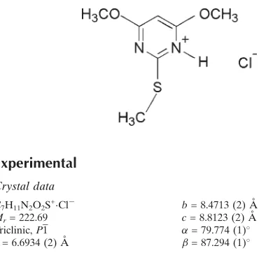
4,6-Dimethoxy-2-(methylsulfanyl)-pyrimidinium chloride
Madhukar Hemamalini and Hoong-Kun Fun*‡
X-ray Crystallography Unit, School of Physics, Universiti Sains Malaysia, 11800 USM, Penang, Malaysia
Correspondence e-mail: hkfun@usm.my
Received 30 December 2009; accepted 30 December 2009
Key indicators: single-crystal X-ray study;T= 100 K; mean(C–C) = 0.002 A˚; Rfactor = 0.027;wRfactor = 0.078; data-to-parameter ratio = 17.0.
In the title compound, C7H11N2O2S +
Cl, the 4,6-dimethoxy-2-(methylsulfanyl)pyrimidinium cation is essentially planar (r.m.s. deviation = 0.043 A˚ ). In the crystal, the anions and cations are connected by intermolecular N—H Cl and C— H Cl hydrogen bonds, forming a two-dimensional network parallel to (011). Adjacent networks are cross-linkedvia– interactions involving the pyrimidinium ring [centroid– centroid distance = 3.5501 (8) A˚ ].
Related literature
For general background to substituted pyrimidines, see: Salas
et al. (1995); Holyet al.(1974); Huntet al. (1980); Baker & Santi (1965); Balasubramani & Fun (2009); For bond-length data, see: Allen et al. (1987). For the stability of the temperature controller used for the data collection, see: Cosier & Glazer (1986).
Experimental
Crystal data
C7H11N2O2S+Cl
Mr= 222.69 Triclinic,P1
a= 6.6934 (2) A˚
b= 8.4713 (2) A˚
c= 8.8123 (2) A˚
= 79.774 (1) = 87.294 (1)
V= 489.24 (2) A˚
Z= 2
MoKradiation
T= 100 K
0.320.220.14 mm
Data collection
Bruker SMART APEXII CCD area-detector diffractometer Absorption correction: multi-scan
(SADABS; Bruker, 2009)
Tmin= 0.836,Tmax= 0.922
9438 measured reflections 2126 independent reflections 1889 reflections withI> 2(I)
Rint= 0.022
Refinement
R[F2> 2(F2)] = 0.027
wR(F2) = 0.078
S= 1.03 2126 reflections 125 parameters
H atoms treated by a mixture of independent and constrained refinement
max= 0.41 e A˚
3 min=0.31 e A˚
[image:1.610.315.565.293.348.2] [image:1.610.50.235.514.695.2]3
Table 1
Hydrogen-bond geometry (A˚ ,).
D—H A D—H H A D A D—H A
N2—H2 Cl1i
0.96 (3) 2.00 (3) 2.9606 (13) 172 (2) C6—H6A Cl1ii
0.96 2.77 3.4896 (16) 132
C6—H6B Cl1 0.96 2.80 3.7002 (15) 157
C7—H7A Cl1iii
0.96 2.76 3.5524 (15) 141
Symmetry codes: (i)x1;y;z; (ii)xþ1;yþ1;z; (iii)x;y;zþ1.
Data collection:APEX2(Bruker, 2009); cell refinement:SAINT
(Bruker, 2009); data reduction:SAINT; program(s) used to solve structure: SHELXTL (Sheldrick, 2008); program(s) used to refine structure:SHELXTL; molecular graphics:SHELXTL; software used to prepare material for publication:SHELXTLandPLATON(Spek, 2009).
MH and HKF thank the Malaysian Government and Universiti Sains Malaysia for the Research University Golden Goose grant No. 1001/PFIZIK/811012. MH thanks Universiti Sains Malaysia for a post-doctoral research fellowship.
Supplementary data and figures for this paper are available from the IUCr electronic archives (Reference: CI5011).
References
Allen, F. H., Kennard, O., Watson, D. G., Brammer, L., Orpen, A. G. & Taylor, R. (1987).J. Chem. Soc. Perkin Trans. 2, pp. S1–19.
Baker, B. R. & Santi, D. V. (1965).J. Pharm. Sci.54, 1252–1257. Balasubramani, K. & Fun, H.-K. (2009).Acta Cryst.E65, o1895.
Bruker (2009).APEX2,SAINTandSADABS. Bruker AXS Inc., Madison, Wisconsin, USA.
Cosier, J. & Glazer, A. M. (1986).J. Appl. Cryst.19, 105–107.
Holy, A., Votruba, I. & Jost, K. (1974).Collect. Czech. Chem. Commun.39, 634–646.
Hunt, W. E., Schwalbe, C. H., Bird, K. & Mallinson, P. D. (1980).Biochem. J.
187, 533–536.
Salas, J. M., Romero, M. A. & Faure, R. (1995).Acta Cryst.C51, 2532–2534. Sheldrick, G. M. (2008).Acta Cryst.A64, 112–122.
Spek, A. L. (2009).Acta Cryst.D65, 148–155. Structure Reports
Online
supporting information
Acta Cryst. (2010). E66, o294 [https://doi.org/10.1107/S1600536809055779]
4,6-Dimethoxy-2-(methylsulfanyl)pyrimidinium chloride
Madhukar Hemamalini and Hoong-Kun Fun
S1. Comment
Pyrimidine and aminopyrimidine derivatives are biologically important compounds as they occur in nature as
components of nucleic acids. Some aminopyrimidine derivatives are used as antifolate drugs (Hunt et al. 1980; Baker &
Santi, 1965). We have recently reported the crystal structure of 4,6-dimethoxy-2(methylsulfanyl)pyrimidine
(Balasubramani & Fun, 2009). In continuation of our studies of pyrimidinium derivatives, the crystal structure
determination of the title compound has been undertaken.
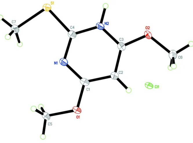
The asymmetric unit of the title compound (Fig. 1) consists of a chloride anion and a
4,6-dimethoxy-2(methyl-sulfanyl)pyridinium cation. Protonation of the pyrimidine base on the N2 site is reflected in a change in the bond angle.
The C4—N1—C1 angle at unprotonated atom N1 is 116.84(13 Å, whereas for protonated atom N2 the C4—N2—C3
angle is 120.03 (13) Å. The bond lengths and angles are normal (Allen et al. 1987).
In the crystal packing (Fig. 2), atoms N2, C7 and C6 act as donors for intermolecular N—H···Cl and C—H···Cl
hydrogen bonds with symmetry related chloride anions (Table 1), forming a two-dimensional network parallel to the
(011). Adjacent networks are cross-linked viaπ–π interactions involving the pyrimidinium ring with centroid···centroid
distance = 3.5501 (8) Å (symmetry code -x, 1-y, 1-z).
S2. Experimental
To a hot methanol solution (20 ml) of 4,6-dimethoxy-2-(methylsulfanyl)pyrimidine (46 mg, Aldrich) was added a few
drops of hydrochloric acid. The solution was warmed over a water bath for a few minutes. The resulting solution was
allowed to cool slowly to room temperature. Crystals of the title compound appeared from the mother liquor after a few
days.
S3. Refinement
Atom H2 was located in a difference Fourier map and refined freely. The remaining H atoms were positioned
geometrically [C–H = 0.93 or 0.96 Å] and were refined using a riding model, with Uiso(H) = 1.2 or 1.5 Ueq(C). A rotating
Figure 1
The molecular structure of the title compound, showing 50% probability displacement ellipsoids and the atom-numbering
Figure 2
The crystal packing of the title compound, viewed along the a axis.
4,6-Dimethoxy-2-(methylsulfanyl)pyrimidinium chloride
Crystal data
C7H11N2O2S+·Cl− Mr = 222.69 Triclinic, P1 Hall symbol: -P 1 a = 6.6934 (2) Å b = 8.4713 (2) Å c = 8.8123 (2) Å α = 79.774 (1)° β = 87.294 (1)° γ = 84.494 (1)° V = 489.24 (2) Å3
Z = 2 F(000) = 232 Dx = 1.512 Mg m−3
Mo Kα radiation, λ = 0.71073 Å Cell parameters from 6382 reflections θ = 2.4–30.1°
µ = 0.57 mm−1 T = 100 K Block, colourless 0.32 × 0.22 × 0.14 mm
Data collection
Bruker SMART APEXII CCD area-detector diffractometer
Radiation source: fine-focus sealed tube Graphite monochromator
φ and ω scans
θmax = 27.0°, θmin = 2.4° h = −7→8
l = −11→11
Refinement
Refinement on F2 Least-squares matrix: full R[F2 > 2σ(F2)] = 0.027 wR(F2) = 0.078 S = 1.03 2126 reflections 125 parameters 0 restraints
Primary atom site location: structure-invariant direct methods
Secondary atom site location: difference Fourier map
Hydrogen site location: inferred from neighbouring sites
H atoms treated by a mixture of independent and constrained refinement
w = 1/[σ2(F
o2) + (0.0453P)2 + 0.2271P] where P = (Fo2 + 2Fc2)/3
(Δ/σ)max = 0.001 Δρmax = 0.41 e Å−3 Δρmin = −0.31 e Å−3
Special details
Experimental. The crystal was placed in the cold stream of an Oxford Cyrosystems Cobra open-flow nitrogen cryostat
(Cosier & Glazer, 1986) operating at 100.0 (1) K.
Geometry. All e.s.d.'s (except the e.s.d. in the dihedral angle between two l.s. planes) are estimated using the full
covariance matrix. The cell e.s.d.'s are taken into account individually in the estimation of e.s.d.'s in distances, angles and torsion angles; correlations between e.s.d.'s in cell parameters are only used when they are defined by crystal symmetry. An approximate (isotropic) treatment of cell e.s.d.'s is used for estimating e.s.d.'s involving l.s. planes.
Refinement. Refinement of F2 against ALL reflections. The weighted R-factor wR and goodness of fit S are based on F2,
conventional R-factors R are based on F, with F set to zero for negative F2. The threshold expression of F2 > σ(F2) is used only for calculating R-factors(gt) etc. and is not relevant to the choice of reflections for refinement. R-factors based on F2 are statistically about twice as large as those based on F, and R- factors based on ALL data will be even larger.
Fractional atomic coordinates and isotropic or equivalent isotropic displacement parameters (Å2)
x y z Uiso*/Ueq
S1 −0.25972 (5) 0.07775 (4) 0.60806 (4) 0.01603 (11)
O1 0.34406 (15) 0.34047 (13) 0.69667 (11) 0.0171 (2)
O2 0.07854 (16) 0.42350 (13) 0.19722 (11) 0.0174 (2)
N1 0.06343 (18) 0.22322 (15) 0.65196 (14) 0.0143 (3)
N2 −0.05961 (19) 0.27213 (15) 0.40095 (14) 0.0145 (3)
C1 0.2115 (2) 0.31501 (18) 0.59794 (17) 0.0146 (3)
C2 0.2360 (2) 0.38881 (18) 0.44398 (17) 0.0154 (3)
H2A 0.3426 0.4501 0.4099 0.018*
C3 0.0918 (2) 0.36459 (17) 0.34645 (16) 0.0144 (3)
C4 −0.0672 (2) 0.20134 (17) 0.55095 (16) 0.0140 (3)
C5 0.3256 (2) 0.2515 (2) 0.85378 (17) 0.0187 (3)
H5A 0.4342 0.2717 0.9131 0.028*
H5B 0.3305 0.1384 0.8513 0.028*
H5C 0.2001 0.2860 0.9002 0.028*
C6 0.2314 (2) 0.52752 (19) 0.12795 (18) 0.0192 (3)
H6A 0.2041 0.5656 0.0211 0.029*
H6B 0.3610 0.4679 0.1365 0.029*
H6C 0.2299 0.6176 0.1806 0.029*
H7A −0.2778 −0.0817 0.8498 0.028*
H7B −0.2161 0.0886 0.8650 0.028*
H7C −0.0563 −0.0391 0.8106 0.028*
Cl1 0.63752 (5) 0.19691 (4) 0.19642 (4) 0.01942 (12)
H2 −0.160 (4) 0.258 (3) 0.331 (3) 0.047 (6)*
Atomic displacement parameters (Å2)
U11 U22 U33 U12 U13 U23
S1 0.0151 (2) 0.0185 (2) 0.01466 (19) −0.00449 (14) −0.00315 (13) −0.00105 (14) O1 0.0169 (5) 0.0222 (6) 0.0127 (5) −0.0044 (4) −0.0054 (4) −0.0020 (4) O2 0.0207 (6) 0.0205 (6) 0.0106 (5) −0.0057 (4) −0.0044 (4) 0.0016 (4) N1 0.0147 (6) 0.0160 (6) 0.0125 (6) −0.0012 (5) −0.0023 (5) −0.0030 (5) N2 0.0153 (6) 0.0164 (6) 0.0123 (6) −0.0027 (5) −0.0045 (5) −0.0018 (5) C1 0.0148 (7) 0.0156 (7) 0.0141 (7) 0.0013 (6) −0.0044 (5) −0.0048 (5) C2 0.0155 (7) 0.0171 (7) 0.0136 (7) −0.0037 (6) −0.0020 (5) −0.0016 (6) C3 0.0166 (7) 0.0140 (7) 0.0123 (7) 0.0000 (6) −0.0018 (5) −0.0019 (5) C4 0.0139 (7) 0.0144 (7) 0.0136 (7) 0.0007 (5) −0.0026 (5) −0.0029 (5) C5 0.0201 (8) 0.0244 (8) 0.0115 (7) −0.0044 (6) −0.0058 (6) −0.0001 (6) C6 0.0225 (8) 0.0189 (8) 0.0157 (7) −0.0048 (6) −0.0003 (6) 0.0002 (6) C7 0.0208 (8) 0.0216 (8) 0.0137 (7) −0.0049 (6) −0.0026 (6) 0.0007 (6) Cl1 0.0191 (2) 0.0229 (2) 0.01680 (19) −0.00531 (15) −0.00808 (14) −0.00133 (14)
Geometric parameters (Å, º)
S1—C4 1.7380 (16) C2—C3 1.375 (2)
S1—C7 1.8113 (15) C2—H2A 0.93
O1—C1 1.3292 (17) C5—H5A 0.96
O1—C5 1.4598 (18) C5—H5B 0.96
O2—C3 1.3251 (17) C5—H5C 0.96
O2—C6 1.4556 (19) C6—H6A 0.96
N1—C4 1.3244 (18) C6—H6B 0.96
N1—C1 1.336 (2) C6—H6C 0.96
N2—C4 1.3519 (19) C7—H7A 0.96
N2—C3 1.358 (2) C7—H7B 0.96
N2—H2 0.97 (3) C7—H7C 0.96
C1—C2 1.399 (2)
C4—S1—C7 99.97 (7) O1—C5—H5A 109.5
C1—O1—C5 116.16 (12) O1—C5—H5B 109.5
C3—O2—C6 116.90 (12) H5A—C5—H5B 109.5
C4—N1—C1 116.84 (13) O1—C5—H5C 109.5
C4—N2—C3 120.03 (13) H5A—C5—H5C 109.5
C4—N2—H2 121.1 (14) H5B—C5—H5C 109.5
C3—N2—H2 118.8 (14) O2—C6—H6A 109.5
O1—C1—N1 118.30 (13) O2—C6—H6B 109.5
O1—C1—C2 117.18 (13) H6A—C6—H6B 109.5
C3—C2—H2A 122.3 H6B—C6—H6C 109.5
C1—C2—H2A 122.3 S1—C7—H7A 109.5
O2—C3—N2 112.49 (12) S1—C7—H7B 109.5
O2—C3—C2 127.29 (14) H7A—C7—H7B 109.5
N2—C3—C2 120.22 (13) S1—C7—H7C 109.5
N1—C4—N2 122.85 (14) H7A—C7—H7C 109.5
N1—C4—S1 120.43 (11) H7B—C7—H7C 109.5
N2—C4—S1 116.73 (11)
C5—O1—C1—N1 5.85 (19) C4—N2—C3—C2 −1.1 (2)
C5—O1—C1—C2 −175.01 (13) C1—C2—C3—O2 178.56 (14)
C4—N1—C1—O1 179.41 (12) C1—C2—C3—N2 −1.0 (2)
C4—N1—C1—C2 0.3 (2) C1—N1—C4—N2 −2.6 (2)
O1—C1—C2—C3 −177.67 (13) C1—N1—C4—S1 177.53 (10)
N1—C1—C2—C3 1.4 (2) C3—N2—C4—N1 3.0 (2)
C6—O2—C3—N2 178.64 (12) C3—N2—C4—S1 −177.09 (10)
C6—O2—C3—C2 −0.9 (2) C7—S1—C4—N1 −4.31 (13)
C4—N2—C3—O2 179.30 (12) C7—S1—C4—N2 175.82 (11)
Hydrogen-bond geometry (Å, º)
D—H···A D—H H···A D···A D—H···A
N2—H2···Cl1i 0.96 (3) 2.00 (3) 2.9606 (13) 172 (2)
C6—H6A···Cl1ii 0.96 2.77 3.4896 (16) 132
C6—H6B···Cl1 0.96 2.80 3.7002 (15) 157
C7—H7A···Cl1iii 0.96 2.76 3.5524 (15) 141