Effect of osmotic stress on the expression of TRPV4 and BK
Cachannels and
possible interaction with ERK1/2 and p38 in cultured equine chondrocytes
Ismail M. Hdud,1Ali Mobasheri,1,2,3,4and Paul T. Loughna1,2
1School of Veterinary Medicine and Science, Faculty of Medicine and Health Sciences, The University of Nottingham, Sutton Bonington Campus, Leicestershire, United Kingdom;2Medical Research Council-Arthritis Research UK Centre for
Musculoskeletal Ageing Research, Nottingham, United Kingdom;3Arthritis Research UK Centre for Sport, Exercise and Osteoarthritis, Arthritis Research UK Pain Centre, Queen’s Medical Centre, Nottingham, United Kingdom; and4Center of Excellence in Genomic Medicine Research (CEGMR), King Fahd Medical Research Center (KFMRC), King AbdulAziz University, Jeddah, Kingdom of Saudi Arabia
Submitted 18 September 2013; accepted in final form 23 March 2014
Hdud IM, Mobasheri A, Loughna PT.Effect of osmotic stress on
the expression of TRPV4 and BKCachannels and possible interaction
with ERK1/2 and p38 in cultured equine chondrocytes.Am J Physiol Cell Physiol 306: C1050 –C1057, 2014. First published March 26, 2014; doi:10.1152/ajpcell.00287.2013.—The metabolic activity of ar-ticular chondrocytes is influenced by osmotic alterations that occur in articular cartilage secondary to mechanical load. The mechanisms that sense and transduce mechanical signals from cell swelling and initiate volume regulation are poorly understood. The purpose of this study was to investigate how the expression of two putative osmolyte channels [transient receptor potential vanilloid 4 (TRPV4) and large-conductance Ca2⫹-activated K⫹(BK
Ca)] in chondrocytes is
modu-lated in different osmotic conditions and to examine a potential role for MAPKs in this process. Isolated equine articular chondrocytes were subjected to anisosmotic conditions, and TRPV4 and BKCa
channel expression and ERK1/2 and p38 MAPK protein phosphory-lation were investigated using Western blotting. Results indicate that the TRPV4 channel contributes to the early stages of hypo-osmotic stress, while the BKCachannel is involved in responding to elevated
intracellular Ca2⫹ and mediating regulatory volume decrease.
ERK1/2 is phosphorylated by hypo-osmotic stress (P⬍0.001), and p38 MAPK is phosphorylated by hyperosmotic stress (P⬍0.001). In addition, this study demonstrates the importance of endogenous ERK1/2 phosphorylation in TRPV4 channel expression, where block-ing ERK1/2 by a specific inhibitor (PD98059) prevented increased levels of the TRPV4 channel in cells exposed to hypo-osmotic stress and decreased TRPV4 channel expression to below control levels in iso-osmotic conditions (P⬍0.001).
cartilage; chondrocyte; mitogen-activated protein kinase; osmotic; transient receptor potential vanilloid 4
ARTICULAR CARTILAGE covers the ends of bones in diarthrodial
joints to provide protection from shearing and compressive forces generated secondary to joint articulation. Cartilage con-sists of extracellular matrix (ECM) and chondrocytes (3, 30). ECM is composed mainly of collagen type II and proteoglycan (PG), as well as other small protein and glycoprotein compo-nents. Chondrocytes are the only resident cells found in artic-ular cartilage. Their metabolic activity is strongly influenced by environmental factors, including soluble mediators, ECM composition, and dynamic changes induced by mechanical loading (13, 46). Mechanical loading of articular cartilage
induces fluid flow, mechanical membrane deformation, hydro-static pressure, and osmotic stress (45).
The osmolarity of the tissue fluid that bathes chondrocytes in the cartilage ECM is different from that of most other tissues and typically exceeds 380 mosM (47). The presence of poly-anionic PG molecules in the ECM attracts cations, such as Na⫹, Ca2⫹, and K⫹, to neutralize the charge, which in turn
increases cartilage osmotic pressure. An increase in interstitial osmolarity increases cartilage hydration (29). In addition, the osmotic pressure of the ECM is disturbed during physiological and pathological conditions. Osmolarity within cartilage has been reported to rise to 480 mosM under loading conditions (45). Osmotic pressure can also be altered during pathological conditions, where damage to the collagen network in the ECM permits PGs to attract water and increase tissue hydration (13). Chondrocytes have been shown to initiate intracellular sig-naling cascades in response to acute volume change to prevent deleterious effects of osmotic alteration followed by regulatory volume pathways involving actin reorganization, as well as solute transport (9, 10, 22). Changes in extracellular osmolarity have been shown to elevate intracellular Ca2⫹ in human, bovine, and porcine articular chondrocytes (9, 55). This in-crease in intracellular Ca2⫹ could be initiated by extracellular influx and augmented by release from intracellular stores (9, 10). Recent studies suggest the transient receptor potential vanilloid (TRPV) 4 channel as a potential cellular osmosensor with possible involvement in mechanotransduction (17, 27, 54) and mediation of Ca2⫹ influx to regulate volume recovery following hypo-osmotic stress in porcine articular chondro-cytes (33). The TRPV4 channel is a Ca2⫹-permeable, nonse-lective cation channel (25, 26). Under physiological condi-tions, Ca2⫹ has priority in crossing the channel; however, in the absence of Ca2⫹, the channel is permeable to Sr2⫹, Ba2⫹,
and Mg2⫹ (37). The TRPV4 channel can be activated by
hypotonicity, moderate heat (⬎27°C), 4␣-phorbol 12,13-dide-conate, and endogenous agonists such as arachidonic acid (14, 51, 53).
Cell swelling induced by exposure of cells to hypotonic stress is followed by initiation of a regulatory volume decrease (RVD) response to restore cell size. The process involves passive loss of Cl⫺and K⫹ via their corresponding channels and osmotically obligated water (4, 5). Investigations of sev-eral other cell types have shown that entry of extracellular Ca2⫹ and consequent activation of large-conductance Ca2⫹
-activated K⫹ (BKCa) channels are essential for initiation of RVD. Expression of TRPV4 and BKCa channels varies be-Address for reprint requests and other correspondence: P. T. Loughna,
School of Veterinary Medicine and Science, Faculty of Medicine and Health Sciences, The Univ. of Nottingham, Sutton Bonington Campus, Leicestershire, LE12 5RD, UK (e-mail: paul.loughna@nottingham.ac.uk).
First published March 26, 2014; doi:10.1152/ajpcell.00287.2013.
by 10.220.32.246 on September 22, 2017
http://ajpcell.physiology.org/
stress, their role in chondrocyte volume regulation has not been elucidated.
In this study we examined the contribution of ERK1/2 and p38 MAPKs to the regulation of TRPV4 and BKCa channel expression in response to osmotic changes.
MATERIALS AND METHODS
Tissue Sources
Equine articular cartilage from load-bearing joints of the metacar-pophalangeal joints of skeletally mature male and female animals (aged 9 –22 yr) was obtained on the day of slaughter from a local abattoir (Nantwich, Cheshire, UK); these animals were euthanized for purposes other than research. All experiments were performed with local institutional ethical approval, in strict accordance with national guidelines.
Chondrocyte Isolation and Culture
Middle and superficial layers (but not full-depth) of equine articular cartilage were rinsed with PBS, and chondrocytes were isolated by overnight incubation with 0.1% type I collagenase fromClostridium histolyticum(Sigma-Aldrich, UK) in serum-free DMEM at 37°C. The filtered chondrocyte suspension was washed three times in PBS supplemented with 10% penicillin-streptomycin (Invitrogen, Paisley, UK), and the cells were cultivated in monolayer culture in DMEM supplemented with 10% FCS until⬃80% confluent. All experiments were conducted on first-passage chondrocytes.
Induction of Osmotic Stress
Medium osmolarity was adjusted using a freezing-point osmometer (Advanced Micro Osmometer model 3300). Medium osmolarity of 380 mosM was used as the iso-osmotic point for chondrocytes (47). Hypo-osmotic medium (280 mosM) was prepared by addition of distilled water and hyperosmotic medium by addition of sucrose to the iso-osmotic medium (33, 38). Chondrocytes were seeded in six-well culture plates at 2⫻105cells/well and maintained until 80%
conflu-ent. Before osmotic stress, the cells were adapted to serum-free medium by 1 h of exposure to iso-osmotic medium (380 mosM). Then the medium was changed to hypo-osmotic, mild hypo-osmotic, and hyperosmotic medium for 90 min, 3 h, and 6 h before chondrocytes were washed in ice using RIPA buffer (150 mM NaCl, 50 mM Tris·HCl, pH 7.5, 5 mM EGTA, 1% Triton, 0.5% sodium deoxy-cholate, and 0.1% SDS) supplemented with protease and phosphatase inhibitor cocktail (Roche Diagnostic, Mannheim, Germany). The whole cell protein lysate was collected, protein concentration was quantified using the Bradford assay, with BSA used as a standard (2), and the lysate was stored at ⫺20°C until use. TRPV4 and BKCa
channel expression and ERK1/2 and p38 MAPK phosphorylation were investigated. All cell culture was maintained at 37°C in 95% air-5% CO2. Medium was changed every other day.
end of the incubation, chondrocytes were washed three times with sterile PBS, whole cell lysate was collected, and protein concen-trations were quantified and used to investigate the influence of ERK1/2 and p38 inhibition on TRPV4 and BKCachannel
expres-sion.
Western Blotting
Total protein lysate was mixed with sample buffer (0.5 M Tris·HCl, pH 6.8, 100% glycerol, 20% SDS, 0.5% bromophenol blue, and 5%
-mercaptoethanol) and denatured at 90°C for 3 min. SDS-PAGE with 4 –10% gels was used to separate 25 g of whole cell lysate under denaturing conditions; then a semidry electroblotting apparatus (Bio-Rad, UK) was used to transfer the lysate to a polyvinylidene difluoride membrane (Invitrogen). The membranes were blocked in 5% (wt/vol) fat-free skimmed milk (Marvel) in TBS-0.1% Tween 20 for 1 h at room temperature and then probed with specific antibodies diluted in blocking reagent at 4°C overnight. After five washes in TBS-0.1% Tween 20, the membranes were incubated with goat anti-rabbit IgG conjugated with horseradish peroxidase (Dako, UK) secondary antibody for 1 h at room temperature. Finally, membranes were washed five times for 5 min each in TBS-0.1% Tween 20 and then developed using the Amersham ECL Western blot enhanced chemiluminescence kit (GE Healthcare, UK) and visualized by expo-sure to X-ray films (Fisher Scientific, UK).
Statistical Analysis
Values are means ⫾ SE. Each experiment was performed in triplicate; relative expression represents the mean of a combination of three experiments. Differences between animals were analyzed utiliz-ing Student’st-test. Statistical analysis was performed with ANOVA followed by Bonferroni’s test.Pⱕ0.05 was considered statistically significant.
RESULTS
Effect of Osmotic Stress on Expression of Ion Channels
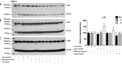
BKCachannel.The expression level of the BKCachannel in EACs following exposure to hypo-osmotic, mild hypo-os-motic, and hyperosmotic stresses was monitored at different time points. Western blotting using a BKCa channel-specific antibody was used to examine the effect of osmotic stress on BKCachannel expression, as previously described (16). There were no significant changes in BKCa channel expression fol-lowing hypo-osmotic and mild hypo-osmotic stress after 90 min and 3 h, whereas 6 h of incubation under hypo-osmotic conditions induced a significant (1.5-fold) increase in BKCa channel expression (P ⬍ 0.01; Fig. 1). In contrast, BKCa channel expression was significantly lower at the early stages (90 min) of hyperosmotic stress than during iso-osmotic stress
by 10.220.32.246 on September 22, 2017
(P⬍0.01). Extending the exposure to hyperosmotic stress for 3 and 6 h returned channel expression to the original level.
TRPV4 channel. A medium osmolarity of 380 mosM was used as the control condition. Western blotting using a TRPV4 channel-specific antibody, as described previously (16), was used to explore TRPV4 channel expression following hypo-osmotic, mild hypo-hypo-osmotic, and hyperosmotic stress. Expo-sure of chondrocytes to hypo-osmotic stress for 6 h (P⬍0.01), 3 h (P ⬍ 0.001), and 90 min (P ⬍ 0.001) increased TRPV4 channel expression by ⬎1.5-fold (Fig. 2). A mild hypo-os-motic environment induced an increase in TRPV4 channel expression at 90 min (P ⬍ 0.001) and 3 h (P ⬍ 0.01), but expression returned to control levels after 6 h (Fig. 2). In EACs exposed to hyperosmotic stress, TRPV4 channel expression was reduced by⬃50% after 3 h (P⬍0.05) and 90 min (P⬍
0.01) but returned to control levels by 6 h (Fig. 2).
Influence of Osmotic Stress on MAPK Phosphorylation
The influence of osmotic stress on activity-related phosphor-ylation of ERK1/2 and p38 MAPKs in chondrocytes was investigated at the protein level, as previously described (1, 39). Specific antibodies for the phosphorylated form of ERK1/2 and p38 MAPKs were used in Western blot experi-ments to investigate the phosphorylation of ERK1/2 and p38 MAPKs following exposure of EACs to osmotic stress for 90 min and 3 h. ERK phosphorylation was significantly (⬎2-fold) increased in response to hypo-osmotic stress at 90 min (P ⬍
0.001) and was reduced at 3 h (Fig. 3). A significant decrease (⬃50%) was induced by exposure of chondrocytes to mild hypo-osmotic and hyperosmotic stress at 90 min and 3 h; however, the greatest decrease was observed in response to hyperosmotic stress at 90 min (P ⬍0.001).
In contrast, p38 MAPK phosphorylation was significantly increased by exposure of EACs to hyperosmotic stress for 90 min and 3 h. Phosphorylation was significantly increased (⬃7-fold) at 90 min (P ⬍ 0.001) but was reduced to
⬃1.5-fold at 3 h (P⬍0.001; Fig. 4). No significant changes in phosphorylation were observed in chondrocytes exposed to hypo-osmotic stress at 90 min and 3 h. Phosphorylation of p38 MAPK was not changed by 90 min of mild hypo-osmotic stress, whereas it was downregulated at 3 h (P ⬍
0.001).
Inhibition of ERK and p38 Activity During Osmotic Loading
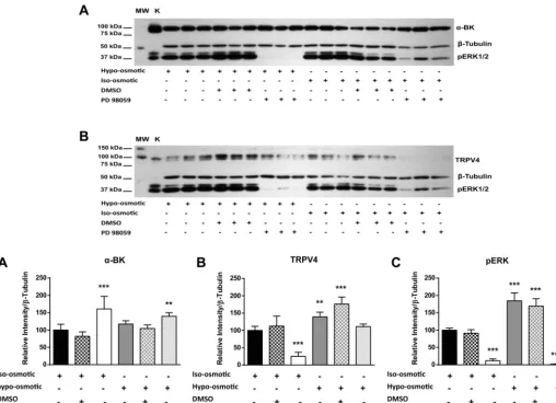
ERK1/2- and p38 MAPK-specific pharmacological inhibi-tors were used to examine the influence of these MAPKs on TRPV4 and BKCa channel expression in EACs. Inhibition of ERK1/2 phosphorylation (by the MEK1/2 inhibitor PD98059) at iso-osmotic conditions for 90 min significantly decreased TRPV4 channel expression to below the endogenous levels (P ⬍ 0.001; Fig. 5). Moreover, inhibition of ERK1/2 phos-phorylation under hypo-osmotic stress for 90 min significantly inhibited the elevation of TRPV4 channel expression induced by hypo-osmotic stress (P⬍0.001; Fig. 5B). In contrast, BKCa channel expression was significantly elevated by inhibition of ERK1/2 phosphorylation under hypo-osmotic (P⬍0.001) and iso-osmotic (P ⬍ 0.01) stress (Fig. 5A). The impact of p38 MAPK phosphorylation on TRPV4 and BKCachannel expres-sion was investigated under hyperosmotic stress for 90 min. Inhibition of p38 MAPK (by the p38 inhibitor SB 203580) significantly elevated BKCa channel expression (P ⬍ 0.001) but did not influence TRPV4 channel expression (Fig. 6). Fig. 1. Influence of hypo-osmotic (280 mosM), mild hypo-osmotic (320 mosM), iso-osmotic (380 mosM), and hyperosmotic (480 mosM) conditions on
large-conductance Ca2⫹-activated K⫹(␣-BKCa) channel expression at 90 min (A), 3 h (B), and 6 h (C) of incubation. K, kidney. Expression relative to-actin
was determined by densitometric analysis of the Western blot. Values are means⫾SE. **P⬍0.01 vs. iso-osmotic control.
by 10.220.32.246 on September 22, 2017
http://ajpcell.physiology.org/
[image:3.612.49.558.70.338.2]DISCUSSION
It has been shown in a number of cell types that the TRPV4 and BKCa ion channels play a role in the regulation of cell volume in altered osmotic environments. It is undoubtedly the case that changes in not only osmotic, but also mechanical and thermal, environments can lead to rapid and probably fluctu-ating changes in the activity of these channels. It is, however, reasonable to suggest that the overall capacity of these chan-nels is dictated, at least in part, by their level of expression. Furthermore, the level of expression of these channels has been shown to differ in pathological cartilage, although whether this
is causative or a result of the disease is unclear (24). In either case, altered expression could lead to progression of the dis-ease and incrdis-eased degeneration of the cartilage.
This study suggests that when chondrocytes are exposed to decreased osmolarity, TRPV4 channel protein expression in-creases rapidly (up to 6 h), whereas BKCa channel expression also increases, but only after 6 h. In contrast, increased osmo-larity initially decreased expression of both channels, but expression levels were restored to the endogenous levels after 90 min for the BKCa channel and after 3 h for the TRPV4 channel. Regulation of cell volume following hypotonic swell-Fig. 2. Influence of hypo-osmotic (280 mosM), mild hypo-osmotic (320 mosM), iso-osmotic (380 mosM), and hyperosmotic (480 mosM) conditions on transient
receptor potential vanilloid 4 (TRPV4) channel expression at 90 min (A), 3 h (B), and 6 h (C) of incubation. Expression relative to-actin was determined by
densitometric analysis of the Western blot. Values are means⫾SE. *P⬍0.5, **P⬍0.01, ***P⬍0.001 vs. iso-osmotic control.
Fig. 3. Influence of hypo-osmotic (280 mosM), mild hypo-osmotic (320 mosM), iso-osmotic (380 mosM), and hyperosmotic (480 mosM) conditions on ERK1/2
phosphorylation at 90 min (A) and 3 h (B) of incubation. Expression relative to-tubulin was determined by densitometric analysis of the Western blot. Values
are means⫾SE. **P⬍0.01, ***P⬍0.001 vs. iso-osmotic control.
by 10.220.32.246 on September 22, 2017
[image:4.612.48.559.73.340.2] [image:4.612.46.564.537.709.2]ing is classically mediated by release of Cl⫺ and K⫹ through activation of coordinated channels (18). In the majority of cell types, including chondrocytes, generation of an intracellular Ca2⫹signal in response to hypotonic stress is followed by the
RVD response, which allows cells to survive (18, 21, 55). This signal is initiated via Ca2⫹entry from the extracellular space
and augmented by Ca2⫹ release from intracellular stores (3). Elevation of intracellular Ca2⫹ induced by cell swelling
acti-Fig. 4. Influence of hypo-osmotic (280 mosM), mild hypo-osmotic (320 mosM), iso-osmotic (380 mosM), and hyperosmotic (480 mosM) conditions on p38
MAPK phosphorylation at 90 min (A) and 3 h (B) of incubation. Expression relative to-tubulin was determined by densitometric analysis of the Western blot.
[image:5.612.47.561.66.232.2]Values are means⫾SE. ***P⬍0.001 vs. iso-osmotic control.
Fig. 5. Inhibitor sensitivity of ERK1/2 osmolarity-dependent activity. Western blot shows effect of hypo-osmotic (280 mosM) and iso-osmotic (380 mosM)
conditions on ERK1/2 phosphorylation (pERK1/2), TRPV4 channel expression, and␣-BKCachannel expression following 90 min of incubation in the absence
(control) and presence of the pERK inhibitor PD98059 and vehicle (DMSO). Values are means⫾SE. **P⬍0.01, ***P⬍0.001 vs. iso-osmotic control.
by 10.220.32.246 on September 22, 2017
http://ajpcell.physiology.org/
[image:5.612.47.556.340.708.2]vates Ca2⫹-activated K⫹channels. Recently, the TRPV4 chan-nel was identified as an osmosensor chanchan-nel that mediates Ca2⫹entry following cell swelling in response to hypo-osmotic challenge. The TRPV4 channel may interact with aquaporins to elicit the RVD response to facilitate rapid movement of water during hypotonic challenge (28). The current study showed an increase in TRPV4 channel expression at the protein level following hypo-osmotic challenge. This finding was in agreement with other reports in bronchial endothelial cells (12) and porcine articular chondrocytes (33). Several hypotheses have been proposed to implicate the BKCachannel in cell volume regulation. The BKCa channel may act as an osmolyte channel (15, 21), where elevation of intracellular Ca2⫹induced by TRPV4 channel activation is sensed by the
Ca2⫹sensor in the BK
Cachannel, leading to its activation and release of K⫹, subsequent decrease in intracellular osmotic potential, and cell volume regulation. The alternative hypoth-esis suggests that BKCa channel activation occurs by sensing membrane stretch, induced by cell swelling or interaction with other mechanoreceptors (31). Differentiating between these two hypotheses is rather difficult, as cell swelling is associated with membrane stretch. Previous studies reported a coupling between TRPV4 and BKCa channels in the vascular smooth muscle response to vasodilatory factors through the ryanodine receptor (RYR) (7), whereas in bronchial endothelial cells the RYR is not involved in the direct coupling between the two channels in response to hypotonic stress (12).
During the course of osmotic challenge in the current study, TRPV4 channel expression increased to allow Ca2⫹ entry at the early phases of challenge followed by increased BKCa channel expression to mediate K⫹efflux and facilitate volume
regulation. As involvement of the RYR is not part of this study, the coupling between the two channels with or without in-volvement of the RYR in chondrocytes is possible.
The current study also showed that changes in cell volume induce MAPK cascades, leading to changes in phosphorylation of ERK1/2 and p38. Hypotonicity induced ERK1/2 phosphor-ylation, whereas hypertonicity provoked p38 phosphorylation during early phases of exposure. Previous studies showed changes in ERK1/2 phosphorylation during osmotic stress [i.e., increased phosphorylation in rat nucleus pulposus cells follow-ing increased osmolarity (44)]. In contrast, phosphorylation of ERK1/2 was increased by hypo-osmotic stress in intestinal 407 cells (48), astrocytes (6), and hepatoma cells (35). Taken together, ERK1/2 phosphorylation following osmotic stress seems to be cell-specific. The role of the ERK1/2 pathway in RVD has not been delineated; however, indirect activation of ERK1/2 via the Ras-Raf-MEK pathway has been suggested in hepatocytes (8). Other studies have linked activation of ERK1/2 to activation of the Cl⫺ channel in corneal epithelial cells (19) and astrocytes (6) and to activation of the K⫹channel in cervical cancer cells (36). Although Cl⫺ and K⫹ play an important role in volume regulation following cell swelling in response to hypo-osmotic stress and elevation of intracellular Ca2⫹(18), the current study suggests a link between ERK1/2
phosphorylation and TRPV4 channel expression, where ERK1/2 phosphorylation regulates endogenous TRPV4 chan-nel expression.
[image:6.612.45.562.74.341.2]Phosphorylation of p38 MAPK following hyperosmotic stress has been shown in several cell types, such as fibroblasts (23), human cervical cells (36), and human articular chondro-cytes (41, 42). In agreement with these studies, we have shown Fig. 6. Inhibitor sensitivity of p38 MAPK osmolarity-dependent activity. Western blot shows effect of the hyperosmotic (480 mosM) condition on TRPV4 and
␣-BKCachannel expression following 90 min of incubation in the absence (control) and presence of the p38 inhibitor SB 203580 and vehicle (DMSO). Values
are means⫾SE. *P⬍0.5, ***P⬍0.001 vs. iso-osmotic control.
by 10.220.32.246 on September 22, 2017
an elevation of p38 MAPK phosphorylation following hyper-osmotic stress at early phases of the exposure. Therefore, activation of p38 could be implicated in the regulatory volume increase response to restore cell volume following hyperos-motic stress. Activation of p38 was strongly associated with upregulation of aggrecan gene expression (52). This was sus-tained by linking p38 pathway activation to elevation of tonic-ity-responsive enhanced binding protein, which in turn acti-vates target genes such as aggrecan (43). Blocking phosphor-ylation of p38 MAPK did not change TRPV4 channel expression, whereas BKCa channel expression was upregu-lated.
In summary, we have shown that TRPV4 and BKCachannel expression in chondrocytes is sensitive to an altered osmotic environment. Furthermore, we have shown that some of these changes may involve activation of ERK and p38. The precise mechanism by which these signaling factors are involved in regulation of this expression is unclear, but further exploration is warranted to understand their role in normal chondrocyte function in healthy cartilage and their potential role in initiation and progression of pathological conditions such as osteoarthritis.
ACKNOWLEDGMENTS
Present address of A. Mobasheri: School of Veterinary Medicine, Faculty of Health and Medical Sciences, University of Surrey, Duke of Kent Building, Guildford, Surrey GU2 7XH, United Kingdom.
DISCLOSURES
No conflicts of interest, financial or otherwise, are declared by the authors.
AUTHOR CONTRIBUTIONS
I.M.H., A.M., and P.T.L. are responsible for conception and design of the research; I.M.H. performed the experiments; I.M.H. and P.T.L. analyzed the data; I.M.H. and P.T.L. interpreted the results of the experiments; I.M.H. prepared the figures; I.M.H. and P.T.L. drafted the manuscript; I.M.H., A.M., and P.T.L. edited and revised the manuscript; I.M.H. and P.T.L. approved the final version of the manuscript.
REFERENCES
1. Atherton PJ, Szewczyk NJ, Selby A, Rankin D, Hillier K, Smith K, Rennie MJ, Loughna PT.Cyclic stretch reduces myofibrillar protein
synthesis despite increases in FAK and anabolic signalling in L6 cells.J
Physiol587: 3719 –3727, 2009.
2. Bradford MM. A rapid and sensitive method for the quantitation of microgram quantities of protein utilizing the principle of protein-dye
binding.Anal Biochem72: 248 –254, 1976.
3. Buckwalter JA, Mankin HJ. Articular cartilage: tissue design and
chondrocyte-matrix interactions.Instr Course Lect47: 477–486, 1998.
4. Bush PG, Hall AC.The osmotic sensitivity of isolated and in situ bovine
articular chondrocytes.J Orthop Res19: 768 –778, 2001.
5. Bush PG, Hall AC.Regulatory volume decrease (RVD) by isolated and
in situ bovine articular chondrocytes.J Cell Physiol187: 304 –314, 2001.
6. Crepel V, Panenka W, Kelly ME, MacVicar BA.Mitogen-activated protein and tyrosine kinases in the activation of astrocyte volume-activated
chloride current.J Neurosci18: 1196 –1206, 1998.
7. Earley S, Heppner TJ, Nelson MT, Brayden JE.TRPV4 forms a novel
Ca2⫹ signaling complex with ryanodine receptors and BKCachannels.
Circ Res97: 1270 –1279, 2005.
8. Ebner HL, Fiechtner B, Pelster B, Krumschnabel G. Extracellular signal regulated MAP-kinase signalling in osmotically stressed trout
hepatocytes.Biochim Biophys Acta1760: 941–950, 2006.
9. Erickson GR, Alexopoulos LG, Guilak F.Hyper-osmotic stress induces volume change and calcium transients in chondrocytes by transmembrane,
phospholipid, and G-protein pathways.J Biomech34: 1527–1535, 2001.
10. Erickson GR, Northrup DL, Guilak F.Hypo-osmotic stress induces
calcium-dependent actin reorganization in articular chondrocytes.
Osteo-arthritis Cartilage11: 187–197, 2003.
11. Everaerts W, Nilius B, Owsianik G.The vanilloid transient receptor
potential channel TRPV4: from structure to disease.Prog Biophys Mol
Biol103: 2–17, 2010.
12. Fernandez-Fernandez JM, Andrade YN, Arniges M, Fernandes J, Plata C, Rubio-Moscardo F, Vazquez E, Valverde MA. Functional coupling of TRPV4 cationic channel and large conductance, calcium-dependent potassium channel in human bronchial epithelial cell lines. Pflügers Arch457: 149 –159, 2008.
13. Guilak F, Hung CT. Physical regulation of cartilage metabolism. In: Basic Orthopaedic Biomechanics. Philadelphia: Lippincott-Raven, 2005, p. 259 –300.
14. Guler AD, Lee H, Iida T, Shimizu I, Tominaga M, Caterina M.
Heat-evoked activation of the ion channel, TRPV4. J Neurosci 22:
6408 –6414, 2002.
15. Hall AC, Starks I, Shoults CL, Rashidbigi S.Pathways for K⫹transport across the bovine articular chondrocyte membrane and their sensitivity to
cell volume.Am J Physiol Cell Physiol270: C1300 –C1310, 1996.
16. Hdud IM, El-Shafei AA, Loughna P, Barrett-Jolley R, Mobasheri A.
Expression of transient receptor potential vanilloid (TRPV) channels in
different passages of articular chondrocytes.Int J Mol Sci13: 4433–4445,
2012.
17. Hdud IM, Mobasheri A, Loughna PT. Effects of cyclic equibiaxial
mechanical stretch on␣-BK and TRPV4 expression in equine
chondro-cytes.Springerplus3: 59, 2014.
18. Hoffmann EK, Lambert IH, Pedersen SF.Physiology of cell volume
regulation in vertebrates.Physiol Rev89: 193–277, 2009.
19. Hsiao HB, Wu JB, Lin H, Lin WC.Kinsenoside isolated from Anoec-tochilus formosanussuppresses LPS-stimulated inflammatory reactions in
macrophages and endotoxin shock in mice.Shock35: 184 –190, 2011.
20. Jung C, Fandos C, Lorenzo IM, Plata C, Fernandes J, Gene GG, Vazquez E, Valverde MA. The progesterone receptor regulates the
expression of TRPV4 channel.Pflügers Arch459: 105–113, 2009.
21. Kerrigan MJ, Hall AC. Control of chondrocyte regulatory volume
decrease (RVD) by [Ca2⫹]iand cell shape.Osteoarthritis Cartilage16:
312–322, 2008.
22. Kerrigan MJ, Hook CS, Qusous A, Hall AC. Regulatory volume
increase (RVI) by in situ and isolated bovine articular chondrocytes.J Cell
Physiol209: 481–492, 2006.
23. Ko BC, Lam AK, Kapus A, Fan L, Chung SK, Chung SS.Fyn and p38 signaling are both required for maximal hypertonic activation of the osmotic response element-binding protein/tonicity-responsive
enhancer-binding protein (OREBP/TonEBP).J Biol Chem277: 46085–46092, 2002.
24. Lewis R, Feetham CH, Barrett-Jolley R. Cell volume regulation in
chondrocytes.Cell Physiol Biochem28: 1111–1122, 2011.
25. Liedtke W.TRPV4 as osmosensor: a transgenic approach.Pflügers Arch 451: 176 –180, 2005.
26. Liedtke W.TRPV4 plays an evolutionary conserved role in the
transduc-tion of osmotic and mechanical stimuli in live animals. J Physiol567:
53–58, 2005.
27. Liedtke W, Tobin DM, Bargmann CI, Friedman JM. Mammalian TRPV4 (VR-OAC) directs behavioral responses to osmotic and
mechan-ical stimuli inCaenorhabditis elegans.Proc Natl Acad Sci USA100Suppl
2: 14531–14536, 2003.
28. Liu X, Bandyopadhyay BC, Nakamoto T, Singh B, Liedtke W, Melvin JE, Ambudkar I.A role for AQP5 in activation of TRPV4 by hypoto-nicity: concerted involvement of AQP5 and TRPV4 in regulation of cell
volume recovery.J Biol Chem281: 15485–15495, 2006.
29. Maroudas A.Physicochemical properties of articular cartilage. In:Adult Articular Cartilage(2nd ed.), edited by Freeman MA. Tunbridge Wells, UK: Pitman Medical, 1979, p. 215–290.
30. Martel-Pelletier J, Boileau C, Pelletier JP, Roughley PJ.Cartilage in
normal and osteoarthritis conditions.Best Pract Res Clin Rheumatol22:
351–384, 2008.
31. Mobasheri A, Carter SD, Martin-Vasallo P, Shakibaei M. Integrins and stretch activated ion channels; putative components of functional cell
surface mechanoreceptors in articular chondrocytes. Cell Biol Int 26:
1–18, 2002.
32. Nilius B, Owsianik G.Channelopathies converge on TRPV4.Nat Genet 42: 98 –100, 2010.
33. Phan MN, Leddy HA, Votta BJ, Kumar S, Levy DS, Lipshutz DB, Lee SH, Liedtke W, Guilak F.Functional characterization of TRPV4 as an
by 10.220.32.246 on September 22, 2017
http://ajpcell.physiology.org/
vertebral disc cells: effect of extracellular osmotic change on
glycosami-noglycan production and cell metabolism.Lab Invest J Neurosurg Spine7:
637–644, 2007.
39. Tarabees R, Hill D, Rauch C, Barrow PA, Loughna PT.Endotoxin transiently inhibits protein synthesis through Akt and MAPK mediating
pathways in C2C12 myotubes.Am J Physiol Cell Physiol 301: C895–
C902, 2011.
40. Tew SR, Hardingham TE. Regulation of SOX9 mRNA in human articular chondrocytes involving p38 MAPK activation and mRNA
stabi-lization.J Biol Chem281: 39471–39479, 2006.
41. Tew SR, Peffers MJ, McKay TR, Lowe ET, Khan WS, Hardingham TE, Clegg PD.Hyperosmolarity regulates SOX9 mRNA
posttranscrip-tionally in human articular chondrocytes.Am J Physiol Cell Physiol297:
C898 –C906, 2009.
42. Tew SR, Vasieva O, Peffers MJ, Clegg PD.Post-transcriptional gene regulation following exposure of osteoarthritic human articular
chondro-cytes to hyperosmotic conditions. Osteoarthritis Cartilage 19: 1036 –
1046, 2011.
43. Tsai TT, Danielson KG, Guttapalli A, Oguz E, Albert TJ, Shapiro IM, Risbud MV. TonEBP/OREBP is a regulator of nucleus pulposus cell
function and survival in the intervertebral disc.J Biol Chem281: 25416 –
25424, 2006.
44. Tsai TT, Guttapalli A, Agrawal A, Albert TJ, Shapiro IM, Risbud
MV.MEK/ERK signaling controls osmoregulation of nucleus pulposus
50. Wang RX, Shi HF, Chai Q, Wu Y, Sun W, Ji Y, Yao Y, Li KL, Zhang CY, Zheng J, Guo SX, Li XR, Lu T.Molecular mechanisms of diabetic
coronary dysfunction due to large conductance Ca2⫹-activated K⫹
chan-nel impairment.Chin Med J125: 2548 –2555, 2012.
51. Watanabe H, Davis JB, Smart D, Jerman JC, Smith GD, Hayes P, Vriens J, Cairns W, Wissenbach U, Prenen J, Flockerzi V, Droogmans G, Benham CD, Nilius B. Activation of TRPV4 channels (hVRL-2/
mTRP12) by phorbol derivatives.J Biol Chem277: 13569 –13577, 2002.
52. Watanabe H, de Caestecker MP, Yamada Y.Transcriptional cross-talk between Smad, ERK1/2, and p38 mitogen-activated protein kinase
path-ways regulates transforming growth factor--induced aggrecan gene
ex-pression in chondrogenic ATDC5 cells.J Biol Chem276: 14466 –14473,
2001.
53. Watanabe H, Vriens J, Suh SH, Benham CD, Droogmans G, Nilius B.
Heat-evoked activation of TRPV4 channels in a HEK293 cell expression
system and in native mouse aorta endothelial cells.J Biol Chem 277:
47044 –47051, 2002.
54. Wu L, Gao X, Brown RC, Heller S, O’Neil RG.Dual role of the TRPV4
channel as a sensor of flow and osmolality in renal epithelial cells.Am J
Physiol Renal Physiol293: F1699 –F1713, 2007.
55. Yellowley CE, Hancox JC, Donahue HJ.Effects of cell swelling on intracellular calcium and membrane currents in bovine articular
chondro-cytes.J Cell Biochem86: 290 –301, 2002.
by 10.220.32.246 on September 22, 2017


