www.wjpr.net Vol 7, Issue 12, 2018. 35
HEPATOPROTECTIVE EFFECT OF PROBIOTIC, CONTAINING
LACTOBACILLUS BULGARICUS DWT1, IN ACUTE
PARACETAMOL-INDUCED LIVER DAMAGE IN RATS
Gabriela Kehayova1*, Marieta Georgieva1 and Kaloyan Georgiev2
1
Department of Pharmacology, Toxicology and Pharmacotherapy, Faculty of Pharmacy,
Medical University, Varna, Bulgaria.
2
Department of Pharmaceutical Technologies, Faculty of Pharmacy, Medical University,
Varna, Bulgaria.
ABSTRACT
Hepatic impairment is one of the most common organ damage and
occurs asymptomatically until damage affects a significant part of the
organ. The aim of the present study was to investigate a
hepatoprotective activity of a new strain of Lactobacillus bulgaricus
DWT1 against paracetamol-induced hepatic damage in Wistar rats.
Laktera Nature, containing Lactobacillus bulgaricus DWT1,
Lactobacillus helveticus DWT2, Lactobacillus lactis DWT3 and
Streptococcus thermophilus DWT 4,5, 6, 7, 8 administered at oral
doses of 800 mg/kg and 1600 mg/kg, showed significant
hepatoprotective effects by decreasing the levels of serum marker
enzymes such as alanine aminotransferase (ALAT), aspartate
aminotransferase (ASAT), alkaline phosphatase (ALP), gama
-glutamiltransferase (GGT), as compared to standard drug (silymarin) and negative control.
Histopathological analysis showed that administration of the probiotic minimized liver
damage, by reducing the level of morphological changes and necrosis. Our findings
demonstrate the possible use of Laktera Nature, containing Lactobacillus bulgaricus DWT1,
Lactobacillus helveticus DWT2, Lactobacillus lactis DWT3 and Streptococcus thermophilus
DWT4,5, 6, 7, 8 for prevention of liver injury.
KEYWORDS: probiotic, paracetamol, liver damage, hepatoprotective effect.
Volume 7, Issue 12, 35-42. Research Article ISSN 2277–7105
Article Received on 24 April 2018,
Revised on 14 May 2018, Accepted on 04 June 2018
DOI: 10.20959/wjpr201812-12610
*Corresponding Author
Dr. Gabriela Kehayova
Department of
Pharmacology, Toxicology
and Pharmacotherapy,
Faculty of Pharmacy,
Medical University, Varna,
www.wjpr.net Vol 7, Issue 12, 2018. 36 INTRODUCTION
Hepatic drug damage is one of the most common side effects when using medicines that are
constantly increasing as frequency. In the United States, drug-related hepatotoxicity is the
leading cause of acute liver failure in patients indicated for hepatic transplantation,
particularly in patients with unintentional or deliberate overdose of paracetamol. Acute
overdose of paracetamol may lead to potentially fatal liver damage.[1, 2]
Probiotics are living microorganisms, which applied in sufficient quantities, provide a health
benefit to the host and contribute to reducing the risk of disease. They are subject of
increasing interest due to their proven immunostimulating, antioxidant and anti-cancer
effect.[3, 4] The use of probiotics is considered as effective and safe alternative treatment for
hepatotoxicity. Detailed studies of the hepatoprotective effect of probiotics on carbon
tetrachloride model in rats were conducted by Georgieva M et al. 2002.[5] As a result of the
systemic toxicological and clinical pharmacology studies carried out on Biostim LBS,
containing Lactobacillus bulgaricus, its extremely hepatoprotective and antioxidant activity
was established.[6] A comparable hepatoprotective effect with silymarin has been
demonstrated.[7]Lactobacillus bulgaricus DWT1 is a neworiginal strain, isolated from spring
water in Bulgaria. In 2015, Georgiev K et al., conducted a study on the antiproliferative effect
of Laktera Nature, containing Lactobacillus bulgaricus DWT1, on a human coloncarcinoma
cell line. The study shows that at high concentration, Laktera Nature inhibits the proliferation
of the HT-29 carcinoma cell line.[8]
In the present study, we aimed to investigate a hepatoprotective effect of a new strain of
Lactobacillus bulgaricus DWT1 in experimental animal model of hepatic damage. We used
the most common drug causing hepatotoxicity – paracetamol, to cause hepatic damage and
for positive control - silymarin. Biochemical markers for hepatotoxicity and histopathology
were included in the study.
MATERIALS AND METHODS
Chemicals
Laktera Nature® was kindly provided by the company Daflorn MLM 5 Ltd. (Sofia, Bulgaria).
1 mg of the substance contained 25 х 106
live and latent CFU Lactobacillus bulgaricus
DWT1, Lactobacillus helveticus DWT2, Lactobacillus lactis DWT3 and Streptococcus
www.wjpr.net Vol 7, Issue 12, 2018. 37
Before administration, the both substances were dissolved in water and administered via
gastric probes.
Animals
Male adult Wistar rats (200 g ±20) received a standard rodent diet and were kept at 12 h
light/dark cycle and constant temperature and humidity. The rats were housed in a controlled
environment (temperature 23±5ºC, humidity 50±10%, and 12 h light/12 h dark cycle) with ad
libitum access to food and water. All procedures concerning animal treatment and
experimentation were conducted in compliance with the national laws and policies, in
conformity with the international guidelines (European Economic Community EEC Council
Directive 86/609, IL 358, 1, December 12, 1987).
Experimental design
The trial was conducted on 30 male Wistar rats which were divided into 5 groups of 6, as
follows: saline-treated healthy controls; a control group treated with an overdose of
paracetamol 1200 mg/kg to create the experimental models; a group of animals pretreated
with Laktera Nature 800 mg/kg for 14 days, with an overdose of paracetamol being
administered; group of animals pre-treated with Laktera Nature 1600 mg/kg for 14 days, with
an overdose of paracetamol being administered; group of animals pretreated with Carsil®
45mg/kg for 14 days, with an overdose of paracetamol being administered. At the end of the
experiment, all animals were sacrificed under diethyl ether anesthesia, blood samples were
collected for biochemical analysis and livers were isolated for histopathological analysis.
Biochemical assays
Blood samples were centrifuged for 10 min at 7000 rpm using micro-centrifuge to separate
the serum. The levels of enzymes, alanine aminotransferase (ALAT), aspartate
aminotransferase (ASAT), alkaline phosphatase (ALP), gama-glutamiltransferase (GGT),
were determined in a licensed hematological research laboratory in Varna/Bulgaria using
Roche COBAS 6000 analyzer and methodologies described on their licensed site.[9]
Histopathology
The liver slides were fixed in 10% neutral formalin, embedded in paraffin, sectioned at a
thickness of 5μm, stained with hematoxylin & eosin or Fouchet van Gieson’s trichrome stain,
according to the methods of Bio-Optica staining kits. The slides were evaluated for any
www.wjpr.net Vol 7, Issue 12, 2018. 38
Statistical analysis
In all experiments, data were presented as means ±SD. One-way analysis of variance
(ANOVA) was used to determine significance between the tested groups. Analysis was
performed using SigmaPlot 11.0 software. A probability level of 0.05 or lower was
considered as statistically significant.
RESULTS
The collected blood after the completion of the animal experiment was given for examination
and the results were obtained.
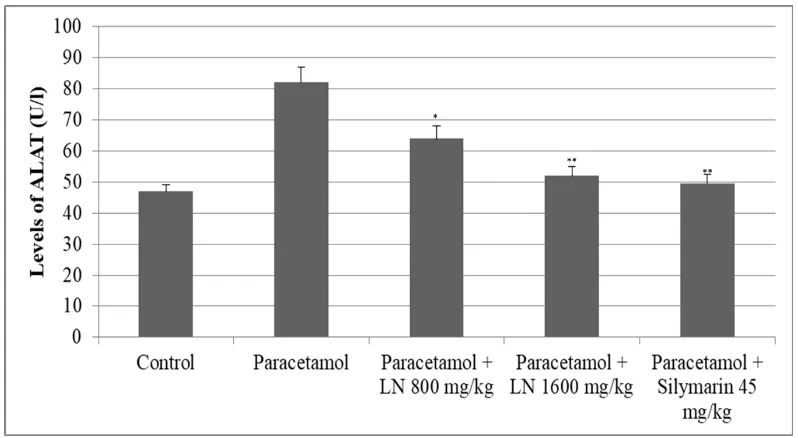
The transaminase enzyme, alanine aminotransferase (ALAT) and aspartate aminotransferase
(ASAT), were significantly elevated after acute overdose of paracetamol. ALAT levels were
almost doubled, while those of ASAT were slightly increased. Pretreatment with Laktera
Nature showed a dose-dependent decrease in transaminase levels (Figure 1 and 2).
Figure 1: Changes of ALAT levels after acute overdose of paracetamol and after
pretreatment with LN 800 mg/kg, 1600 mg/kg and Silymarin 45 mg/kg in rats. *p<0.05,
[image:4.595.98.497.359.578.2]www.wjpr.net Vol 7, Issue 12, 2018. 39 Figure 2: Changes of ASAT levels after acute overdose of paracetamol and after
pretreatment with LN 800 mg/kg, 1600 mg/kg and Silymarin 45 mg/kg in rats. *p<0.05,
**p<0.01, ***p<0,001.
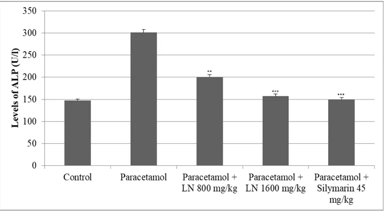
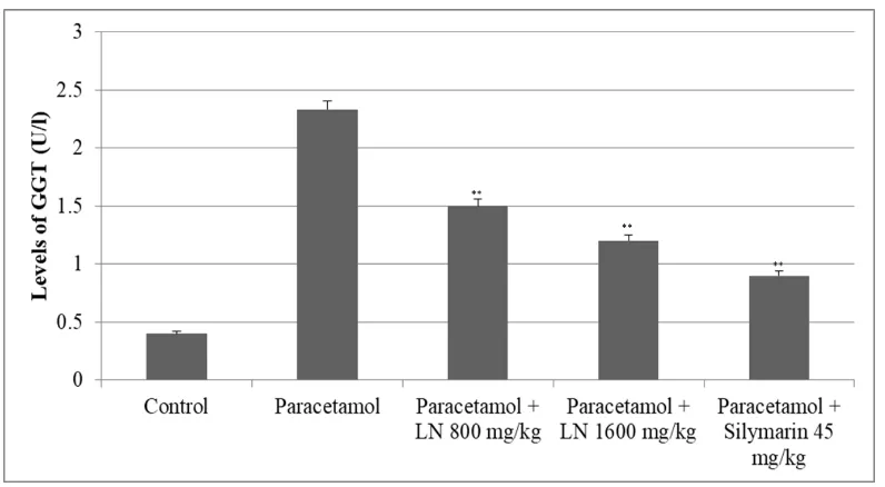
The other two enzymes, alkaline phosphatase (ALP) and gama-glutamyltransferase (GGT),
were elevated significantly as well. The levels of alkaline phosphatase (ALP) were doubled,
while those of GGT were eight times higher than the control group (Figure 3 and 4).
Figure 3. Changes of ALP levels after acute overdose of paracetamol and after
pretreatment with LN 800 mg/kg, 1600 mg/kg and Silymarin 45 mg/kg in rats. *p<0.05,
[image:5.595.107.490.67.287.2] [image:5.595.107.490.445.655.2]www.wjpr.net Vol 7, Issue 12, 2018. 40 Figure 4: Changes of GGT levels after acute overdose of paracetamol and after
pretreatment with LN 800 mg/kg, 1600 mg/kg and Silymarin 45 mg/kg in rats. *p<0.05,
**p<0.01, ***p<0,001
The histopathological examination showed significant impairments in the use of paracetamol
alone (e.g. necrosis), whereas pretreatment with the Laktera Nature 800 and 1600 mg/kg,
showed normal hepatocellular architecture, similar to pretreatment with silymarin (results not
showed).
DISCUSSION
In many liver diseases, the desired therapeutic effect of the standard therapy is not achieved.
There is a significant percentage of treatment side effects and the treatment of patients with
toxic liver damage is becoming an extremely challenging. The addition of the Bulgarian
probiotic containing Lactobacillus bulgaricus DWT1, thanks to its proven hepatoprotective
and anti-tumor effect, categorically provides a "powerful weapon" in both the therapeutic
approach and the prevention of liver diseases. Paracetamol is one of the most commonly used
analgesics-antipyretics worldwide. At therapeutic doses, paracetamol is safe, but if overdosed
it can cause liver necrosis in both human and rat. The reason for this is the formation of a
highly reactive metabolite under the action of cytochrome P450 enzymes.[10] The induction of
these enzymes on the one hand and the depletion of hepatic glutathione on the other hand, are
the basis of developing liver necrosis. Relating a hepatocyte lesion, a cellular leakage and a
[image:6.595.100.498.68.287.2]www.wjpr.net Vol 7, Issue 12, 2018. 41
In the present study, paracetamol has caused significant elevation in the levels of ALAT,
ASAT, ALP and GGT. Pretreatment with Laktera Nature in dosage 800 mg/kg and 1600
mg/kg, was found to be significantly reversing the changes induced by paracetamol. The
hepatoprotective effect of lactobacilli is probably due to their elevated glutathione
concentration in the liver, which is involved in the detoxification of endogenous and
exogenous carcinogens and free radicals and modulates the immune function.[12, 13] The
immune system plays an important role in the detoxification of the body, and the
predominance of lactobacilli has an immunostimulating effect. Many studies have shown that
the potential therapeutic effect of lactic acid bacteria, including their immunostimulating
effect, is mainly due to their induced changes in gastrointestinal microeconomics. After
entering the intestine, living or biologically active lactobacilli may activate the specific and
non-specific immune response of the gastrointestinal lymphoid tissue and the systemic
immune response. Reduction in the levels of ALAT and ASAT, in the larger dose 1600
mg/kg LN, is an indication of a possible regeneration process. From the histological analysis
of the paracetamol overdosage group, extensive coagulation necrosis zones affecting
hepatocytes from the central and intermedicinal areas of the liver pads were established. In
many places, necrotic stretches merged into so-called "bridge necrosis." In the group
pretreated with Laktera Nature at a dose of 800 mg/kg, there were significantly less
pronounced necrotic liver changes. In the group pretreated with Laktera Nature at a dose of
1600mg/kg, the probiotic showed a significant hepatoprotective effect similar to that of
silymarin, resulting in a reduction of affected liver fragments and lack of necrotic changes in
hepatocytes. The use of probiotics minimizes hepatic impairment by reducing morphological
changes and necrosis.
CONCLUSION
An extremely high hepato-prophylactic activity of Laktera Nature, containing Lactobacillus
bulgaricus DWT1, Lactobacillus helveticus DWT2, Lactobacillus lactis DWT3 and
Streptococcus thermophilus DWT 4, 5, 6, 7, 8 was observed for the first time in acute
paracetamol-induced liver toxicity in rats. The Laktera Nature showed a pronounced
hepatoprotective effect in paracetamol-induced acute liver toxicity in rats, expressed in
preventing paracetamol-induced severe degenerative and necrotic liver changes, and
decreasing paracetamol-elevated liver enzymes ASAT, ALT, ALP, GGT. The probiotic does
not affect the serum transaminase values of healthy controls and can be used in the
www.wjpr.net Vol 7, Issue 12, 2018. 42 REFERENCES
1. Suk KT, Kim DJ. Drug-induced liver injury: present and future. Clin Mol Hepatol, 2012
Sep; 18(3): 249-57.
2. Yoon E, Babar A, Choudhary M, Kutner M, Pyrsopoulos N. Acetaminophen-Induced
Hepatotoxicity: a Comprehensive Update. J Clin Transl Hepatol, 2016 Jun 28; 4(2):
131-42.
3. Iannitti T, Palmieri B. Therapeutical use of probiotic formulations in clinical practice.
Clin Nutr, 2010 Dec; 29(6): 701-25.
4. Georgieva M, Georgiev K, Dobromirov P. Probiotics and Immunity, Immunopathology
and Immunomodulation, Intech Open, 2015; DOI: 10.5772/61337.
5. Georgieva M, Eliseev V, Borissova P. Study of the effects of "BIOMILK" and Silymarin
on an experimental model of carbontetrachloride induced hepatotoxicity in rats, Medicine
and Pharmacy, 2002; 7-8,12, 23-24.
6. Georgieva M. Toxicological and Clinical Pharmacological Study of Bulgarian
Low-Lactose Lactate Probiotic Biostim LBS, Autoreferat, Medical University - Sofia, 2006.
7. Georgieva, M., Paskalev, D., Aleksandrov, N. Probiotics: The Good Bacteria of
Bulgarian Yoghurt, Historical Medical Collection, Varna, 2003; 2: 47-51.
8. Georgiev K, Georgieva M, Iliev I, Peneva M, Alexandrov G. Antiproliferative effects of
Bulgarian spring water probiotics (Laktera Nature Probiotic®) against human colon
carcinoma cell line, WJPPS 2015; 4(06): 130-136.
9.
https://usdiagnostics.roche.com/en/core_laboratory/instrument/cobas-6000-analyzer-series.html#menu.
10.Mitchell JR, Jollow DJ, Potter WZ, Davis DC, Gillette JR, Brodie BB.
Acetaminophen-induced hepatic necrosis. I. Role of drug metabolism. J Pharmacol Exp Ther, 1973; 187:
185–94.
11.Drotman RB, Lawhorn GT. Serum enzymes as indicators of chemically induced liver
damage. Drug Chem Toxicol, 1978; 1: 163–71.
12.Yamauchi A, Bloom ET. Requirement of thiol compounds as reducing agents for
IL-2-mediated induction of LAK activity and proliferation of human NK cells. J Immunol,
1993; 151: 5535–44.
13.McIntosh GH, Regester GO, Le Leu RK, Royle PJ, Smithers GW. Dairy proteins protect