http://www.scirp.org/journal/ojog ISSN Online: 2160-8806
ISSN Print: 2160-8792
Can We Predict Menorrhagia with Intrauterine
Contraceptive Device (IUCD) Insertion?
Ghada M. Mansour1*, Sherif H. Hussein1, Haitham F. Mohammed1, Sherif F. El Mekkawy1,
Sherif A. Akl1, Asmaa A. Abd El Dayem2
1Department of Obstetrics and Gynecology, Ain Shams University, Cairo, Egypt 2Ashmoun Hospital, Ministry of Health Hospitals, Cairo, Egypt
Abstract
Objective: Studying sub endometrial vascularity and blood flow in cases using intrauterine contraceptive devices for contraception with and without me-norrhagia compared to cases not using intrauterine contraceptive devices. Methods: Three hundred and fifteen women attending gynecology and family planning outpatient clinics in the maternity hospital, Ain Shams University were included in the study. They were classified into three groups, 105 women using IUCD with menorrhagia (group I), 105 women using IUCD without menorrhagia (group II), and 105 normal controls not using IUCD (group III). After excluding local causes for bleeding, blood disease or any medical dis-orders, transvaginal ultrasound including three dimensional power Doppler (3DPD) ultrasound was done for all women. Right and left uterine artery pul-satility index (PI) and resistance index (RI) were calculated, subednometrial blood flow RI and PI were obtained then 3DPD Vascular indices (VI, FI and VFI) of subendometrial blood flow were obtained for all cases. Statistical analysis was done to compare between the three groups. Results: A significant statistical difference was found as regards subendometrial vascularity indices, while there was no difference as regards bilateral uterine arteries Doppler in-dices in the three groups. Conclusion: Subendometrial vascularity in cases of menorrhagia with IUCD was markedly higher than in cases without menorr-hagia and cases with no IUCD. 3DPD may be used for selection of cases prior to insertion of IUCD.
Keywords
Intrauterine Contraceptive Devices, IUCD, 3D Power Doppler Ultrasound, Menorrhagia
1. Introduction
An intrauterine contraceptive device (IUCD) is one of the most frequently used How to cite this paper: Mansour, G.M.,
Hussein, S.H., Mohammed, H.F., El Mek-kawy, S.F., Akl, S.A. and Abd El Dayem, A.A. (2017) Can We Predict Menorrhagia with Intrauterine Contraceptive Device (IUCD) In- sertion?. Open Journal of Obstetrics and Gy-necology, 7, 753-766.
https://doi.org/10.4236/ojog.2017.77077
Received: June 3, 2017 Accepted: July 28, 2017 Published: July 31, 2017
Copyright © 2017 by authors and Scientific Research Publishing Inc. This work is licensed under the Creative Commons Attribution International License (CC BY 4.0).
methods for birth control around the world [1]. However, menorrhagia is among its side effects. Menorrhagia may cause iron deficiency anemia and usually ends by removing the IUCD in the first year after its insertion in many cases.
There are several possible mechanisms that explain the cause of menorrhagia in patients using IUCD. Several studies reported that IUCD insertion increases the production of prostaglandins in the endometrium which cause an increase in vascularity, vascular permeability and inhibit platelet activity and therefore, in-crease menstrual bleeding.
Probably there is a relation between IUCD adverse effects and uterine vascu-larization. However, this association is neither well-known nor well-studied. Three dimensional power Doppler (3DPD) was widely introduced for studying subendometrial vascularity by quantitative indices, vascularization index (VI), flow index (FI), and vascularization flow index (VFI).
The most important copper intrauterine contraceptive device (IUCD) related side effects are excessive uterine bleeding and menstrual pain. The menstrual blood may be excessive to the extent of causing iron deficiency anemia [2].
These side effects are responsible for a removal rate of 5% to 15% during the first year after intrauterine contraceptive device (I.U.C.D) insertion [3].
Several possible mechanisms may explain the cause of menorrhagia in patients using IUD. Among these theories, the increased production of prostaglandins in the endometrium which causes an increase in vascularity, vascular permeability, inhibit platelet activity and therefore, increase menstrual bleeding [4].
Probably there is a relation between IUD adverse effects and uterine vascula-rization. However, this association is neither well-known nor well-studied [3].
Only a few studies have demonstrated an increase in subendometrial vascula-rization in patients with IUD induced menorrhagia [5].
The three-dimensional power Doppler is based on three-dimensional recon-struction of vessels image, received from power Doppler system. The intensity of three-dimensional angiography Doppler signal is directly dependent from blood flow velocity. It enables quantitative evaluation of vessels in the area studied due to the use of angio histogram function in which 3-dimensional vascularization and blood flow indices [Vascular Index (VI)], Flow Index (FI) and Vascular Flow Index (VFI), are counted automatically [6].
2. Subjects and Methods
This cross sectional case control study was performed in Ain Shams University Maternity Hospital. Three hundred and fifteen women attending gynecology and family planning outpatient clinics were included in the study.
to feel the threads of IUCD or requesting IUCD removal, Group (III): was a control group which included 105 women without IUCD who attended outpa-tient clinic requesting copper IUCD insertion and not complaining of abnormal uterine bleeding.
Cases with acute or chronic pelvic pain, cases with pelvic inflammatory dis-ease, benign or malignant genital tumors or any uterine congenital anomalies, cases with bleeding tendency, patients on anticoagulants, non steroidal anti in-flammatory drugs and patients on hormonal therapy in the previous 3 months were all excluded from the study.
The study protocol was approved by the hospital research ethics board. Study participants were counseled and informed consent was obtained.
A complete history was taken including menstrual history before and after IUCD insertion, including duration and amount of menstrual flow, regularity and length of the cycle, inter menstrual bleeding or spotting or contact bleeding. In addition, history of any drug intake, blood disease or any medical disorders were considered.
Clinical examination was done including general, abdominal and pelvic ex-amination which included bimanual exex-amination to detect any abnormal find-ings and speculum examination to detect the threads of the IUCD and exclude any local cause of bleeding as polyp and erosion.
3. Ultrasound Examination
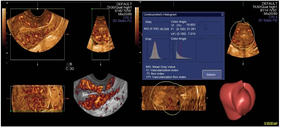
The ultrasound equipment used was Voluson E6 B12 system with 6 - 9 MHz transvaginal transducer. A 2-dimensional B-mode real-time sonographic ex-amination of the uterus and adnexae was initially carried out to study uterine size and shape and exclude any uterine or ovarian pathology. The color pulsed Doppler was activated in the 2D mode, the right and left uterine artery pulsatili-ty index (PI) and resistance index (RI) were calculated. Then, the ultrasound machine was switched to the 3D mode with power Doppler and 3D multiplanar view was seen.
Figure 1. 3D power Doppler indices of subendometrium of a case of menorrhagia: VI: 18.8, FI: 37.28, VFI: 7.01. VI: Vasculariza-tion index, FI: flow index, VFI: VascularizaVasculariza-tion flow index.
4. Statistical Methods
Statistical analysis was done using IBM© SPSS© Statistics version 22 (IBM© Corp., Armonk, NY, USA) and MedCalc© version 13 (MedCalc© Software bvba, Ostend, Belgium).
Continuous numerical data were presented as mean and SD, and one-way analysis of variance (ANOVA) was used for comparison of the three study groups with application of the Student-Newman-Keuls test for post hoc pairwise comparisons, whenever the ANOVA revealed statistically significant differences among the groups.
Qualitative variables were presented as number and percentage and inter-group differences were compared using the Pearson chi-squared test.
Receiver-operating characteristic (ROC) curve analysis was used to examine the value of ultrasonic indices for prediction of menorrhagia in women using IUCD. The DeLong method was used for comparison of the areas under various ROC curves.
P < 0.05 was considered statistically significant.
5. Results
There was no significant difference between patients in the three study groups as regards age, body mass index and parity.
The mean uterine artery PI was significantly lower in women of group I (1.78 ± 0.33) than in women of groups II and III, (2.28 ± 0.35 and 2.33 ± 0.41), p-value < 0.001 (Table 1).
The mean uterine artery RI was significantly lower in women of group I (0.68 ± 0.09) than in women of groups II and III (0.87 ± 0.13 and 0.85 ± 0.11), p-value < 0.001 (Table 2).
of group I (1.84 ± 0.56) than in women of groups II and III (1.64 ± 0.41 and 1.93 ± 0.49), p-value < 0.001 (Table 3).
Mean subendometrial resistance index (RI) was significantly lower in group I (0.59 ± 0.12) than in women of groups II and III (0.87 ± 0.27 and 0.85 ± 0.32), p-value < 0.001 (Table 4).
Mean subendometrial vascularization index (VI) was significantly higher in women of group I (6.86 ± 2.9) than in women of groups II and III (2.61 ± 0.97 and 2.77 ± 1.5) p-value < 0.001 (Table 5).
Mean subendometrial flow index was higher in women of group I (36 ± 3.52) than in women of group II (29.26 ± 3.3) and III (28.8 ± 3.32), p-value < 0.001 (Table 6).
Mean subendometrial vascular flow index (VFI) was higher in women of group I (1.93 ± 1.03) than in women in group II (0.8 ± 0.21) and III (0.93 ± 0.57) p-value < 0.001 (Table 7).
[image:5.595.208.539.330.434.2]Receiver-operating characteristic (ROC) curve was used for analysis of the
Table 1. Comparison of uterine artery pulsatility index (PI) in the three study groups.
p-value¶ F statistic
Group III (n = 105) Group II
(n = 105) Group I
(n = 105) Variable <0.001 32.097 2.22 (0.54) 2.26 (0.43) 1.77 (0.36)* Right Uterine artery PI <0.001 29.072 2.45 (0.52) 2.30 (0.68)†
1.79 (0.52)* Left uterine
artery P I
<0.001 77.986
2.33 (0.41) 2.28 (0.35)
1.78 (0.33)* Average UA PI
[image:5.595.209.540.501.609.2]Data are presented as mean (SD). ¶One-way analysis of variance (ANOVA). *p-value < 0.05 vs. IUD-O & Control groups (Student-Newman-Keuls test). †p-value < 0.05 vs. Control group (Student-Newman-Keuls test).
Table 2. Comparison of uterine artery RI in the three study groups.
p-value¶ F statistic
Group III (n = 105) Group II
(n = 105) Group I
(n = 105) Variable <0.001 59.686 0.86 (0.17) 0.89 (0.20) 0.66 (0.09)* Right Uterine artery RI <0.001 58.018 0.85 (0.11) 0.86 (0.12) 0.70 (0.13)* Left uterine
artery R I
<0.001 96.986
0.85 (0.11) 0.87 (0.13)
0.68 (0.09)* Average UA RI
Data are presented as mean (SD). ¶One-way analysis of variance (ANOVA). *p-value < 0.05 vs. IUD-O & Control groups (Student-Newman-Keuls test).
Table 3. Comparison of subendometrial PI in the three study groups.
p-value¶ F statistic
Group III (n = 105) Group II
(n = 105) Group I
(n = 105) Variable <0.001 10.081 1.93 (0.49) 1.84 (0.56) 1.64 (0.41)* Subendometrial PI
[image:5.595.207.541.665.711.2]data of all cases included in the study. Cut off values for prediction of menorr-hagia in women with IUCD using uterine artery PI, RI, subendometrial blood flow PI, RI, VI, FI and VFI are mentioned in Figures 2-8, Table 8.
6. Discussion
[image:6.595.207.539.333.378.2]The first published paper on actual IUCD insertions was made by Dr. Richard Richter in 1909 in Germany. The device he inserted was a ring made of silkworm gut, with 2 ends which protruded from the cervical os enabling him both to check the device and remove it. In the mid 1920s, Ernest Graefenberg added coiled metal ring made of an alloy of copper, nickel, and zinc. Graefenberg ring was widely used, however it was considered risky in continental Europe and in the U.S.. In 1959, Dr. Alan Guttmacher co-authored a paper in which the IUD was condemned for its ineffectiveness, potential source of infection, and its car-cinogenic potential. Since 1960, various kinds of IUDs have been developed and renewed interest in IUCD [7].
Table 4. Comparison of subendometrial RI in the three study groups.
p-value¶ F statistic
Group III (n = 105) Group II
(n = 105) Group I
(n = 105) Variable <0.001 81.968 0.85 (0.32) 0.87 (0.27) 0.59 (0.12)* Subendometrial RI
Data are presented as mean (SD). ¶One-way analysis of variance (ANOVA). *p-value < 0.05 vs. IUD-O & Control groups (Student-Newman-Keuls test).
Table 5. Comparison of subendometrial vascularization index (VI) in the three study groups.
p-value¶ F statistic
Group III (n = 105) Group II
(n = 105) Group I
(n = 105) Variable <0.001 107.929 2.77 (1.53) 2.61 (0.97) 6.86 (2.90)* Subendometrial VI
[image:6.595.207.538.448.493.2]Data are presented as mean (SD). ¶One-way analysis of variance (ANOVA). *p-value < 0.05 vs. IUD-O & Control groups (Student-Newman-Keuls test).
Table 6. Comparison of subendometrial flow index (FI) in the three study groups.
p-value¶ F statistic
Group III (n = 105) Group II
(n = 105) Group I
(n = 105) Variable <0.001 170.574 28.83 (3.32) 29.26 (3.39) 36.56 (3.52)* Subendometrial FI
Data are presented as mean (SD). ¶One-way analysis of variance (ANOVA). *p-value < 0.05 vs. IUD-O & Control groups (Student-Newman-Keuls test).
Table 7. Comparison of subendometrial vascularization flow index (VFI) in the three study groups.
p-value¶ F statistic
Group III (n = 105) Group II
(n = 105) Group I
(n = 105) Variable <0.001 86.074 0.93 (0.57) 0.80 (0.21) 1.93 (1.03)* Subendometrial VFI
[image:6.595.205.540.548.595.2] [image:6.595.208.539.666.711.2]Table 8. Doppler indices highly suggestive of menorrhagia after IUCD insertion. Specificity Sensitivity
Cut-off point Variable
87.6% 80.95%
≤1.9 Average uterine artery PI
90.4% 85.7%
≤0.76 Average uterine artery RI
64.7% 67.6%
≤1.83 Subendometrial PI
91.4% 80.9%
≤0.67 Subendometrial RI
96% 87.62%
≥4.1 Subendometrial VI
95.24% 89.5%
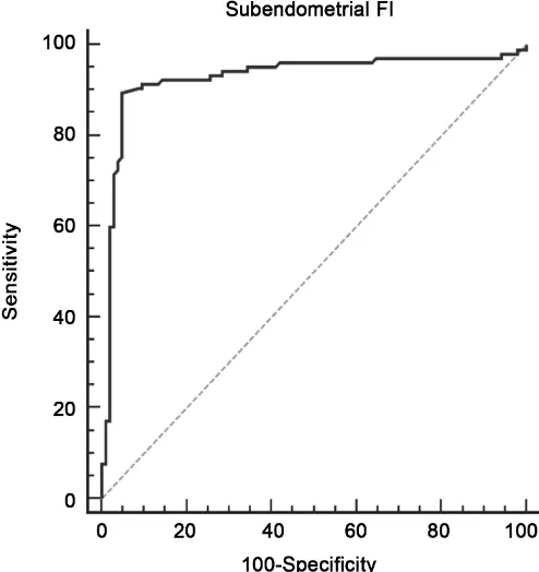
≥34 Subendometrial FI
87.6% 85.7%
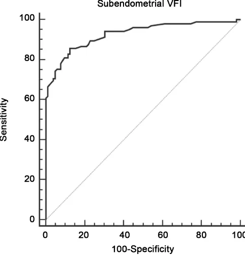
[image:7.595.252.473.123.401.2]≥0.98 Subendometrial VFI
Figure 2. Receiver-operating characteristic (ROC) curve for predic-tion of menorrhagia in women with IUCD using UA PI. Area under the curve (AUC) = 0.869 (95% CI 0.816 to 0.911). Best cutoff point is PI of ≤1.9. This has a sensitivity of 80.95% and specificity of 87.62%. (UA PI: uterine artery pulsatility index).
[image:7.595.281.462.478.668.2]Figure 4. Receiver-operating characteristic (ROC) curve for pre-diction of menorrhagia in women with IUCD using subendome-trial PI. Area under the curve (AUC) = 0.653 (95% CI, 0.585 to 0.718) of low predictive value. Best cutoff point of subendometri-al PI is <1.83. This is has a sensitivity of 67.6% and specificity of 64.7%. (PI: pulsatility index).
[image:8.595.254.491.411.654.2]Figure 6. Receiver-operating characteristic (ROC) curve for predic-tion of menorrhagia in women with IUCD using subendometrial VI. Area under the curve (AUC) = 0.953 (95% CI, 0.915 to 0.977). Best cutoff point of VI is >4.1. This has a sensitivity 87.6% and spe-cificity of 96.19%. (VI: Vacularization index).
[image:9.595.249.496.403.665.2]Figure 8. Receiver-operating characteristic (ROC) curve for predic-tion of menorrhagia in women with IUCD using subendometrial VFI. Area under the curve (AUC) = 0.929 (95% CI, 0.885 to 0.960). Best cutoff point of VFI is >0.98. This has a sensitivity of 85.7% and specificity of 87.6%. (VFI: Vascularization flow index).
Globally, 14.3% of women of reproductive age use intrauterine contraception (IUCD). In some countries, the percentage of women using IUC is <2%, whereas in other countries, it is >40% [8]. It was a widelyaccepted method for many women avoiding the systemic effects of hormonal contraception and the lower efficacy of the local methods or other physiological methods. However, IUCDs had their side effects, including the most common cause for discontinuation of using the method: menorrhagia. Excessive uterine bleeding up to iron deficiency anemia is among the most common causes of discontinuation of using the de-vices [9].
Trying to study the vasculature of the uterus and endometrium by assessment of uterine artery Doppler and subendometrial blood flow vascualrity by 2D and 3DPD, 315 cases were included in the current study, 105 with IUCD and me-norrhagia, 105 with IUCD without meme-norrhagia, and 105 controls. The result of the current study showed that women in group I with menorrhagia had a signif-icant increase in subendometrial vascularization index (VI), flow index (FI) and vascular flow index (VFI), and a significant decrease in subendometrial pulsitili-ty index (PI), resistance index (RI) and uterine artery (PI) and (RI), in compari-son with women in group II using copper IUD and not complaining of abnor-mal uterine bleeding and women in group III (control group). According to the results obtained in this study, it seems that the uterine arteries and subendome-trial blood flow is significantly higher in women with copper IUD and com-plaining of menorrhagia than women using copper IUD with normal menstrual flow or women with normal menstruation and not using any contraceptive me-thod.
Usama et al. measured PI and RI of uterine arteries in 93 women divided into three groups, group I included 32 women complaining of menorrhagia with IUCD, group II including 30 women not complaining of abnormal uterine bleeding with IUCD and control group including 31 women not using IUCDs. The uterine artery PI and RI were significantly lower in group I compared to group II and group III. [2] The total number in their groups was 93; the total number in the current study was 315. They did not assess the sub endometrial vasuclarity, in the current study sub endometrial blood flow 2D and 3DPD pa-rameters were assessed and revealed increased vascularity in cases of IUCD with menorrhagia.
El-Mazny et al., measured PI and RI of uterine arteries, endometrial and sub-endometrial VI, FI, VFI in 120 women before and three months after the copper IUD insertion, concluded that there was a significant increase in the endometrial and subendometrial VI, FI and VFI in 47 women who had menorrhagia after IUD insertion compared to 73 women who were not complaining of abnormal uterine bleeding after insertion. Whereas the uterine artery PI and RI were not significantly different before and after IUD insertion [5]. The current study agree with the study of El-mazny et al. as regards the increase in the microvas-cularization in cases after inserting IUCDs, however they stated that it was not predictive for menorrhagia. In the current study, uterine artery and subendo-mertial vascularity Doppler parameters cut off values for prediction of menorr-hagia were suggested. The current study included a larger number of total cases and larger number of menorrhagia cases, while their study—as a prospective study—depended only on the number of cases of menorrhagia among all the cases with IUCD, the number of menorrhagia cases in their study, forty seven cases may not express the whole population.
However uterine artery PI and RI were not altered after IUD insertion [3]. Frajndlich et al. in 2000 measured uterine artery RI and PI in 101 women, 74 women who were using copper IUCD and 27 controls who were not using any contraceptive method.
The intrauterine contraceptive device users were divided into three groups; those with normal bleeding n = 34, those with abnormal uterine bleeding with-out medication n = 16 and those with abnormal bleeding corrected with use of prostaglandin inhibitors n = 24. PI and RI were significantly lower in the group of women using intrauterine contraceptive devices who had abnormal bleeding than in all other groups [11], in the current study we preferred to include ge-nuine cases presenting with menorrhagia not on medical treatment to avoid any effect on the results of Doppler of the endometrium.
In agreement also with the results of Momtaz et al., they measured PI and RI of uterine arteries in 68 women, including 44 using intrauterine contraceptive device and 24 control, women who were not using a method of contraception. Both PI and RI were significantly lower in women with copper IUD-induced bleeding than in those using IUCD and not complaining of abnormal vaginal bleeding. In addition, there were no statistically significant differences in PI and RI in women using IUCD without complaining of abnormal vaginal bleeding and women in control group [12].
Yigit et al. found that the PI and systole/diastole ratio in the uterine artery in-creased significantly 3 - 5 month after the insertion of a copper IUD. However, patients with increased bleeding scores had significantly lower uterine artery PI compared with those without increased bleeding scores [13]. Although their re-sults of decreasing RI and PI agree with the rere-sults of the current study and many other studies, but their note of increasing PI and RI after the insertion of IUCD is of debate.
In contrary to results of the current study, desauza and Geber, measured the uterine artery PI and RI in both sides in 100 patients before and 30 days after insertion of Copper IUD, no statistically significant changes in PI and RI values were detected [1].It should be mentioned that they compared the group before and after insertion after 30 days, and that duration may not be enough for de-tectable changes by Doppler ultrasound. And that they did not select cases with menorrhagia, in the current study 105 cases presented with menorrhagia were included in the study, and all cases passed 3 months duration of insertion.
In contrary to the results of the current study, Jamenez et al.found no statis-tically significant differences in uterine artery PI and RI between women with IUD induced bleeding and women using IUD with normal menstruation [3]. El-Mazny et al. also reported that were no statistically significant differences in uterine artery PI and RI before and after Copper IUD insertion in patients with IUD induced menorrhagia [5].
production of prostaglandins leading to increase in subendometrial, and uterine artery blood flow.
Cut off values for uterine artery RI, PI, subendometrial RI, PI, VI, FI and VFI in the current study may be used for prediction of menorrhagia with IUCD in-sertion, however, further studies are needed with larger numbers and follow up pre and post insertion of IUCDs for a proper duration, as some cases develop menorrhagia after a longer duration.
7. Conclusion
Subendometrial vascularity in cases of menorrhagia with IUCD was markedly higher than in cases without menorrhagia and cases with no IUCD. 3DPD may be used for selection of cases prior to insertion of IUCD.
Conflicts of Interests
Authors state that there is no conflict of interest.
References
[1] De Souza, M.A. and Geber, S. (2006) Doppler Color Flow Analysis of the Uterine Arteries before and after Intrauterine Device Insertion: A Prospective Study. Jour-nal of Ultrasound in Medicine, 25, 153. https://doi.org/10.7863/jum.2006.25.2.153
[2] Fouda, U.M., Yossef, D. and Gaafar, H.M. (2010) Uterine Artery Blood Flow in Pa-tients with Copper Intra Uterine Device-Induced Abnormal Uterine Bleeding. Mid-dle East Fertility Society Journal, 15, 168-173.
https://doi.org/10.1016/j.mefs.2010.07.003
[3] Jimenez, M.F., Vetori. D., Fagundes, P.A., Freitas, F.M. and Arbo, E. (1998) Effect of the Copper—Intrauterine Device (TCU 380A) on Subendometrial Micro-Vascula- rization and Uterine Artery Blood Flow. Fertility Sterility, 86, 1780-1782.
https://doi.org/10.1016/j.fertnstert.2006.04.036
[4] Xin, Z.M., Cao, L.M., Xie, Q.Z., Sun, Y., Su, Y.C. and Guo, Y.H. (2009) Effects of the Copper Intrauterine Device on the Expression of Cyclooxygenase-1 and -2 in the Endometrium. International Journal of Gynecology & Obstetrics, 105, 166-168.
https://doi.org/10.1016/j.ijgo.2008.12.018
[5] El-Mazny, A., Abou-Salem, N. and Elshenoufy, H. (2013) Three-Dimensional Pow-er DopplPow-er Study of Endometrial and Subendometrial Microvascularization in Women with Intrauterine Device-Induced Menorrhagia. Fertility Sterility, 99, 1912- 1915. https://doi.org/10.1016/j.fertnstert.2013.01.151
[6] Dubiel, M., Moszczynska, K., Breborowicz, G., et al. (2010) Three-Dimensional Power Doppler in Obstetrics and Gynecology. Archives of Perinatal Medicine, 16, 109-112.
[7] Margulies, L. (1975) History of Intrauterine Devices. Bulletin of the New York Aca- demy of Medicine, 51, 662-667.
[8] Buhling, K.J., Zite, N.B., Lotke, P. and Black, K. (2014) INTRA Writing Group. Worldwide Use of Intrauterine Contraception: A Review. Contraception, 89, 162- 173. https://doi.org/10.1016/j.contraception.2013.11.011
https://doi.org/10.1080/ejc.7.3.178.183
[10] El-Sahwi, S., Toppozada, M., Kamel, M., Gaweesh, S., Riad, W., Ibrahim, I., et al. (1987) Prostaglandins and Cellular Reaction in Uterine Flushings. Advances in Contraception, 3, 291-302. https://doi.org/10.1007/BF01849285
[11] Frajndlich, R., von Eye Corleta, H. and Frantz, N. (2000) Color Doppler Sono-graphic Study of the Uterine Artery in Patients Using Intrauterine Contraceptive Devices. Journal of Ultrasound in Medicine, 19, 577-579.
https://doi.org/10.7863/jum.2000.19.8.577
[12] Momtaz, M., Zayed, M., Rashid, K. and Idriss, O. (1994) Doppler Study of the Ute-rine Artery in Patients Using an IntrauteUte-rine Contraceptive Device. Ultrasound in Obstetrics & Gynecology, 4, 231-234.
https://doi.org/10.1046/j.1469-0705.1994.04030231.x
[13] Yigit, N., Kacar, M., Yigit, H., Kosar, P. and Kosar, U. (2009) The Effects of Copper Contraceptive Intrauterine Device on the Uterine Blood Flow: A Prospective Transvaginal Doppler Study. Journal of Clinical Ultrasound, 37, 380-384.
https://doi.org/10.1002/jcu.20587
Submit or recommend next manuscript to SCIRP and we will provide best service for you:
Accepting pre-submission inquiries through Email, Facebook, LinkedIn, Twitter, etc. A wide selection of journals (inclusive of 9 subjects, more than 200 journals)
Providing 24-hour high-quality service User-friendly online submission system Fair and swift peer-review system
Efficient typesetting and proofreading procedure
Display of the result of downloads and visits, as well as the number of cited articles Maximum dissemination of your research work