http://www.scirp.org/journal/ym ISSN Online: 2475-7349
ISSN Print: 2475-7330
Meta-Analysis of Atrial Electro-Gram Applied to
PICC Catheter Tip Positioning
Jian Song
1, ChunLin Wang
2, Jun-E Hu
21Department of Medical College, Yangtze University, Jingzhou, China
2Department of oncology, Jing Zhou Central Hospital, Jingzhou, China
Abstract
Objective: Assess the location effect of the atrial electrogram in the PICC ca-theter tip. Method: Make online research in the CNKI, CBM, Wanfang, VIP, Database, Cochrane Library, JBI Library, PubMed, Ovid, Springer, Web of science, MEDLINE database to collect the randomized controlled trial data of using the atrial electrogram to locate the PICC catheter tip. When making the online research, only the data to November 20, 2016 can be searched out. Two researchers independently select the literature and information, make quality assessment, and then use the RevMan 5.2 software to make Meta analysis. Re-sult: 16 random tests are done. The Meta analysis results show that compared with the traditional X ray chest radiograph location method, the atrial electro-gram can increase the accuracy rate of the location of the catheter tip [RR = 1.13, 95%CI (1.07, 1.19), P < 0.00001]; it can decrease the incidence rate of re-lated complications [RR = 0.37, 95%CI (0.21, 0.66), P < 0.0008]; and it can save the catheter placing time [RR = −12.56, 95%CI (−18.56, −6.57), P < 0.0001]. Conclusion: The atrial electrogram can increase the accuracy rate of the loca-tion of the catheter tip, decrease the incidence rate of related complicaloca-tions and save the catheter placing time. Therefore, it is worthy of clinical application.
Keywords
Atrial Electrogram, PICC Catheter, Meta
1. Introduction
Peripherally inserted central catheter (PICC) has been widely applied in our country due to its advantages, namely, long-term use, reduced puncture pain, safety, efficiency, resistance to hypertonic and intense irritant drugs as well as fewer complications [1]. In order to achieve the effect of peripheral venous How to cite this paper: Song, J., Wang,
C.L. and Hu, J.-E. (2017) Meta-Analysis of Atrial Electro-Gram Applied to PICC Ca-theter Tip Positioning. Yangtze Medicine, 1, 65-76.
https://doi.org/10.4236/ym.2017.12007
Received: March 9, 2017 Accepted: June 18, 2017 Published: June 21, 2017
Copyright © 2017 by authors and Scientific Research Publishing Inc. This work is licensed under the Creative Commons Attribution International License (CC BY 4.0).
puncture and central venous therapy, the position of PICC tip is essential. American Society for Intravenous Infusion (INS) recommended PICC tip should be located at the lower 1/3 of patient’s superior vena cava [2]. At present, X-ray film combined with in vitro measurement method is adopted for the location of PICC tip in domestic. With the progress of the discipline, X-ray positioning method, as the “gold standard” of PICC tip location shows its hysteretic nature
[3], and it is gradually replaced by atrial electro-gram due to its drawback of radiation damage for pregnant women, children and other special groups. In clinical work, there are many studies regarding the application of atrial elec-tro-gram on PICC catheter tip location, but the majority of them are lack of evi-dences and clinical practice guidelines to confirm the accuracy and superiority
[4] [5]. The aim of this study is to investigate the accuracy and advantages of atrial electro-gram in the location of the tip of PICC catheters, with a view to providing references for clinical care and the clinical practice guidelines of se-lecting a tip location method after PICC catheterization.
2. Materials and Methods
2.1. Document Inclusion and Exclusion Criteria (PICOD
Principles)
2.1.1. Study Subject (Population)
1) PICC catheterized patients aged over 18 and voluntary to participate in this research. 2) Patients with body surface electrocardiograms being normal before catheterizing. 3) Patients whose catheter head ends were localized through in-tra-atrial electrocardiogram technology. 4) Patients who didn’t have heart dis-eases or previous operation history of heart disdis-eases. Exclusion criterion: 1) Pa-tients whose electrocardiographic examination results were normal before ca-theterizing such as patients with atrial fibrillation and ventricular tachycardia and those carrying cardiac pacemakers.
2.1.2. Intervening Measure (Intervention)
In the control group: The PICC catheter was implanted with the predicted length according to the length of the in vitro measurement. X-ray examination was performed to determine that the tip of the catheter was located in the lower 1/3 of the superior vena cava.
2.1.3. Control Measure (Control)
Pick up the surface ECG, observe the ECG P wave changes during the catheteri-zation process in real time, and then determine the catheter tip should be in the lower 1/3 of the superior vena cava based on the specificities of P wave, further X-ray examination should be performed to verify the location of the tip of PICC cathether [6].
2.1.4. Outcome Indicator (Outcome)
indicators mainly include occurrence rate catheter-related complications and the time taken by catheterizing.
2.1.5. Study Design (Design)
Randomized Controlled Trial (RCT) of atrial electro-gram applied to PICC ca-theter head-head location. Exclusion criteria: non-randomized controlled trials, clinical controlled trials, systematic review.
2.2. Literature Retrieval Strategy
Using “Peripherally inserted central catheter”, “PICC”, “Catheter tip location”, “Atrial electro-gram positioning”, “Electrocardiography”, “ECG”, “EKG” as key words ,the authors searched the CNKI, CBM, WanFang ,VIP databases, Coch-rane Library, JBI Library, PubMed, Ovid, Springer, Web of science, and MED- LINE databases. The time limit of these databases is from the establishment to April, 2016, and the retrieval is conducted in the form of free words with a key-word. In this study, CNKI and PubMed are taken as an example to introduce specific retrieval strategy [7] [8].
2.3. Literature Screening and Data Extraction
Two researchers independently screened the literature according to the inclusion and exclusion criteria set out in this study. By reading the literature title and ab-stract, the literature that clearly did not conform to the study was excluded. The controversial literature shall be discussed to decide whether or not to be in-cluded. If the result of the discussion is not consistent, a third party shall be asked to decide. For the literatures that meet the inclusion criteria, the informa-tion needed for this study, including the author’s name, the time of publicainforma-tion, the source of the literature, the baseline data comparability, the sample size, as well as the intervening measures and outcome indicators of intervention group and the control group, were included in the study.
2.4. Document Quality Evaluation
The literature quality for randomized controlled trials and semi-randomized controlled trials is evaluated according to the quality criteria of Cochrane Re-view's Handbook [9]. The evaluation criteria specifically include: 1) whether the random order is generated correctly; 2) whether the allocation of random pro-grams is hidden; 3) whether the blind method is used; 4) reports about the with-drawal and loss to follow-up; 5) whether the outcome indicators are complete; 6) baseline data comparability. The quality of the literature is divided into A, B, C three grades; Grade A fully meets the above criteria, Grade A meets part of the above criteria; and Grade C does not meet any of the above criteria.
2.5. Statistical Methods
(odds risk) as effective size. The continuous variable was expressed by using the weighted mean difference MD as the effect size, and the first thing was to deter-mine whether there is heterogeneity in the study. If there is no heterogeneity in different studies (P > 0.1, I2 < 50%) the fixed effect model is selected; if hetero-geneity exists in different studies. (P < 0.1, I2 > 50%), then it is necessary to ana-lyze the reason for the heterogeneity. If there is statistical heterogeneity but no clinical heterogeneity among the various studies, random effects model will be adopted [7]; if the source of heterogeneity can’t be determined, descriptive anal-ysis will be adopted.
3. Results
3.1. Literature Retrieval Results
576 articles were retrieved in the relevant databases, after reading the title and abstract, 531 articles of repeated publication or obviously do not meet the inclu-sion criteria were excluded, so 45 articles were included initially. In the follow-ing, 29 articles with unclear interventions, no outcome evaluation indicators or low quality were also excluded after reading the full text, and 16 articles were fi-nally included [10]-[25]. Meta-analysis was performed on 16 articles incorpo-rating qualitative synthesis.
3.2. General Condition of Included Literature
A total of 16 randomized controlled trials were included. The total sample size was 2996. In the experimental group, atrial electro-gram real time location was used as an intervention measure during the catheterization, and then X-ray ex-amination was used to confirm the accuracy of atrial electro-gram positioning; In the control group, X-ray examination was used as an intervention measure to have the catheter tip location after the end of the catheterization. The observed indicators were catheter tip arrival rate, complication rate, catheterization time- consuming. The general situation of the study literature is shown in Table 1.
3.3. Evaluation of Literature Quality
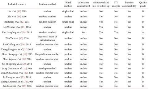
The 16 studies included were evaluated according to the Cochrane Review's Handbook quality assessment criteria, among the 16 studies that included in Meta-analysis, 10 studies [11] [12] [14] [15] [16] [18] [19] [21] [22] [25] specifi-cally described the grouping method, and six studies [10] [13] [17] [20] [23] [24]
reported only the using of random allocation, but not specifically described the grouping method; 3 studies [10] [12] [14] used single-blind, 1 study [14] used the envelope drawing method to follow the allocation of concealment; 5 studies
Table 1. General condition of the 16 literatures included.
Included research
Sample size Intervention measure
Outcome indicator Experimental
group Control group Experimental group Control group
Liu et al. [10] 2015 85 85 I II a
Elli et al. [11] 2016 77 44 I II a
Baldinelli et al. [12] 2015 42 48 I II a
Lei Wenlan et al. [13] 2016 130 130 I II a
Pan Longfang et al. [14] 2015 90 90 I II a, b, c
Zhu Ya et al. [15] 2010 65 64 I II a
Lin Cuifang et al. [16] 2015 60 60 I II a, b
Zhang Ronghua et al. [17] 2015 45 45 I II a
Zhou Lianqing et al. [18] 2013 51 49 I II a, c
Zhuo Yajuan et al. [19] 2014 60 60 I II a, b
Xu Mingming et al. [20] 2012 70 70 I II a, b
Jiang Guichun et al. [21] 2016 500 500 I II a
Wang Chaohong et al. [22] 2016 43 40 I II a, b
Li Xiangjun et al. [23] 2016 98 106 I II a
Zheng Chunhua et al. [24] 2016 60 60 I II a
Ren Xiaomin et al. [25] 2016 35 35 I II a, c
Note: 1) indicates the verified catheter tip position after the atrial electro-gram and X-ray chest positioning; indicates the verified catheter tip position after traditional catheterization and X-ray chest positioning. 2) a indicates the Arrival rate of catheter tip, b indicates the complication rate, c indicates the catheterization time-consuming.
Table 2. Quality evaluation results of 16 literatures included.
Included research Random method method Blind concealment Allocation loss to follow-up Withdrawal and analysis ITI comparability Baseline Quality grade
Liu et al. [10] 2015 unclear single-blind unclear No No Yes B
Elli et al. [11] 2016 random number unclear unclear Yes No Yes B
Baldinelli et al. [12] 2015 random number single-blind unclear Yes No Yes B
Lei Wenlan et al. [13] 2016 unclear unclear unclear Yes No Yes B
Pan Longfang et al. [14] 2015 random number single-blind Yes Yes Yes Yes A
Zhu Ya et al. [15] 2010 sequential order of catheterization unclear unclear No No Yes B
Lin Cuifang et al. [16] 2015 random number table unclear unclear No No Yes B
Zhang Ronghua et al. [17] 2015 unclear unclear unclear No No Yes B
Zhou Lianqing et al. [18] 2013 Hospitalization number unclear unclear No No Yes B
Zhuo Yajuan et al. [19] 2014 random number table unclear unclear No No Yes B
Xu Mingming et al. [20] 2012 unclear unclear unclear No No Yes B
Jiang Guichun et al. [21] 2016 envelope method unclear unclear No No Yes B
Wang Chaohong et al. [22] 2016 random number table unclear unclear No No Yes B
Li Xiangjun et al. [23] 2016 unclear unclear unclear No No Yes B
Zheng Chunhua et al. [24] 2016 unclear unclear unclear Yes No Yes B
[image:5.595.59.542.442.730.2]3.4. Meta Analysis Results
3.4.1. The Accuracy of Applying Atrial Electro-Gram Technique to PICC Catheter Tip Location
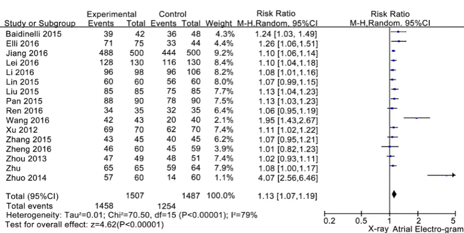
The accuracy of applying atrial electro-gram technique to PICC catheter tip lo-cation was analyzed in 16 randomized controlled studies, and the total sample size was 2996 (there was 1507 cases in atrial electro-gram positioning group, and 1489 cases in X-ray chest positioning group), statistical heterogeneity exists among various studies (RR = 1.13, I2 = 79%, P < 0.00001), so the random effect model was used for meta-analysis (P < 0.05). The Meta-analysis showed that the accuracy of the PICC catheter at the lower 1/3 of the superior vena cava in the atrial electro-gram positioning group was higher than that in the X-ray chest po-sitioning group. [RR = 1.13, 95% CI (1.07, 1.19), P < 0.00001]. See Figure 1. 3.4.2. Effects on the Complications of Applying Atrial Electro-Gram on
PICC Catheter Tip Location.
The results of two studies showed the effect of two different positioning methods on the incidence of complications in PICC catheter. The total number of sam-ples was 643 (323 cases in atrial electro-gram group, 320 cases in X-ray chest group), and there was no heterogeneity among various studies (RR = 0.41, I2 = 32%, P < 0.21). The restorative model was used for meta-analysis. Meta-analysis showed that PICC catheter-related complications in the atrial electro-gram posi-tioning group were lower (RR = 0.37, 95% CI (0.21, 0.66), P < 0.0008] compared with the X-ray chest positioning group. See Figure 2.
3.4.3. Effects on Catheterization Time-Consuming of Applying Atrial Electro-Gram on PICC Catheter Tip Location
Two studies compared the time consumed in two different positioning methods. The total number of samples was 350 (174 cases in atrial electro-gram group, and 176 cases in X-ray chest positioning group), statistical heterogeneity exists between two studies (RR = −12.56, I2 = 99%, P < 0.00001), and the random ef-fects model was used. Meta-analysis showed that PICC catheterization in atrial electro-gram positioning group took less time than in X-ray chest positioning group [RR = −12.56, 95% CI (−18.56, −6.57), P < 0.0001]. See Figure 3.
4. Discussion
4.1. The Application of Atrial Electro-Gram to PICC Catheter Tip
Location has Improved the Positioning Accuracy.
Figure 1. Meta-analysis forest map of applying atrial electro-gram technology in the accuracy of PICC catheter tip positioning.
Figure 2. Meta-analysis forest map of applying atrial electro-gram technology in the complications of PICC catheter tip posi- tioning.
Figure 3. Meta-analysis forest map of applying atrial electro-gram technology in the catheterization time-consuming of PICC catheter tip positioning.
[image:7.595.66.537.343.469.2] [image:7.595.70.538.517.610.2]head end is located in the right atrium, leads to the biphasic P wave; when the catheter tip was located in the right atrium, diphasic P wave was introduced; when the catheter tip was located in the ventriculus dexter, negative P wave that showed double peaks was introduced [26] [27] [28]. During the catheterization process, the nurse can determine the position of the catheter tip according to the change of the P wave, and timely debug the catheter until a specific P wave ap-pears. The traditional X-ray chest positioning mainly based on in-vitro predic-tion length before puncture, which is susceptible to be affected by puncture measurement method, puncture point changes and puncture subjective factors. As a result, the catheter tip can be too deep, too shallow or ectopic. The above situation can’t be identified by the naked eye, which can only be determined re-lying on the chest radiography after the catheterization, so the PICC catheter tip accuracy is greatly reduced. In the early 1990s, atrial electro-gram techniques have been applied to central venous catheters tip positioning in European coun-tries [29]. Over the past decade, many clinical studies have further confirmed that atrial electro-gram location is more accurate than traditional X-ray posi-tioning [30], Gebhard [31] and other studies also show that atrial electro-gram is more secure, more convenient and more reliable than X-ray positioning.
3.3. The Application of Atrial Electro-Gram to PICC Catheter Tip
Location has Reduced the Catheterization Time
The results of this study (Figure 3) showed that atrial electro-gram positioning method can save the catheterization time compared with the traditional X-ray positioning method. When the X-ray positioning method is used for PICC ca-theter tip positioning, the location of the tip can only be pre-determined based on the length of in vitro measurement and it is also necessary to go to the radi-ology department for routine filming. [34] [35]. If the ectopia occurs, you need to re-establish the aseptic area and film again, until the catheter tip location is correct. As we can see this process wastes a lot of valuable time of not only pa-tients but also nurses. Atrial electro-gram positioning method is able to monitor the catheter tip position in real time during the process of catheterization, and adjust any ectopia in time to achieve the integration between catheter and posi-tioning. In addition, it is not necessary for patients to have X-ray examination before the usage of medication, so its advantages are quite prominent [36] for critical patients. In this way, a lot of unnecessary troubles can be saved to ensure the timeliness of treatment. A large sample study carried out by Mourean [6]
and other researchers confirmed that the use of atrial electro-gram catheter tip positioning is able to reduce the catheterization time; generally, about 30 mi-nutes of operation time can be saved on average, which is consistent with the results of this study.
3.4. Limitations of the Study
Since the literature included in this study included only published Chinese and English literature, publication bias may exist for incomplete search; the me-ta-analysis included 16 randomized controlled trials, and the sample sizes range from 70 to 500 cases, so methodological and sample heterogeneity exists; the quality of the 16 literature included are graded to be lower-intermediate, which is not high. Only one literature [14] uses randomized grouping, strictly follows the blind method, sticks to the allocation concealment and focuses on the loss to follow-up, as well as reports the outcome indicators completely; while the re-maining 15 studies all have flaws in the methodology. The detailed grouping methods fail to be described in six studies [10] [13] [17] [20] [23] [24], thus pa-tients in both groups may have no comparability in baseline information and result differences will be large. In addition, Thirteen studies [11] [13] [15]-[25]
position-ing. Meanwhile, literature quality evaluation is intermediate level or below. In the future, it is necessary to popularize and generalize randomized controlled trial, improve clinical researchers’ research quality in the randomized controlled trial, and control bias through rigorous experimental design, so as to ensure re-search quality. It can be seen that there is few randomized controlled trials for atrial electro-gram to be actually conducted applied to PICC catheter tip posi-tioning. In the future, we should strengthen the popularization and promotion of randomized controlled research methods, improve the quality of randomized controlled trials of clinical researchers, and control the bias caused by various reasons through rigorous experimental design, so as to ensure the quality of re-search.In addition, literatures in the study give no explanations to the position-ing security of ECG in atriums and it is short of some evaluations that whether its security will affect final indicators, thus the future study should pay more at-tention to the positioning security of ECG in atriums. Limited by literature qual-ity and quantqual-ity, the study still has some limitations, thus more samples should be used for the randomized controlled trial with the longer duration in the fu-ture, so as to further explore and verify superiority and comprehensive effects of positioning PICC conductor housing of ECG in atriums.
In summary, the existing studies show that atrial electro-gram applied to PICC catheter tip positioning can improve the arrival accuracy of catheter tip, reduce the incidence of related complications and save the catheterization time, which can also provide scientific basis for further promotion of the atrial elec-tro-gram positioning method. However, in view of the quality limitations of the literature included in this study, multi-center and large sample of randomized controlled studies are still required in the future to further explore and confirm the superiority of applying atrial electro-gram to PICC catheter tip positioning.
References
[1] Yao, H. (2012) Clinical Research of Intravenous Electro-Gram on PICC Tip Loca-tion. Liaoning Medical College, Jinzhou.
[2] Deng, Z.Y., Chen, M., Huang, M.Y., et al. (2012) Effect Analysis of Intravenous Electro-Gram on PICC Tip Location. Hainan Medical Journal, 23, 116-117.
[3] Oliver, G. and Jones, M. (2014) ECG or X-Ray as the “Gold Standard” for Estab-lishing PICC-Tip Location. British Journal of Nursing, 23, S10-S16.
https://doi.org/10.12968/bjon.2014.23.Sup19.S10
[4] Liu, H.M. and Tan, J. (2012) Application of New Catheter Technique in PICC Ca-theterization Patients Guided by Atrial Electro-Gram. Journal of Qilu Nursing, 18, 43-44.
[5] Li, Q.L., Yan, M.Q., Zhang, X.J., et al. (2013) Study on the Effect of Different PICC Catheters on the Complications. Chinese Journal of Nursing, 48, 390-395.
[6] Mourean, N.L., Dennis, G.L., Ames, E., et al. (2010) Electorcardiogram (EKG) Guided Peripherally Inserted Central Catheter Placement and Tip Position: Results of Trial to Radiological Confirmation. Journal of the Association for Vascular Access, 15, 8-14.https://doi.org/10.2309/java.15-1-3
Pro-mote Neonatal Breastfeeding. Nursing Journal, 23, 9-13.
[8] Kuang, Y.H., Xu, Y.L., Yi, H.L., et.al. (2016) Meta-Analysis of Health Education In-tervention on Treatment Compliance of CPAP Patients. Nursing Journal, 23, 13-18. [9] Hu, Y. (2012) Evidence-Based Nursing. People's Health Publishing House, Beijing,
73-74.
[10] Liu, Y.J., Dong, L., Lou, X.P., et al. (2015) Evaluating ECG-Aided Tip Localization of Peripherally Inserted Central Catheter in Patients with Cancer. International Journal of Clinical and Experimental Medicine, 8, 14127-14129.
[11] Elli, S., Cannizzo, L., Marini, A., et al. (2016) Evaluation of Tip Location Reliability of Intraprocedural ECG vs. Chest X-Rays in PICC Placement. Assistenza Infermie-ristica E Ricerca, 35, 174-179.
[12] Baldinelli, F., Capozzoli, G., Pedrazzoli, R., et al. (2015) Evaluation of the Correct Position of Peripherally Inserted Central Catheters: Anatomical Landmark vs. Elec-trocardiographic Technique. The Journal of Vascular Access, 16, 394-398. https://doi.org/10.5301/jva.5000431
[13] Lei, W. and Yan, X. (2016) Application Effect Observation on Central Venous Ca-theter Tip Positioning under the Guidance of Intraline Electro-Gram. West China Medical, 31, 1-3.
[14] Pan, L., Hong, Y., Tang, L., et al. (2015) Clinical Application Research of Atrial Electro-Gram Positioning of PICC Puncture Technique. Journal of Chongqing Medical University, 40, 472-476.
[15] Zhu, Y., Ge, X., Huang, Y., et al. (2010) Effects of Intravenous Venous Catheter Vein Catheter on Ectopia Cordis by ECG. Nursing Journal of Chinese People’s Armed Police Forces, 11, 1711-1712.
[16] Lin, C., Xiao, Y. and Liu, Y. (2015) Effects of Electrocardiogram on PICC Tip Posi-tion and CatheterizaPosi-tion ComplicaPosi-tions. Journal of Nursing, 30, 38-40.
[17] Zhang, R., Zhang, J. and Qiu, J. (2015) Clinical Practice and Analysis of ECG Guided PICC Catheter Tip Positioning. Chinese and Foreign Medicine, No. 34, 68- 70.
[18] Zhou, L., Chen, Y., Wang, J., et al. (2013) Clinical Application Research of Atrial Electro-Gram Guided PICC Tip Positioning Method. Journal of Nurses Training, 28, 2021-2023.
[19] Zhuo, Y., Feng, L., Jiang, J., et al. (2014) Clinical Application of New Intravenous Electro-Gram Guided PICC Catheter Tip Positioning. Rehabilitation, 13, 868-870. [20] Xu, M., Wang, T., Yan, J., et al. (2012) Effect Observation of Applying ECG
Guid-ance Method to Improve the Success Rate of PICC Catheterization. Journal of Nursing, 19, 66-67.
[21] Jiang, G. (2016) Effect Evaluation of Applying Atrial Electro-Gram to Assist PICC Catheter Tip Positioning. Chinese Nursing Management, 16, 743-746.
[22] Wang, C., Wang, Z., Hou, X., et al. (2016) Application of Atrial Electro-Gram in PICC Catheterization in Cancer Patients. Medical Equipment, 29, 21-22.
[23] Li, X., Li, L. and Zhang, Y. (2016) Effect Research of Atrial Electro-Gram Position-ing on ReducPosition-ing PICC Catheter Ectopia. Journal of Qiqihar Medical College, 37, 3723-3725.
[24] Zheng, C. and Wu, X. (2016) Clinical Application of Intracardiac Electro-Gram Locates PICC the Catheter Tip Position. Nursing Research, 30, 4208-4209.
[26] Yao, H., Song, M. and Liu, Y. (2011) Clinical Study of Intravenous Electro-Gram Guided PICC Tip Positioning. Chinese Journal of Nursing, 46, 26-28.
[27] Feng, B., Yao, S., Zhou, S., et al. (2010) Intracardiac Electro-Gram Changes in the Process of Catheterization and the Guidance Role on Catheter Manipulation. Chi-nese Journal of Nursing, 45, 26-28.
[28] Guo, L., Qin, Y., Wang, G., et al. (2015) The Stability and Influencing Factors of Intracardiac Electro-Gram in Adult Venous Catheter. Chinese Journal of Nursing, 50, 724-727.
[29] Schummer, W., Schummer, C., Schelenz, C., et al. (2004) Central Venous Cathe-ters—The Inability of “Intra-Atrial ECG” to Prove Adequate Positioning. British Journal of Anaesthesia, 93, 193-198. https://doi.org/10.1093/bja/aeh191
[30] Schummer, W., Schummer, C., Schelenz, C., et al. (2005) Modified ECG-Guidance for Optimal Central Venous Catheter Tip Positioning. A Transesophageal Echocar-diography Controlled Study. Anaesthesist, 54, 983-990.
https://doi.org/10.1007/s00101-005-0886-2
[31] Gebhard, R.E., Szmuk, P., Pivalizza, E.G., et al. (2007) The Accuracy of Electrocar-diogram-Controlled Central Line Placement. Anesthesia & Analgesia, 104, 65-70. https://doi.org/10.1213/01.ane.0000250224.02440.fe
[32] Capozzoli, G., Accinelli, G., Fabbro, L., et al. (2012) Intra-Cavitary ECG Is an Effec-tive Method for Correct Positioning the Tip of Tunneled Groshong Catheters. The Journal of Vascular Access, 13, 393-396.https://doi.org/10.5301/jva.5000065 [33] Walker, G., Chan, R.J., Alexandrou, E., et al. (2015) Effectiveness of
Electrocardio-graphic Guidance in CVAD Tip Placement. British Journal of Nursing, 24, 28-30. https://doi.org/10.12968/bjon.2015.24.sup14.s4
[34] Chen, G., Wang, H. and Zhao, R. (2011) Progress in the Positioning of the Central Venous Catheter by Peripheral Vein Puncture. Chinese Journal of Nursing, 46, 1037-1039.
[35] Peng, X., Wen, Q. and Yao, Q. (2012) Progress on PICC Tip Positioning Method. Nursing Management Journal, 12, 861-862.
[36] Vesely, T.M. (2003) Central Venous Catheter Tip Position: A Continuing Contro-versy. Journal of Vascular and Interventional Radiology, 14, 527-534.
Submit or recommend next manuscript to SCIRP and we will provide best service for you:
Accepting pre-submission inquiries through Email, Facebook, LinkedIn, Twitter, etc. A wide selection of journals (inclusive of 9 subjects, more than 200 journals)
Providing 24-hour high-quality service User-friendly online submission system Fair and swift peer-review system
Efficient typesetting and proofreading procedure
Display of the result of downloads and visits, as well as the number of cited articles Maximum dissemination of your research work