warwick.ac.uk/lib-publications
A Thesis Submitted for the Degree of PhD at the University of Warwick
Permanent WRAP URL:
http://wrap.warwick.ac.uk/98579/
Copyright and reuse:
This thesis is made available online and is protected by original copyright. Please scroll down to view the document itself.
Please refer to the repository record for this item for information to help you to cite it. Our policy information is available from the repository home page.
2
Laser Doppler Imager Flare (LDIflare) small fibre
function
by
Prashanth Roshan Joseph Vas
MBBS MRCP (UK)
Thesis submitted for consideration
for the degree of
Doctor of Philosophy by published work
Division of Health Sciences
Warwick Medical School
University of Warwick
United Kingdom
3 Table of Contents
List of Tables... 4
List of Figures ... 4
Appendix A ... 4
Appendix B ... 4
Acknowledgement... 5
Submission Declaration ... 6
Word Count ... 6
Abbreviations used: ... 9
Abstract: ... 11
1.0 Introduction ... 15
2.0 Small Fibre Neuropathy in Diabetic Neuropathy : Significance and Emerging focus ... 18
2.1 The nerve fibres ... 18
2.2 Diabetic Neuropathy and Small Nerve Fibres ... 20
2.3 Diagnostic Criteria for Small Fibre Neuropathy ... 22
2.4 Techniques for Small Nerve Fibre Assessment... 22
2.4.1 The case for newer small fibre techniques ... 24
3.0 The LDIflare technique ... 31
3.1 Principle of LDIflare technique ... 32
3.2 Interpreting the results ... 34
3.3 LDIflare and Small Fibre Function ... 37
4.0 Synopsis of Studies included in this submission ... 38
4.1 Modification and validation of the LDIflare technique. ... 38
4.2 Estimating the rate of decline in LDIflare small fibre function assessed and developing age-related centile values in the detection of clinical neuropathy. ... 39
4.3 Additional validation of the LDIflare ... 40
4.3.1 LDIflare and point-of-care nerve conduction device (NC-stat®|DPNCheck™) for the measurement of early diabetic Neuropathy ... 40
4.3.2 The LDIflare in chemotherapy-induced peripheral neuropathy ... 41
4.4 LDIflare small fibre function in normal glucose tolerant subjects with and without hypertriglyceridaemia ... 42
4
5.0 Discussion ... 46
6.0 Limitations of the papers presented: ... 53
7.0 Conclusions and future outlook ... 57
7.1 LDIflare as a validated marker of small fibre function ... 57
7.2 Small fibre nerves and pathogenesis of diabetic neuropathy ... 58
7.3 Future Outlook for the LDIflare ... 60
List of Tables Table 1. Classification of Nerve Fibres ... 19
Table 2. Overview of the main tests used in small fibre assessment currently. ... 26
Table 3: The area under the curve for LDIflare for detection of DSPN categories. ... 41
List of Figures Figure 1: The Lewis Triple Response. ... 32
Figure 2: Principle of the LDIflare technique. ... 33
Figure 3: Baseline perfusion in the foot skin prior to a skin stimulus being applied. .. 34
Figure 4: post stimulation (skin heating) laser Doppler image acquired using a LDI. ... 35
Figure 5: Calculation of the LDIflare area. ... 36
Figure 6: Normogram of the LDIflare. ... 48
Appendix A: Statement of Author Contributions... A
5 Acknowledgements
Firstly, I would like to acknowledge and thank all the patients at the Ipswich Hospital and volunteers from the local community in Ipswich who agreed to participate in the research. Prof. Gerry Rayman, who encouraged me to pursue this research, and taught me the importance of lateral thought. During my research time he walked a perfect line between supervisor and collaborator, guiding and providing support when needed.
Prof Sudhesh Kumar, for his excellent support, guidance and for providing me an opportunity to complete my thesis.
My thanks go to my colleagues who have helped me with my research, Dr Sanjeev Sharma, Dr Al Green, Ms Helen Atkins, Mrs Ann Scott, Dr Krish Singham, Dr Ketan Dhatariya and Prof Mike Edmonds.
6 Submission Declaration
I hereby declare that I am the sole author of this thesis. The published works which this thesis is based are presented alongside and I can confirm that I have been closely involved in authoring them.
I declare that the submitted material as a whole is not substantially the same as published or unpublished material that I have previously submitted, or am currently submitting, for a degree, diploma, or similar qualification at any university or similar institution. No parts of the works submitted have been submitted previously for any aforementioned qualification.
7
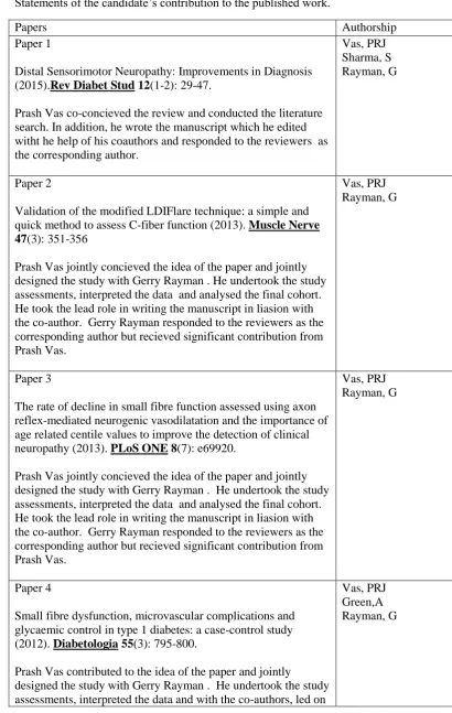
Table of publications submitted for consideration for the degree of Doctor of Philosophy by Published Work
Statements of the candidate’s contribution to the published work.
Papers Authorship
Paper 1
Distal Sensorimotor Neuropathy: Improvements in Diagnosis (2015).Rev Diabet Stud 12(1-2): 29-47.
Prash Vas co-concieved the review and conducted the literature search. In addition, he wrote the manuscript which he edited witht he help of his coauthors and responded to the reviewers as the corresponding author.
Vas, PRJ Sharma, S Rayman, G
Paper 2
Validation of the modified LDIFlare technique: a simple and quick method to assess C-fiber function (2013). Muscle Nerve 47(3): 351-356
Prash Vas jointly concieved the idea of the paper and jointly designed the study with Gerry Rayman . He undertook the study assessments, interpreted the data and analysed the final cohort. He took the lead role in writing the manuscript in liasion with the co-author. Gerry Rayman responded to the reviewers as the corresponding author but recieved significant contribution from Prash Vas.
Vas, PRJ Rayman, G
Paper 3
The rate of decline in small fibre function assessed using axon reflex-mediated neurogenic vasodilatation and the importance of age related centile values to improve the detection of clinical neuropathy (2013). PLoS ONE 8(7): e69920.
Prash Vas jointly concieved the idea of the paper and jointly designed the study with Gerry Rayman . He undertook the study assessments, interpreted the data and analysed the final cohort. He took the lead role in writing the manuscript in liasion with the co-author. Gerry Rayman responded to the reviewers as the corresponding author but recieved significant contribution from Prash Vas.
Vas, PRJ Rayman, G
Paper 4
Small fibre dysfunction, microvascular complications and glycaemic control in type 1 diabetes: a case-control study (2012). Diabetologia 55(3): 795-800.
Prash Vas contributed to the idea of the paper and jointly
designed the study with Gerry Rayman . He undertook the study assessments, interpreted the data and with the co-authors, led on
8
writing the manuscript. Gerry Rayman responded to the reviewers as the corresponding author but recieved significant contribution from Prash Vas.
Paper 5
LDIflare small fiber function in normal glucose tolerant subjects with and without hypertriglyceridemia (2015). Muscle Nerve 52(1): 113-119.
Prash Vas contributed to the idea of the paper and jointly
designed the study with Gerry Rayman . He undertook the study assessments, interpreted the data and with the co-authors, led on writing the manuscript. Gerry Rayman responded to the
reviewers as the corresponding author but recieved significant contribution from Prash Vas
Vas, PRJ Sharma, S Rayman, G
Paper 6
Early recognition of diabetic peripheral neuropathy and the need for one-stop microvascular assessment (2016). Lancet Diabetes Endocrinol. doi: 10.1016/S2213-8587(16)30063-8.
Prash Vas concieved the idea of this commentary and wrote the manuscript with contribution from Prof Edmonds. Prash Vas responded to the reviewers as the corresponding author.
Vas, PRJ Edmonds, M
9 Abbreviations used
:
CBNFD Corneal branch nerve fibre density
CHEPs Contact evoked potentials
CIPN Chemotherapy induced peripheral neuropathy
CNFD Corneal nerve fibre density
CNFL Corneal nerve fibre length
CPT Current perception thresholds
DPN Diabetic Peripheral Neuropathy
DSPN Diabetic Sensorimotor Polyneuropathy
EMG Electromyography
HBA1c Glycated haemoglobin
HiTG Triglycerides > 2.25 mmol/L
HTG Hyperriglyceridaemia
IENFD Intra epidermal nerve fibre density
IVCCM In-vivo Corneal Confocal Microscopy
LDIflare Laser Doppler Imager flare, area expressed in cm2
LDL Low density lipoprotein
LEPs Laser evoked potentials
MiTG Triglycerides between 1-7 and 2.25 mmol/L
MV- Microvascular disease Negative
MV+ Microvascular disease Positive
NCS NDS
Nerve conduction studies
Neuropathy disability score (scored out of 10) NIS-LL Neuropathy Impairment Score in the Lower Limbs
QSART Quantitative Sudomotor Axon Reflex
QST Quantitative Sensory Tests
SFF Small fibre function
SFN Small fibre neuropathy
SNAP Sural nerve action potential
SNCV Sural nerve conduction velocity
SNMFD Sural nerve myelinated fibre density
11 Abstract:
Distal sensorimotor peripheral neuropathy (DSPN), the classical length dependent
symmetrical neuropathy of diabetes, can affect up to 50% of those with diabetes
leading to significant morbidity, mortality and healthcare costs. There is
increasing recognition that small nerve fibres, mediating pain, temperature and
autonomic function, are involved early in the course of diabetic neuropathy,
preceding large fibre involvement. However, assessment small fibre neuropathy
(SFN), continues to be a significant challenge. The currently available options are
either invasive, subjective with poor reproducibility, may not directly assess the
region of interest or are still in a research phase. Thus, there is an ongoing need
for simple, non-invasive and reproducible techniques for the evaluation of SFN.
The laser Doppler imager flare (LDIflare) is one such novel, non-invasive
technique of assessing small fibre function. It has been shown to be a reliable
indicator of small fibre neuropathy, even when other SFN markers are either
inconclusive or normal. However, the original methodology, took over an hour to
complete, which limited its use as a clinical tool. The LDIflare methodology was
therefore modified to overcome this limitation by incorporating an accelerated
acclimatisation phase and a shorter duration of skin heating but at a higher final
temperature reducing the total procedure time to under 30 minutes. The size of the
resultant flares was nearly twice as large compared to the older method while
demonstrating similar group differences in those with and without clinical
neuropathy. Assessment of the LDIflare in healthy volunteers (n=94)
demonstrated significant inverse relationship of LDIflare size to age (r=-0.42,
p<0.0001) but not with other anthropometric or metabolic factors except for
12
sensitivity of 77%, specificity of 90%, positive predictive value (PPV) of 82% and
negative predictive value of (NPV) 87% for the detection of clinical neuropathy.
Recent observations, exploring into the aetiopathogenesis of diabetic neuropathy,
have suggested that triggers for neuropathy development in the two main forms of
diabetes may be different. Small fibre function (SFF) in individuals with type 1
diabetes with (MV+, n=24) and without (MV-, n=24) renal and retinal
microvascular disease, but all without clinical neuropathy was assessed using the
LDIflare. The finding of abnormal SFF only in the MV- group suggests that
direct microvascular damage is an early aetiopathogenic factor in type 1 diabetes.
Furthermore, in another study of normal glucose tolerant individuals not meeting
the criteria for metabolic syndrome, SFF was observed to be abnormal with
increasing fasting triglyceride concentrations. This is suggestive that
hypertriglyceridaemia may play a pathogenic role in the development of neural
dysfunction, and may partly explain the presence of neuropathy early in the
course of type 2 diabetes, when significant hyperglycaemia is not a factor.
The LDIflare in its current modification is a novel, reliable, non-invasive measure
and objective method of detecting small fibre neuropathy. It has good
reproducibility and offers excellent accuracy for the detection of clinical
neuropathy. The age based normative values allow for a clear distinction of
abnormal results. While further comparative studies between the LDIflare and
modern markers of SFN are desired, the studies included in this submission
support the use of the LDIflare technique to investigate abnormalities in the
peripheral nervous system, in particular small nerve fibres, in research but also in
14
15 1.0 Introduction
Diabetes Mellitus is a complex metabolic disorder characterised by defects in
insulin action, insulin secretion or both, resulting in disturbances of carbohydrate,
fat and protein metabolism [1]. The burden of diabetes has been rapidly
increasing, affecting up to 347 million individuals worldwide, with prevalence
rates reaching 9.8% in men and 9.3% in women [2] . In addition, a significant
number live with the condition undiagnosed and it is projected that with an aging
population and the continued rise in obesity, the global prevalence of diabetes
will reach 530 million individuals by the year 2035. At the same time, an
increasing number of individuals are being diagnosed with prediabetes, a
biochemical state where the glycaemic variables that are higher than normal, but
lower than the thresholds for diabetes, many of whom will eventually develop
diabetes [3]. The prevalence of prediabetes is projected to reach 470 million by
year 2030, a number not dissimilar to projected diabetes figures for same time
period [3-4]. Given the enormity of the numbers affected, diabetes is perceived as
a major public health challenge with substantial healthcare costs associated with
managing the condition and its complications [5-6]. A recent study from the
United Kingdom (UK) put the total cost of managing diabetes in the year
2010/2011 at around £24 billion while estimating that by the year 2036/2037,
these costs will rise to nearly £40 billion [5]. Figures emerging from other
countries also demonstrate a similar or higher degree of economic burden and the
overall picture is one of high cost with continued individual and societal suffering
[6-7].
The chronic complications accompanying diabetes can be divided into
16
infarction, stroke or peripheral vascular disease and, microvascular – those
affecting the small blood vessels, leading to the development of neuropathy,
nephropathy and retinopathy. Neuropathy is perhaps the most common
microvascular complication, ultimately affecting more than 50% of those with
diabetes [8]. Diabetic Neuropathy (DN) is a collective term for a heterogeneous
group of conditions that affect different parts of the nervous system and present
with diverse clinical manifestations [9]. Although any part of the nervous system
can be affected, the most common presentation is the length dependent distal
sensory predominant sensorimotor neuropathy (DSPN) which accounts for over
80% of the cases.
The consequences of DN are significant – it can lead to considerable morbidity
[10] and is increasingly recognised to confer an increased mortality risk [11-12].
Development of DSPN in particular, may lead to neuropathic pain, foot
deformities, loss of protective pain sensation and foot ulceration. In the latter
group, development of infection may increase the risk of a future lower extremity
amputation [13]. The life expectancy of patients with neuropathic foot ulceration
is approximately 50% at 5 years, an outcome worse than many of the major
cancers including breast, colon and prostate [14]. Like diabetes in general, the
healthcare costs associated with DN and its associated complications are
significant. In the year 2003, it was estimated that the direct cost of managing
DSPN in the UK was £252 million[15], but a more recent estimate for the year
2010/2011, commissioned by the charity Diabetes UK, which included the cost of
all diabetes related neurological conditions and foot disease, has put the figure
closer to £1.2 billion [6]. At the moment, there are no pathogenesis-based
17
Although many putative agents with disease modifying potential have been
investigated, none are currently licensed for the treatment of DN/DSPN [16].
Therefore, early recognition is vital. Findings from the Diabetes Control and
Complications Trial/Epidemiology of Diabetes Interventions and Complications
(DCCT/EDIC) studies have demonstrated that the clinical course of diabetic
complications in type 1 diabetes, DSPN in particular, may be influenced by
improved glycaemic control [17]. It is thought that the development of DSPN has
a temporal trend resulting in a clinical spectrum ranging from early, almost
undetectable, asymptomatic changes to an advanced stage with loss of protective
sensation [18-20]. The early neuropathic changes by virtue of being asymptomatic
are unlikely to be recognised by patients and clinicians alike. Unlike retinal
imaging for diabetic retinopathy and microalbuminuria for nephropathy, no
simple marker indicative of early neural damage exists for DSPN. Commonly
used neuropathy screening devices such as the 10gm monofilament or the 128Hz
tuning fork only detect advanced neuropathy and lack sensitivity for early
neuropathy [21]. It has been suggested by experts that more advanced neuropathy
is less likely to be amenable to therapy [16, 22]. Recognising neuropathy earlier
may thus increase the 'window of opportunity' - allowing for intensification of
diabetes therapy, management of associated vascular risk factors and for
institution of interventions aimed at arresting the progression to more advanced
neuropathy. Therefore, research aimed at recognition of early diabetic neuropathy
18
2.0 Small Fibre Neuropathy in Diabetic Neuropathy : Significance and Emerging focus
2.1 The nerve fibres
The Nobel laureates Gasser and his mentor Erlanger in studies of mammalian
neural tissue, observed neurologic structural deficits by cautious sectioning of
nerve fibres and subsequently classified nerve fibres into A, B and C groups.
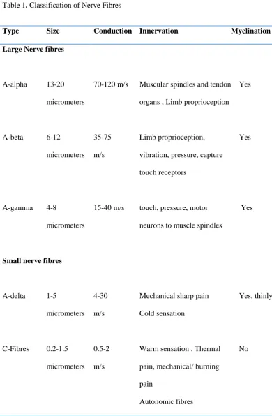
Subtypes of the A-fibres - A -alpha (afferent or efferent fibres), A -beta (afferent
or efferent fibres), A -gamma (efferent fibres) along with B- fibres are considered
as large fibres on account of their generous myelination. These are fast conducting
and mediate sensory modalities such as proprioception, touch, pressure and
vibration alongside mediating afferent and efferent motor function. Small nerve
fibres are slow conducting and comprise of the thinly myelinated A -delta and the
non-myelinated C-fibres (Table 1). Overall, the small fibres make up between
55% - 70% of the peripheral nerves; the A-delta fibres constitute ∼80% of
primary sensory nerves sprouting from dorsal root ganglia, whereas the C -fibres
make up ∼20% of the primary afferents [23]. Together, these nerve fibres
mediate pain, temperature perception and autonomic function. Thus, small fibre
neuropathy (SFN) may lead to generation of nociceptive pain (neuropathic pain),
abnormalities of temperature perception, impairment of vasodilatory responses
and also impaired sweating, heart rate variability, abnormal blood pressure
19
Table 1. Classification of Nerve Fibres
m/s= metres/second
Type Size Conduction Innervation Myelination
Large Nerve fibres
A-alpha 13-20 micrometers
70-120 m/s Muscular spindles and tendon organs , Limb proprioception
Yes
A-beta 6-12
micrometers
35-75 m/s
Limb proprioception, vibration, pressure, capture touch receptors
Yes
A-gamma 4-8
micrometers
15-40 m/s touch, pressure, motor neurons to muscle spindles
Yes
Small nerve fibres
A-delta 1-5
micrometers
4-30 m/s
Mechanical sharp pain Cold sensation
Yes, thinly
C-Fibres 0.2-1.5 micrometers
0.5-2 m/s
Warm sensation , Thermal pain, mechanical/ burning pain
Autonomic fibres
20
2.2 Diabetic Neuropathy and Small Nerve Fibres
There is increasing recognition that small nerve fibres are involved early in the
course of neuropathy development in both forms of diabetes [24-27] and also in
prediabetic states [28-29] . Although the precise sequence of nerve fibre damage
remains unclear, mainly because of the lack of longitudinal studies, assessment of
small fibre parameters in crossectional cohorts has suggested that small fibre
change may precede large fibre involvement [27, 30-32]; however these findings are not consistent [18]. Therefore, small fibre neuropathy (SFN) assessment
techniques have an important role in not only in characterising the presence of
diabetic neuropathy but also in the recognition of early diabetic neuropathy.
In addition to the potential role in the diagnosis of early diabetic neuropathy,
understanding small fibre neuropathy may be important in unravelling the
aetiopathogenesis of diabetic neuropathy. There is emerging evidence that the
pathogenesis of neuropathy differs between type 1 and type 2 diabetes.
Ultrastructural studies have suggested that paranodal degeneration seen in
neuropathy of type 1 diabetes is not seen in type 2 diabetes, with axonal loss and
atrophy expressed to a larger extent in the former [33]. While improvement in
glycaemic control has shown to significantly reduce the risk of diabetic
neuropathy in type 1 diabetes [17, 34], there is only a modest improvement in
neuropathy risk reduction in type 2 diabetes [9, 34]. Therefore, factors in addition
to hyperglycaemia are thought to potentially contribute diabetic neuropathy,
especially in type 2 diabetes, and may also be responsible for driving the earliest
damage. This is supported by a growing body of literature linking prediabetes and
neuropathy, which is typically of the small fibre type [35]. The metabolic,
21
neuropathy develops and may also in time allow for the development of
interventions designed to alter the course of neuropathy.
At the same time, it is felt by experts that small fibre assessments may be better
suited as future surrogate endpoints in neuropathy clinical trials than the current
large fibre based measures [9, 22]. Longitudinal follow up of diabetic subjects has
suggested the progression of large fibre neuropathy is minimal during a 1-5 year
follow up period, typical of most studies, but that the progression of small fibres
may be more rapid [36-38]. However, at what stage such a progression in a
particular small fibre marker constitutes a meaningful, clinically relevant change
is also unclear. This particular area needs attention as until this occurs, their
relevance as true 'clinical endpoints' for research will remain challenged.
Despite significant research and many molecules with putative disease
modification potential entering into phase III trials, there are currently no licensed
treatments for diabetic neuropathy. The most significant reason for failure in
clinical trials has been the lack of demonstrable treatment efficacy. While the
reasons for this have been greatly debated including poor subject selection, short
duration on intervention, and lack of homogeneity between study groups [16, 22,
39-40], a recent line of enquiry has been questioning the appropriateness of study
endpoints recommended by regulatory authorities, which are predominantly
clinical and large fibre based by which time the neuropathy may not be reversible
[41-42]. It has been advocated by some that small fibre based endpoints may be
superior, as these fibres are damaged early, and may be therefore, potentially more
amenable to regenerative or corrective therapy [22, 43]. However, there is no
current consensus on which of the small fibre techniques are best suited. As a
22
emerging into the understanding of small fibre neuropathy and into its detection
techniques.
2.3 Diagnostic Criteria for Small Fibre Neuropathy
The diagnostic criteria of SFN in diabetes mellitus were reviewed by the Toronto
Consensus on Diabetic Neuropathy panel, who proposed a grading of SFN into : i)
Possible SFN - length dependent symptoms and/or clinical signs ii) Probable
SFN - symptoms and signs as above and normal sural nerve electrophysiology,
iii) Definite SFN - symptoms and signs as above, normal sural nerve
electrophysiology and altered intra-epidermal nerve fibre density at the ankle
and/or abnormal quantitative sensory testing of thermal thresholds at the foot [31].
'Subclinical SFN' was considered if there were abnormalities on a validated SFN
marker but no symptoms and signs.
2.4 Techniques for Small Nerve Fibre Assessment
Detection of small fibre neuropathy, however, continues to be a significant
challenge. DSPN in particular, is typically suspected when symptoms such as
numbness, burning/throbbing pain, hyperaesthesia, hyperalgesia or parasthaesia
are reported. However, early diabetic neuropathy can be asymptomatic and typical
symptoms are often not recognised until the abnormalities are well advanced.
Clinical examination for reflexes, proprioception, light touch, vibratory perception
and motor strength is normal in pure SFN. Therefore in the absence of clinical
signs and symptoms to guide the diagnosis, there is a need for precise and reliable
testing methodologies for detecting and confirming SFN. Nerve conduction
studies are widely used to assess and quantitate nerve function but they detect
23
of SFN. Their role in SFN is complementary - to detect or exclude any associated
large fibre component [31, 44]. Recognising the importance of detecting such
early neuropathy, the Toronto Diabetic Neuropathy Consensus document has
recommended the use of small fibre testing, using a validated marker, when nerve
conduction assessment is normal [31].
Until recently, most diabetic neuropathy guidelines and position statements paid
limited cognizance to the importance of small fibre neuropathy. This was
primarily because most small fibre techniques were still in a research phase with
limited clinical utility. The 2017 position statement on diabetic neuropathy by the
American Diabetes Association has recently recommended using pinprick and
temperature sensation as bedside screening tests for small fibre function [9].
However, over the last three decades, significant progress has been made in the
development of techniques which allow for more detailed, clinically valuable,
small fibre characterisation and measurement [45]. They are divided into two
main groups:
I) Structural methods - allowing for morphometric analysis or quantitative
measurement of the small nerves. Examples include skin biopsy and in-vivo
corneal confocal microscopy (IVCCM).
and
II) Functional markers - these assess function of the small fibres, mainly
indirectly. Many functional techniques are qualitative but quantitative
methodologies are available and beginning to be more widely utilised. Examples
24
contact heat evoked potentials, sudomotor assessment techniques and the laser
Doppler imager flare (LDIflare) [45].
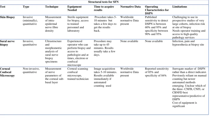
Table 2 summarises the characteristics, principles and limitations of some of the
main small fibre assessment tools.
2.4.1 The case for newer small fibre techniques
Skin biopsy with measurement of the intra-epidermal nerve fibre density (IENFD)
has been recently proposed as a 'gold standard' technique for the assessment of
SFN [46-47]. It allows for precise structural quantification and has well
established worldwide normative data [48]. However, its invasive nature is a
limitation, especially for application in large longitudinal studies requiring
repetitive assessments. Quantitative sensory testing (QST) of temperature and
pain thresholds is a non-invasive technique widely used in clinical practice as
well as research studies [46, 49-51]. Well recognised limitations include the
subjective, psychophysical nature of testing and its inability to distinguish
between a peripheral and a central lesion. Additionally and specifically in
relevance to diabetic neuropathy, it has been shown that there is poor relationship
between quantitative sensory tests and morphometric indicators of small nerve
fibre damage and repair [52]. In-vivo corneal confocal microscopy (IVCCM) with
visualisation small nerve fibres in the subbasal nerve layer of the Bowman layer
of the cornea has received significant recent attention [53-54]. It is easy to
acquire the images, non-invasive, has been validated against IENFD [55-56] and
possesses good sensitivity and specificity for the detection of DSPN [56-57] . In
addition, multi-centre reference values have been recently established [58]. A
25
away' from the distal leg. Moreover, morphometric analysis of the corneal
epithelial nerve terminals is not possible and variation in data has been reported,
especially in cloudy and fibrotic corneas [59]. Furthermore, it is unclear which
among the three parameters measured using IVCCM are best suited for
neuropathy assessment. While other SFN techniques are available and
summarised in Table 2, many are still in a research validation phase with limited
clinical experience.
Another challenge for SFN techniques is their ability to detect meaningful
improvement with treatment. Although IENFD has been shown to improve in
subjects receiving intensive lifestyle treatment for impaired glucose tolerance [60]
and IVCCM has shown improvement in treated type 2 diabetes [61] and after
pancreatic transplantation [62-63], the cohorts studied were small or the within group changes not significant. Thus, there is an overall need for newer SFN
measures that are simple, non-invasive, reliable and with an ability to detect a
26 Table 2. Overview of the main tests used in small fibre assessment currently.
Tests for Small fibre Neuropathy
Structural tests for SFN
Test Type Technique Equipment
Needed
Time to acquire results
Normative Data Operating
Characteristics for DSPN
Limitations
Skin Biopsy Invasive
(minimally), quantitative Measurement of intra-epidermal nerve fibre density Sterile equipment for biopsy, access to trained personnel and laboratory
Procedure takes 5-10 minutes but takes a few days to get the results back.
Worldwide normative Data present
Published
sensitivity to detect DSPN is between 60% and 95% and specificity between 90% and 95%
Challenging to use in prospective studies of very large cohorts, infection risk at site of biopsy
Needs operator training and access to high quality pathology services Sural nerve biopsy Invasive, quantitative Ultrastructure and morphometric analysis of sural nerve biopsy specimens Experienced operator who can perform biopsy and access to
pathologist and at times, electron or confocal
microscope
Procedure may take up to 45 minutes. Results usually take a few days.
None available None available Infection, pain and hypoesthesia at biopsy site
Corneal Confocal Microscopy Non-invasive, quantitative Measurement of nerve parameters of the corneal sub-basal layer Corneal scanning confocal microscope, trained technician Image acquisition takes 5-10 minutes. Results available immediately if automated counting used
Worldwide normative Data present
Reported sensitivity of 85% and specificity of 84%
Surrogate marker of DSPN rather than a direct indicator. Previously reliant on manual counting but newer
automated methods
emerging. Unclear which of the three- CNFB, CNFL or CBNFD best
representative/predictive of DSPN
27
Functional tests for SFN
Quantitative sensory testing (QST) Non-invasive, quantitative Computerised measurement of thermal thresholds and heat pain thresholds Computerised assessment device, temperature controlled laboratory and a trained technician
Takes about half an hour but could take longer, depending on subject concentration Commercial normative data present from the bigger
manufacturers.
Psychophysical test- results are dependent on subject compliance and attention. Complex testing protocols present . Varying
reproducibility depending on experience of the unit undertaking testing. Laser Doppler imager Flare (LDIflare) Non-invasive, quantitative Measurement of the axon-reflex mediated flare response as a marker of small fibre function Laser Doppler imager, temperature controlled room, operator with experience Image acquisition took ~1 hour with the older method. Newer method takes
approximately 25 minutes (discussed within the thesis). Results available immediately
One site
normative values determined at a single centre. Larger data set of normative valves desired
For the newer technique: Sensitivity of 70-75%, specificity of 66-85% , positive predictive value of 74%, and negative predictive value of 86%
Dependent on the microcirculation. Patients need to have no significant macrovascular distal circulatory impairment. Current perception threshold (CPT) Non-invasive, quantitative Low current intensity stimulation of the small nerve fibres at frequency of 250 Hz for A-delta fibres and the
5 Hz for C-fibres.
Neurometer device temperature controlled room and a trained technician
Takes about half an hour but could take longer, depending on subject concentration None available. Most studies have included age matched controls for comparison.
28 Contact Heat Evoked Potentials (CHEPs) Non-invasive, quantitative Measure cerebral responses to thermal stimuli mediated by A-delta fibres
Needs thermal threshold testing first. Then small discs are placed on the head to record signals received to the brain from application of 10 to 20 short (a fraction of a second) heat or cold
stimuli at a particular point of interest (face, arm or leg)
Takes about half an hour but could take longer, depending on subject concentration
Multicentre normative data on 226 adult
subjects are available
The AUC for DSPN detection in a small sample has been estimated at 0.778.
Requires active patient co-operation. Like QST and CPT, therefore
reproducibility has been a challenge. Not widely available. Also unclear if both A-delta and C-fibres are assessed.
Participant discomfort is another major limitation of use. Microneuro-graphy Minimally invasive, semi quantitative Measurement of Single fibre recordings from peripheral axons Skilled operator and extensive equipment list. Preserve of a large neurophysiology lab rather than clinic based procedure.
May take up to 3 hours to get a satisfactory recording.
None available None available. Considered by EFNS to possess grade A evidence for assessing function of the A-delta fibre pathways in patients with neuropathic pain
Still primarily a research tool. May have a role in assessment of neuropathic pain rather than early neuropathy. Expensive and needs skill to elicit responses. Patient cooperation is also extensively required. Laser Evoked Potentials (LEPs) Non-invasive, quantitative Radiant heat generate by laser selectively excites free nerve
endings in the superficial skin layers
activating nearby A-delta and C -fibre nociceptors
CO2-laser stimulator, technician with experience and a temperature controlled room ideally.
May take up to 1 hour to complete the procedure and ensure no artefacts presents in readings gained
Single centre normative values available on 100 subjects. No decade specific data reported.
None available. Studies have used age matched control data.
Limited availability. May be useful in demonstrating reduced function but unable to detect enhanced
29 Quantitative
sudomotor axon reflex test (QSART) Sudomotor Non-invasive, quantitative Information on skin autonomic function and evaluation of postganglionic sudomotor function using acetylcholine iontophoresis Purpose built lab, iontophoresis and sudomotor quantification equipment.
45-60 minutes to complete.
Normative data available from specific centres for QSART. A commercially available device QSWEAT is also available
No specific data available for DSPN but has been widely used, especially in the Rochester Diabetic
Neuropathy study
Requires precautions for electrical safety and small risk of minor local injury to the skin Thermo-regulatory sweat test (TST) Sudomotor Non-invasive, semi-quantitative When core temperature rises beyond a hypothalamic thermoregulatory set point (>38°C), sweating occurs
Needs a laboratory and a digital camera
90-120 minutes to perform correctly. Maximal sweating is achieved within 30–65 minutes.
Unclear Helpful data on the TST available in DSPN mainly from the autonomic lab at the Mayo Clinic, Rochester USA.
Patients may not be able to tolerate 60 minutes of warming up Sympathetic Skin Response Sudomotor Non-invasive, quantitative Information on skin autonomic function and evaluation of postganglionic sudomotor function using electrodermal activity Purpose built lab, SSR equipment includes electrodes.
45-60 minutes to complete.
Normative data available from specific centres but usually has been derived from a small normative group
Minimal data only available in DSPN. Some helpful data in diabetic autonomic neuropathy and bladder dysfunction.
Limited availability, needs expertise and experience to test correctly. Popular in Japan.
Sudoscan® Sudomotor
Non-invasive, quantitative
Testing is based on stimulation of sweat glands by a low-voltage current (<4volts) representing a electrochemical reaction between electrodes and chloride ions, Just the Sudoscan® device
Takes less than 5 minutes
Comes with inbuilt normative data. Limited experience at the moment
Increasing literature now available of its use in DSPN. Similar AUC as IENFD (0.761) in one study. For Cardiac autonomic neuropathy sensitivity was 65%, specificity 80%.
Still limited availability . Needs more detailed validation work for different ethnicities.
Reproducibility is yet to be established.
30
Neuropad® Simple
qualitative indicator of sudomotor dysfunction Simple sticker which changes colour in the presence of sweating.
Relatively cheap and easy to avail. Cost is approximately £8/pad.
Takes less than 10 minutes
Qualitative, does not need normative data.
Lots of available literature and has been validated against IENFD. In one study, Neuropad had a sensitivity 85% and specificity of 45% for detection of clinical DSPN.
Difficult to interpret when there is partial change in colour though. One centre has published data on semi-quantification using digital imaging of the Neuropad®.
Cardiovascular Autonomic tests Quantitative and assess cardiac autonomic neuropathy Complex laboratory based testing protocols Special labs, equipment and expertise in testing. Usually undertaken by
Neurophysiologists
Takes up to 90-120 minutes.
Simpler algorithms may take less time (30-45 minutes)
No worldwide reference values, each laboratory tends to have its own values
Not easily available to frontline clinicians.
Mainly a tool for research in DSPN or helpful in
31 3.0 The LDIflare technique
The laser Doppler imager flare (LDIflare) is a novel non-invasive method for the
detection of small fibre neuropathy, based on measurement of the axon-reflex
mediated vasodilatory flare response, using skin heating as the nociceptive stimulus.
This neurogenic vasodilatory response has been shown to be directly related to small
nerve fibre and in particular, c-fibre function [64-66]. The axon reflex is part of the
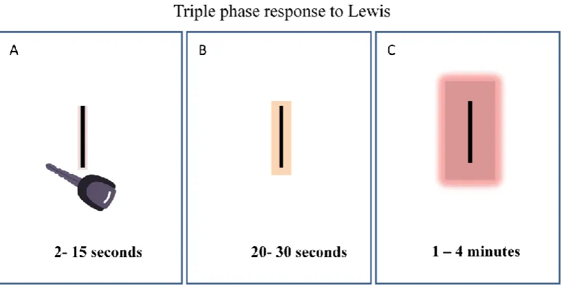
Lewis triple response which was first described by Sir Thomas Lewis in 1924 [67].
This response can be elicited by drawing a sharp object (a key) across the skin and
comprises of three phases:
1) Red reaction or the Flush: This occurs in the first few seconds post injury and
visually appears as a red line at the site of injury. It is due to capillary dilatation
secondary to histamine release.
2) Wheal: This is localized ( to around the region of the redline) and is due to
increased capillary permeability and exudation of fluid from dilated arterioles,
capillaries and venules driven by the local histamine release and appears
approximately 30 seconds to 1 minute after the initial injury.
3) Flare: Spreading redness, extending beyond the redline, secondary to the axon
32 Figure 1: The Lewis Triple Response.
In panel: A) the skin is injured with a key resulting in a pink flush; B) Wheal
developing immediately as a direct response and C) Flare response secondary to
activation of the local small fibre network.
3.1 Principle of LDIflare technique
Stimulation of nociceptive C-fibres leads to the simultaneous conduction of impulses
orthodromically to the spinal cord and antidromically to the nerve endings abutting
arterioles, where the release of neurovascular transmitters causes vasodilatation and
increased blood flow to injured tissues [68]. Substance P and Calcitonin Gene
Related Peptide (CGRP) are the two main neurovascular transmitters that are
released from C-fibres and cause vasodilatation of the arteriolar blood vessels
innervated by the neural network [69]. Substance P acts on its preferred receptor, the
NK1 receptor and on endothelial cells to cause plasma leakage [70]. Substance P
33
with the stimulatory activity of CGRP acting on its receptor contribute to arteriolar
dilatation [71-72]. The neural course of the axon- reflex is illustrated in Figure 2.
The resultant flare response may be detected visually, and objectively quantified
[image:34.595.139.495.278.502.2]using the laser Doppler imager (LDI).
Figure 2: Principle of the LDIflare technique.
The course of the nerve axon-reflex and the antidromic conduction into the adjacent
34 3.2 Interpreting the results
[image:35.595.114.499.160.375.2]Baseline perfusion
Figure 3: Baseline perfusion in the foot skin prior to a skin stimulus being applied.
On the right, is a representative B&W image of the scanned area. Using the palette
provided one can see that the perfusion in this image is <100PU in most of the
35
[image:36.595.114.500.146.361.2]Post Stimulation change in perfusion and flare response
Figure 4: The post stimulation (skin heating) laser Doppler image acquired using a
LDI.
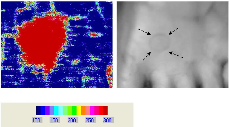
The image analysis palate has been set such that flux < 100PU appears as dark blue
(representing standard blood flow) and anything >300PU appears as bright red. The
36 Measurement of the LDIflare area.
Figure 5: Calculation of the LDIflare area.
The white freeform constitutes the area of the post skin heating resultant flare
(LDIflare) and is expressed in cm2. The area under the heater (black circle)
represents the area directly under the heater probe and the change in perfusion is
non-neurogenic and endothelium dependent. It is termed LDImax and is a measure
of endothelial function. The area outside the skin heating is the true axon-reflex
mediated flare (LDIflare).
37 3.3 LDIflare and Small Fibre Function
The original methodology of the LDIflare (oLDIflare) was described in 2004 [73] .
Initial validation demonstrated good reproducibility along with an ability to detect
early neuropathic abnormalities, even when QST's were normal [73-74]. In a select
cohort of impaired glucose tolerant individuals without clinical neuropathy, the
oLDIflare demonstrated evidence of small nerve fibre dysfunction [75]. At the same
time, a group of patients with longstanding type 1 diabetes, specifically selected for
the absence of any micro/macrovascular complications demonstrated normal small
fibre function. Discussing these findings in an editorial, Prof. Boulton and Prof.
Malik, highlighted the oLDIflare as a emerging marker of SFN with potential for
application in longitudinal cohorts and neuropathy drug trials [76]. The Toronto
Consensus on Diabetic Neuropathy also provided further recognition but suggested
that further studies were required to validate the technique and develop its diagnostic
potential [31]. In order to achieve this, the methodology needed addressing to make
the oLDIflare more practical and accessible - testing took 90 minutes to complete -
making examination of large patient numbers difficult. It was unclear if the original
heating temperature of 44ᵒC fully stimulated the small nerve fibres and there were
additional validity concerns regarding its specificity as a marker of small fibre
function. Furthermore, it lacked normative data and precise operating characteristics
for the detection of diabetic neuropathy - important attributes required for clinical
application.
In order to overcome these challenges, modifications were made to the methodology
to allow the testing procedure to be completed faster and further validation studies
38
4.0 Synopsis of Studies included in this submission
In this section, I have provided a brief synopsis of the studies included in this
submission. They include experiments conducted to validate the LDIflare
methodology, determination of normative values and operating characteristics such
as sensitivity and specificity for the detection of clinical diabetic neuropathy. A
further study examining the relationship of fasting triglycerides in normal glucose
tolerant on small fibre function is described. In addition, in a group of individuals
with type 1 diabetes, normal small fibre function is demonstrated in the absence of
clinical microvascular disease elsewhere.
4.1 Modification and validation of the LDIflare technique.
The primary modifications to the LDIflare methodology were: 1) the shortening of
the acclimatisation period, 2) reducing the duration of skin heating required to elicit
a nociceptive response, thus allowing the process to be completed faster, and 3)
utilising a higher final skin heating temperature. The methodology is detailed in the
papers submitted. The neurogenic nature of the mLDIflare was confirmed in a study
in which a local anaesthetic cream (EMLA; lidocaine 2.5% and prilocaine 2.5%;
AstraZeneca, Luton, UK) was applied over the heating area prior to eliciting the
mLDIflare. The axon-mediated flare response was near completely obliterated
(9.3±3.0 cm2 before and 1.7±0.3 cm2 after; p < 0.0001), confirming the neurogenic
nature of the LDIflare. In another study it was shown that there was near perfect
correlation of flare sizes between the right and left feet (Pearson’s coefficient r=
0.95, p<0.0001). Finally, and in agreement with the original technique, the
mLDIflare was significantly lower in the group with diabetic neuropathy (DN+)
39
when the DN+ group was compared to those without diabetic neuropathy (DN-)
(1.99±1.1 cm2 v 6.78±2.78 cm2; p<0.001) [73].
4.2 Estimating the rate of decline in LDIflare small fibre function assessed
and developing age-related centile values in the detection of clinical
neuropathy.
The peripheral nervous system, both somatic and autonomic, changes with age [48,
58, 77-78] . Determination of accurate normative values underpins the diagnostic
validity and overall performance of a test [79]. Given the potential role of the
LDIflare in future testing of early DSPN, it was essential to understand the variables,
both clinical and metabolic, that influence it. It was also important to establish
operating characteristics, such as sensitivity and specificity to determine what values
can be confidently defined as 'abnormal'. Therefore, studies were undertaken to
establish determinants of small fibre function as assessed by the LDIflare method
and derive age-related centile values. An additional aim was to determine which of
the two analytical techniques, receiver -operator derived cut-off values or centile
based values, were superior in assessing the operating characteristics of the LDIflare.
Therefore, across-sectional cohort of 94 healthy volunteers was recruited to
understand the variables that influence the LDIflare and develop age-specific
normative values. Glucose dysregulation was excluded using a composite of fasting
40 4.3 Additional validation of the LDIflare
The two studies described below were not specifically designed to validate the
LDIflare. They provide crucial supportive literature evidence for the LDIflare as a
useful SFN marker, but are not part of the thesis submission.
4.3.1 LDIflare and point-of-care nerve conduction device
(NC-stat®|DPNCheck™) for the measurement of early diabetic Neuropathy
More recently, a simple point-of-care device (POCD) has been developed and
validated for the detection of sensory changes in the sural nerve and to serve as an
acceptable proxy to NCS [80]. The hand-held device: NC-stat®|DPNCheck™
system (developed by Neurometrix, Waltham, MA) allows the quantitative
measurement of sural nerve conduction velocity (SNCV) and amplitude of the
sensory nerve action potential (SNAP). Validation studies have suggested that it has
excellent correlation with conventional NCS (r=0.95, p<0.001) and is reliable
(inter-rater reproducibility values of 0.83 for SNAP and 0.79 for SNCV) [80-81]. In
addition, the sensitivity and specificity for identification of DSPN defined by
electrophysiological criteria was 95% and 71% respectively [81].
The accuracy of the NC-Stat®|DPNCheck™ was compared with the modified
LDIflare technique for the detection of DSPN categorised into no (0-2), mild (3-5),
moderate (6-8) and severe (9-10) DSPN using the neuropathy disability score (NDS)
[82]. A total of 80 healthy controls (HC) alongside 162 diabetic individuals (50%
type 1 diabetes) were recruited. All groups were age (p=0.22) and gender (p=0.57)
matched. Among HC, the LDIflare correlated strongly with both SNAP (r=0.88,
41
the LDIflare and POCD measures were noted within the no, mild, moderate and
severe DSPN groups [82]. The LDIflare was significantly smaller among those with
DM when compared to HC (5.81±2.09 vs. 9.11± 2.17 cm2, p<0.0001). The results
with the POCD were similarly significantly lower in the DM cohort - SNCV
(42.04±9.11 vs. 50.24±5.69 m/s; p=<0.0001) and SNAP (10.13±3.12 vs. 18.49±4.13
µV; p=<0.0001). In addition, the LDIflare and POCD both demonstrated individual
reduction in mean values across the DSPN categories [82]. However, while there
was a highly significant difference in the LDIflare sizes between the HC and the
diabetic control group (those with NDS 0-2) (9.11± 2.17 vs 7.52 ± 2.59 cm2, p <
0.0001) there was no such difference between the same groups for SNAP (18.49 ±
4.13 vs 16.61± 8.45, p=0.15) and SNCV (50.22 ± 5.69 vs 48.95 ± 12.70, p=0.11)
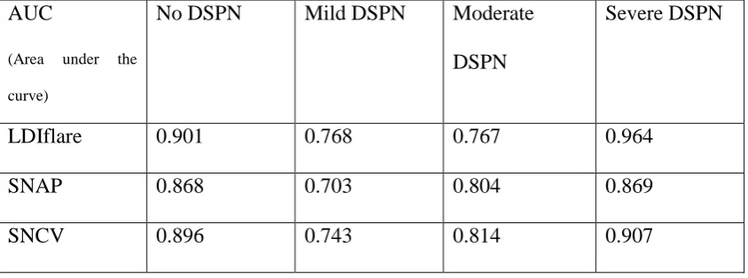
[82]. The AUC for the LDIflare and POCD were as follows (Table 3, data extracted
from Sharma et al [82]).
AUC
(Area under the
curve)
No DSPN Mild DSPN Moderate
DSPN
Severe DSPN
LDIflare 0.901 0.768 0.767 0.964
SNAP 0.868 0.703 0.804 0.869
[image:42.595.109.531.439.596.2]SNCV 0.896 0.743 0.814 0.907
Table 3: The area under the curve for LDIflare for detection of DSPN categories.
4.3.2 The LDIflare in chemotherapy-induced peripheral neuropathy
A common, unpredictable and at times dose limiting complication of cancer drug
42
symptoms of CIPN such as paraesthesia, dysaesthesia, hyperalgesia, hypoalgesia
and patients are similar to those described in DSPN. Patients may also experience at
the same burning, shooting or electric-shock type discomfort described in diabetic
neuropathic pain [83]. Furthermore, like DSPN, the distribution of symptoms is in a
“stocking and glove” manner, due to the vulnerability of the long nerves [84]. The
diagnosis of CIPN is clinical as there is no specific test. However, it is understood
that IENFD depletion occurs early despite normal peripheral nerve axon counts and
preserved nerve conduction results and may at times be the only hallmark of CIPN
[83, 85].
The LDIflare was evaluated 24 patients with established CIPN and distal sensory
symptoms to determine its utility in assessing the diagnosis [86]. Of these, 12 were
CIPN secondary to platinum analogues and 12 with damage from taxanes. An
additional 24 age, gender and BMI matched healthy controls were also studied.
Apart from the LDIflare, additional neurological assessments included determination
of vibration perception thresholds (VPT), sural nerve amplitude (SNAP) and
conduction velocity (SNCV) [86]. The LDIflare was significantly reduced in CIPN
group compared to HC (3.75 ±1.68 vs 6.53 ±0.75 cm2 , p<0.001) while SNAP (p=
0.06 and SNCV (P = 0.09) were not [86]. Additionally, the LDIflare was the only
neurological marker to correlate with European Organisation for Research and
Treatment of Cancer - QLQ-CIPN20 disease severity questionnaire [86].
4.4 LDIflare small fibre function in normal glucose tolerant subjects with
and without hypertriglyceridaemia
The relationship between early diabetic neuropathy and dyslipidemia, and
hypertriglyceridaemia in particular, is receiving increasing attention. However, the
43
asymptomatic glucose tolerant individuals remains unclear. Neuropathy studies in
non-diabetic individuals have either focussed on small cohorts with marked
hypertriglyceridaemia (HTG) [87-89], or on those referred to hospital with overt positive sensory neurological symptoms [88, 90-91]. Furthermore, such studies have
typically utilised large fibre assessments. Therefore, we explored the relationship of
TG's on small fibre function, measured using the LDIflare, in groups with
normotriglyceridaemia (HC), mild hypertriglyceridemia (MiTG, TG=1.7 and 2.25
mmol/L) and significant hypertriglyceridemia (HiTG , TG=>2.25 mmol/L) [92]. A
total of 79 age-matched subjects (HC=43, MitG=17 and HiTG=19) and were
selected for normal glucose tolerance and absence of metabolic syndrome (Met-S)
as defined by International Diabetes Federation (IDF) [92].
Analysis of variance (ANOVA) between the 3 groups showed that total cholesterol,
Log10 converted triglycerides and TC/HDL ratio differed significantly (F-statistic -
8.6, 110.0, 15.9 respectively, p<0.0001). LDL-C also differed between the groups
(F-statistic 4.6, p=0.01). Post-hoc analysis showed no significant difference in total
cholesterol, LDL-C, HDL-C or TC/HDL ratio between MiTG and HiTG groups.
However, the difference in Log10 converted triglycerides remained significant [92].
For all 3 groups combined, there was a significant inverse association between
LDIflare and age ( r=-0.42, p<0.0001), BMI (-0.51, p<0.0001), log10 triglycerides
(r=-0.66,p<0.0001,), total cholesterol (r=-0.26, p=0.02) and TC/HDL ratio (r=-0.37,
p=0.002). However, on multivariate regression analysis, only log10 triglycerides (B
coefficient 5.8, 95% CI 7.5 to 4.2, p<0.0001) and age (0.08, 95% CI 0.13 to
-0.03, p=0.003) were independently associated . A similar association for age (B-
44
coefficient -5.0, 95% CI -7.9 to -2.1, p=0.01) was noted when the MiTG and HiTG
groups were combined.
4.5 Small fibre dysfunction, microvascular complications and glycaemic
control in type 1 diabetes: a case-control study
The relationship of microvascular disease and small fibre function was investigated
using the LDIflare in a group of type 1 diabetic individuals of moderate duration of
disease with (MV+) and without (MV-) microvascular complications and compared
to healthy controls (HC) [93]. Each group consisted of 24 individuals without overt
clinical neuropathy (neuropathy disability score, NDS <3 and Toronto clinical
neuropathy score, TCNS <5). Subjects with type 1 diabetes were considered to have
microvascular disease if they had either confirmed diabetic retinopathy for at least 2
years' duration as per the English National Screening Programme for Diabetic
Retinopathy [94] and/or microalbuminuria (urinary albumin to creatinine ratio >2.5
mg/mmol for males and >3.5 mg/mmol for females). In addition, duration-averaged
HbA1C was calculated in all participants by calculating the mean of their HbA1C
recorded at annual review assessments over the period they had diabetes [74].
The 3 groups (HC, MV- and MV+) did not differ for sex, age, height, weight or
BMI. The duration of diabetes was not significantly different between the two type 1
groups (MV- 17.7±5.7 years versus MV+ 20.1±5.2 years, p=0.21). The HbA1C
values at the point of LDIflare estimation were also similar (MV- 8.0±1.2% [64±10
mmol/mol] versus MV+ 8.0±0.9% [64±9 mmol/mol], p=0.53). Neither of the above
HbA1C correlated with LDIflare. However, the duration-averaged HbA1C was
significantly higher in the MV+ group ( MV+ 8.6±0.9% [70±9 mmol/mol] versus
45
there was a significant inverse correlation between the LDIflare size and
duration-averaged HbA1C, (r= -0.50, p<0.001) which persisted after adjustment for age, BMI,
and lipids [93].
The LDIflare size did not differ between the HC and MV- groups ( HC 10.0±3.09
cm2 versus MV- 9.9±2.9 cm2, p=0.55). However, the LDIflare size was significantly
lower in the MV+ group (5.1±1.8 cm2) when compared to HC (p<0.0001) and MV-
(p<0.001) groups. The LDImax, which relates to the area directly under the heater
probe and a measure of non-neurogenic vasodilatation, was not significantly
different between the MV- and MV+ groups (MV- 685±141 PU versus 632±156 PU,
p=0.10) [74]. The MV+ demonstrated a higher fasting triglyceride concentration
compared to the MV- group ( MV+ 1.23±0.12 mmol/l versus MV− 0.92±0.07,
p=0.04). There was no intra-group correlation between LDIflare size and any of the
lipid parameters including triglycerides. However, when the MV- and MV+ groups
were combined, after adjusting for age, gender and BMI, the LDIflare inversely
46 5.0 Discussion
Measurement of small fibre nerve impairment is a major challenge. While studies in
diabetic retinopathy and nephropathy have had access to clinically relevant
indicators of early abnormalities such as digital retinal images and urine albumin
excretion rate estimation, no such reliable marker of early damage exists in
neuropathy. Although, small fibre abnormalities are increasingly recognised as the
earliest to be acquired and may potentially present a higher regenerative ability, the
lack of accessible tools to quantitate such damage has been a limitation. Hence,
studies looking into fundamental clinical questions such as - does good glycaemic
control impact on neural function - have had to rely on indicators of more established
peripheral nerve involvement such as vibratory sensation, light touch or ankle
reflexes [95]. This may be a major reason for the failure of putative pharmacological
interventions to demonstrate a reduction in relative risk for nerve events, especially
in type 2 diabetes [34, 95]. It also has meant that the natural course and history of
diabetic neuropathy continues to remain poorly understood.
The data presented in this thesis validates the modifications to the LDIflare and
confirms that it is a novel, non-invasive, reproducible and importantly, an objective
marker of small fibre function thereby fulfilling an important niche in small fibre
testing. The coefficient of variation (CoV) was 11% and compared well with the
original methodology which was between 12 and 15% [75]. The testing process can
be completed in less than 30 minutes, thus allowing subjects to be assessed relatively
quickly and is similar to the testing time required to perform a nerve conduction
study. This specification, in particular, will allow for its application in studies with
larger cohorts. Additionally, the testing site is directly representative of the most
47
straightforward and unlike IVCCM or CHEPS, there is only one neurogenic variable
to analyse. The non-invasive nature allows for the test to be repeated as many times
as required. Finally, the simplicity of the testing process, allows for the procedure to
be carried out in a clinic environment without the need for complex, purpose built
laboratory space. It is of note that after a short period of training all 5 researchers in
the Ipswich Diabetes Research unit have achieved CoV of less than 15%. A potential
disadvantage of the method is the cost of the LDI scanning device, however this is
on par with the cost of a IVCCM or a CASE-IV™ QST device.
The clinical translation of any technique is dependent on the definition of clear
normative values, allowing for the determination of whether a condition is present or
absent. It is also important to understand the various factors that influence a
particular technique, as statistical adjustments may need to be undertaken, especially
in longitudinal studies, to evaluate potential epoch and cohort effects [79]. If age, as
a dependent variable plays a significant role, then it is important that the test results
are interpreted accordingly. For example, if there is a significant inverse relationship
with age, what may be abnormal value for a young individual, may be well within
the normal range in the elderly. Age was a major determinant of small fibre function
as assessed by the LDIflare [78](Figure 6). In addition, fasting triglycerides but not
total cholesterol or LDL-Cholesterol was an independent predictor.
The relationship of small fibre indices with age are in keeping with findings reported
by the worldwide normative reference study on IENFD [48] as well as the recently
published multinational normative data on IVCCM values [58]. Normative values of
small fibre function markers such as contact heat evoked potential (CHEPS) [96] as
well as laser evoked potentials (LEPs) [97] have also demonstrated a similar
48
(BMI), HbA1C (within the normal range) on SFN indices has also been reported
previously [48, 58]. Other normative datasets have noticed a gender association [48,
96, 98], however, we did not find such an association. Although the LDIflare
elicited larger flare sizes in females, the difference did not reach statistical
significance [78]. This may be partly explained by the relatively moderate size of our
normative sample and similar sized cohorts in IVCCM and laser evoked potential
studies have not demonstrated a gender effect [99-100]. Importantly, the consistent
influence of age in almost every published study underscores the need for all small
[image:49.595.113.566.361.601.2]fibre markers to have clear age-determined normative values [101].
Figure 6: Normogram of the LDIflare
.
The LDIflare demonstrated excellent sensitivity and specificity for the detection of
DSPN and compares favourably with other established small fibre markers. Using
NDS>3 to stratify the presence of DSPN, IVCCM has a reported sensitivity of 82%
0 2 4 6 8 10 12 14 16
Under 30 30-45 45-60 Over 60
L
DIfla
re
(
cm
2)
Age groups (years)
5th Centile
25th
50th
75th