A Thesis Submitted for the Degree of PhD at the University of Warwick
Permanent WRAP URL:
http://wrap.warwick.ac.uk/96350
Copyright and reuse:
This thesis is made available online and is protected by original copyright. Please scroll down to view the document itself.
Please refer to the repository record for this item for information to help you to cite it. Our policy information is available from the repository home page.
CONTROVERSIES IN THE INVESTIGATION AND MANAGEMENT OF
SUBARACHNOID HAEMORRHAGE AND ITS COMPLICATIONS:
IS A MORE PRAGMATIC APPROACH AND COMPREHENSIVE
ENDOVASCULAR TREATMENT JUSTIFIED?
Dr Alexander Mortimer
BSc (Hons), MBChB (Hons), MRCS, FRCR
Submitted for the degree of Doctor of Philosophy by Published Work
University of Warwick Medical School
TABLE OF CONTENTS
ACKNOWLEDGEMENTS
DECLARATION
8
9
ABSTRACT 10
GLOSSARY OF TERMS 11
CHAPTER 1: INTRODUCTION AND SUMMARY OF RESEARCH 13
1.1 Subarachnoid haemorrhage 14
1.1.1 Definition 14
1.1.2 Aetiology 14
1.2 Cerebral Aneurysms 15
1.2.1 Pathophysiology of cerebral aneurysm formation 15
1.2.2 Aneurysm rupture 15
1.2.3 Complications of aneurysm rupture and subarachnoid
haemorrhage
16
1.2.3.1 Clinical and radiological grading systems 17
1.2.3.2 Rehaemorrhage 19
1.2.3.3 Early ischaemic brain injury 19
1.2.3.4 Hydrocephalus 19
1.2.3.5 Non-neurological complications 20
1.2.3.6 Delayed cerebral ischaemia 20
1.3 Prognosis 22
1.3.1 Outcome scales 22
1.3.2 Factors impacting on patient outcome 23
1.4 Aneurysm treatment modality 24
1.4.1 A brief history of aneurysm treatment 24
1.4.2 Controversies 24
1.6 Brain injury following treatment of anterior communicating artery aneurysms
29
1.7 Physiological derangement and choice of treatment modality 32
1.8 Invasive imaging in patients with perimesencephalic subarachnoid
haemorrhage
34
1.9 Delayed cerebral ischaemia, cerebral vasospasm and endovascular
approaches to prevent delayed infarction
35
1.10 The need for long term follow-up of coiled aneurysms 41
1.11 Conclusion 42
CHAPTER 2: ENDOVASCULAR TREATMENT OF 300 CONSECUTIVE
MIDDLE CEREBRAL ARTERY ANEURYSMS: CLINICAL AND RADIOLOGIC OUTCOMES
44
2.1 Abstract 45
2.1.1 Background 45
2.1.2 Materials and methods 45
2.1.3 Results 45
2.1.4 Conclusion 45
2.2 Introduction 46
2.3 Materials and methods 47
2.3.1 Patient population 47
2.3.2 Endovascular procedure 47
2.3.3 Aneurysms treated 48
2.3.4 Clinical and radiologic follow-up 48
2.4 Results 50
2.4.1 Patient population and aneurysm characteristics 50
2.4.2 Procedural success 51
2.4.3 Complications 52
2.4.3.1 Aneurysmal perforation 53
2.4.3.3 Rebleeds 53
2.4.3.4 Other complications 53
2.4.4 Clinical outcome 53
2.4.5 Anatomic outcome 54
2.4.6 Retreatment 56
2.5 Discussion 57
2.5.1 Clinical outcome in patients with ruptured aneurysms 57
2.5.2 Coiling in the presence of intracerebral haematoma 58
2.5.3 Technical success 59
2.5.4 Short-term follow-up 60
2.5.5 Adjunctive therapies 60
2.5.6 Long-term follow-up, retreatment, and surgical occlusion rates: do they confer a long-term advantage?
61
2.5.7 Morbidity and mortality outcomes 63
2.5.8 Surgical morbidity and mortality 64
2.5.9 Surgical and endovascular treatment of unruptured MCA aneurysms
65
2.6 Conclusion 67
CHAPTER 3: RATES OF LOCAL PROCEDURAL-RELATED
STRUCTURAL INJURY FOLLOWING CLIPPING OR COILING OF ANTERIOR COMMUNICATING ARTERY ANEURYSMS
68
3.1 Abstract 69
3.1.1 Background 69
3.1.2 Methods 69
3.1.3 Results 69
3.1.4 Conclusion 69
3.2 Introduction 70
3.3 Methods 71
3.6 Discussion 82
3.7 Limitations of the study 87
3.8 Conclusion 88
CHAPTER 4: SHORT TERM OUTCOMES FOLLOWING CLIPPING AND COILING OF RUPTURED INTRACRANIAL ANEURYSMS: DOES SOME OF THE BENEFIT OF COILING STEM FROM LESS PROCEDURAL IMPACT ON DERANGED PHYSIOLOGY AT PRESENTATION?
89
4.1 Abstract 90
4.1.1 Background 90
4.1.2 Methods 90
4.1.3 Results 90
4.1.4 Conclusion 90
4.2 Introduction 91
4.3 Methods 92
4.3.1 Study design and inclusion/exclusion criteria 92
4.3.2 Patient management 92
4.3.3 Data collected and analysis 92
4.4 Results 94
4.5 Discussion 102
4.6 Conclusion 107
CHAPTER 5: THE NEGATIVE PREDICTIVE VALUE OF CT ANGIOGRAPHY IN THE SETTING OF PERIMESENCEPHALIC SUBARACHNOID HAEMORRHAGE
108
5.1 Abstract 109
5.1.1 Background 109
5.1.2 Methods 109
5.1.3 Results 109
5.2 Introduction 110
5.3 Methods 111
5.3.1 Definition of perimesencephalic subarachnoid haemorrhage 111
5.3.2 Imaging protocol 112
5.4 Results 113
5.5 Discussion 114
5.6 Conclusion 118
CHAPTER 6: INSTITUTION OF SUSTAINED ENDOVASCULAR TREATMENT PRIOR TO CLINICAL DETERIORATION IN PATIENTS WITH SEVERE ANGIOGRAPHIC VASOSPASM: A RETROSPECTIVE OBSERVATIONAL STUDY OF CLINICO-RADIOLOGICAL OUTCOMES
119
6.1 Abstract 120
6.1.1 Background 120
6.1.2 Methods 120
6.1.3 Results 120
6.1.4 Conclusion 120
6.2 Introduction 122
6.3 Methods 124
6.4 Results 126
6.4.1 Distribution of angiographic vasospasm 126
6.4.2 Endovascular treatment 127
6.4.3 Complications 128
6.4.4 Vasospasm-related infarction 129
6.4.5 Clinical outcomes 129
6.5 Discussion 132
6.6 Conclusion 137
CHAPTER 7: THE DETRIMENTAL CLINICAL IMPACT OF SEVERE ANGIOGRAPHIC VASOSPASM MAY BE DIMINISHED BY MAXIMAL MEDICAL THERAPY AND INTENSIVE ENDOVASCULAR
TREATMENT
138
7.1 Abstract 139
7.1.1 Background 139
7.1.2 Methods 139
7.1.3 Results 139
7.1.4 Conclusion 139
7.2 Introduction 140
7. 3 Methods 142
7.3.1 Study design and inclusion/exclusion criteria 142
7.3.2 Patient management 142
7.3.3 Endovascular technique 143
7.3.4 Data collected and analysis 144
7.4 Results 145
7.5 Discussion 152
7.6 Conclusion 158
CHAPTER 8: IS LONG-TERM FOLLOW-UP OF ADEQUATELY COIL-OCCLUDED RUPTURED CEREBRAL ANEURYSMS ALWAYS
NECESSARY? A SINGLE-CENTRE STUDY OF RECURRENCES AFTER ENDOVASCULAR TREATMENT
159
8.1 Abstract 160
8.1.1 Background 160
8.1.2 Methods 160
8.1.3 Results 160
8.1.4 Conclusion 160
8.2 Introduction 161
8.3.1 The endovascular procedure 164
8.3.2 Imaging follow-up protocol 164
8.3.4 Statistical analysis 165
8.4 Results 166
8.4.1 Patients not included in the primary study population 166
8.4.2 Outcomes for completely occluded aneurysms 169
8.4.3 Comparison of anatomical outcomes over time 170
8.5 Discussion 173
8.6 Conclusion 178
CHAPTER 9: AVENUES FOR FUTURE RESEARCH 179
9.1 Introduction 180
9.2 Treatment of middle cerebral and anterior communicating artery
aneurysms
180
9.3 Aneurysm treatment modality and physiological status 181
9.4 Invasive imaging in patients with perimesencephalic haemorrhage 182
9.5 Treatment of cerebral vasospasm using endovascular techniques 182
9.6 The necessity of long term follow-up of coiled cerebral aneurysms 183
REFERENCES 184
APPENDIX 1: SUPPLEMENTARY IMAGES FOR CHAPTER 5
APPENDIX 2: AUTHOR’S PUBLICATIONS
219
221
APPENDIX 3: American Journal of Neuroradiology (2014);35:706-714 226
APPENDIX 4: Journal of Neurointerventional Surgery (2016);8:256-264 235
APPENDIX 5: Journal of Neurointerventional Surgery (2016);8:145-151 244
APPENDIX 6: Journal of Neurointerventional Surgery (2016);8:728-731 251
APPENDIX 7: Journal of Neuroradiology (2015);42:176-183 255
APPENDIX 8: Journal of Neurointerventional Surgery (2015);7:881-887 263
APPENDIX 9: Journal of Neurointerventional Surgery (2015);7:373-379 270
ACKNOWLEDGEMENTS
I would like to acknowledge the co-authors of the original papers featured in
this thesis for their input. In particular, I would like to thank Drs Renowden and
Bradley, Bristol, and Drs Bradford, Steinfort, Faulder and Harrington, Sydney,
Australia, for imparting their knowledge, their guidance and their encouragement.
I would like to thank Drs Renowden and Bradley for proposing certain
research topics and Dr Bradford, for releasing data from the induced
hypermagnasaemia in subarachnoid haemorrhage trial for further analysis.
I would like to thank my supervisor, Professor Charles Hutchinson, Warwick
DECLARATION
I declare that this thesis is an accurate record of results obtained by myself
following work at North Bristol NHS Trust, United Kingdom and Royal North Shore
and Westmead Hospitals, Sydney, Australia. The work has previously been
published in peer reviewed journals and the original papers are presented in the
appendices. All sources of support and assistance have been stated in the text of the
acknowledgments. None of the work has been previously submitted for a higher
degree. All sources have been specifically acknowledged by means of reference.
ABSTRACT
There are multiple factors that may impact on the outcome of a patient with
subarachnoid haemorrhage (SAH). As well as the acute injury, these include the
mode of treatment: surgical clipping (SC) or endovascular coiling (EVC); the
complications and durability of investigations and treatment and the delayed
ischaemic processes/cerebral vasospasm. The International Subarachnoid Aneurysm
Trial (ISAT) demonstrated a significant reduction in the rate of death or dependency
for patients treated with EVC rather than SC but ISAT did not address certain issues
that remain controversial. This research focuses on some of the reasons for the ISAT
findings and investigates areas that were not specifically investigated in the trial
including the role of EVC of middle cerebral artery (MCA) aneurysms; cognition
and the brain injury incurred through SC and EVC of anterior communicating artery
(ACOM) aneurysms; the interaction between SC and EVC and the physiological
derangement of the patient; the potential use of non-invasive imaging only in the
investigation of more benign perimesencephalic subarachnoid haemorrhage
(PMSAH); endovascular treatment of severe vasospasm and finally, the follow-up
necessary for adequately coiled aneurysms.
The findings of this thesis suggest that EVC is a suitable first line treatment
for MCA aneurysms; that the injury sustained by brain regions heavily involved with
cognition is significantly greater in patients with ACOM aneurysms treated with SC;
that clinical outcomes in patients with more severe physiological derangement are
superior for coiled patients; that invasive imaging may be unnecessary in patients
with PMSAH; that endovascular treatment of vasospasm results in low rates of
cerebral infarction and may negate its clinical impact; and finally that long term
GLOSSARY OF TERMS
ACA, anterior cerebral artery
ACOM artery, anterior communicating artery
APACHE II, Acute Physiology and Chronic Health Evaluation II score
ATENA, Analysis of Treatment by Endovascular Approach of Nonruptured
Aneurysms (study)
aVSP, angiographic vasospasm
BA, basilar artery
BRAT, Berrow Ruptured Aneurysm Trial
CARAT, Cerebral Aneurysm Rerupture After Treatment
CBF, cerebral blood flow
CI, confidence interval
CLARITY, Clinical and Anatomical Results in the Treatment of Ruptured
Intracranial Aneurysms
CT, computed tomography
CTA, CT angiography
DSA, digital subtraction angiography
EVC, endovascular coiling
FLAIR, Fluid-attenuated inversion recovery
GCS, Glasgow Coma Scale
GOS, Glasgow Outcome Score
HELPS, HydroCoil Endovascular Aneurysm Occlusion and Packing Study
IA, intra-arterial
ICA, internal carotid artery
ISAT, International Subarachnoid Aneurysm Trial
ISUIA, International Study of Unruptured Intracranial Aneurysms
MCA, middle cerebral artery
MRA, magnetic resonance angiography
mRS, modified Rankin Scale
NCCT, non-contrast CT
NPV, negative predictive value
OR, odds ratio
PCA, posterior cerebral artery
PCOM artery, posterior communicating artery
PMSAH, perimesencephalic subarachnoid haemorrhage
RAH, recurrent artery of Heubner
TBA, transluminal balloon angioplasty
TCD, transcranial Doppler
SAH, subarachnoid haemorrhage
SC, surgical clipping
VA, vertebral artery
CHAPTER 1
1.1 Subarachnoid haemorrhage
1.1.1 Definition
Subarachnoid haemorrhage (SAH) describes extravasation of blood into the
subarachnoid space (the cerebro-spinal fluid-filled area lying between pia mater
deeply and arachnoid membrane more superficially). This can be as a result of head
trauma or can be spontaneous and in the latter case an underlying neurovascular
abnormality is commonly identified. Spontaneous SAH was first described in detail
in the British medical literature in the late 19th century (Bramwell, 1886) and, by the
1920’s there was considerable understanding of the clinical and pathological features
of the condition (Symonds, 1924).
1.1.2 Aetiology
When spontaneous, SAH usually lies within the cisterns at the base of the
brain and most commonly results from the rupture of a cerebral aneurysm, a focal
abnormal dilatation in the wall of a cerebral artery. Indeed, aneurysmal SAH
accounts for approximately 85% of all SAH, whilst perimesencephalic haemorrhage
(which may have a venous origin and represents a distinct clinic-radiological entity)
accounts for approximately 5% (van Gijn et al, 2001, Flaherty et al, 2005). There are
a number of other pathological processes that can result in spontaneous SAH
including dural arteriovenous fistula, arteriovenous malformations, cavernous
angiomata, reversible cerebral vasoconstriction syndrome, vasculitis, venous
sinus/cortical vein thrombosis and amyloid angiopathy. In these cases, there may be
a distinct clinical course and the SAH is usually of a more peripheral distribution and
often present in combination with intra-parenchymal haemorrhage (reviewed by
Mortimer et al, 2013).
Although a minority of aneurysms may be the result of an arterial dissection (and
in that instance are usually fusiform), the vast majority are saccular in morphology.
A systematic review of 68 studies, which reported on 83 study populations and 1450
unruptured aneurysms in 94, 912 patients from 21 countries, estimated that the
1.1 Cerebral Aneurysms
1.2.1 Pathophysiology of cerebral aneurysm formation
It is likely that aneurysm formation is a result of a complex interaction
between inherent and environmental factors. Family history, connective tissue
disease and smoking predispose to aneurysm formation (Ronkainen et al, 1997;
Raaymakers, 1999; Wardlaw et al, 2000; Juvela et al, 2001; Kissela et al, 2002).
Arteriosclerosis and hypertension may be associated (Hokari et al, 2014) and
developmental deficiency of the media and internal elastic lamina has also been cited
as an aetiological factor (Crawford et al, 1959).
Aneurysm formation most commonly occurs at outer curvatures,
bifurcations, or branching points of cerebral arteries; points that have been shown to
suffer the highest wall shear stress (Kulcsar et al, 2011). In turn, this stress may
induce pathological vascular remodeling of the vessel transmitted by endothelial
cells (Resnick et al, 2003) and vessel wall inflammation which appears to be a
dominant mechanism in cerebral aneurysm formation and has been the subject of a
considerable body of aneurysm research (reviewed by Chalouhi et al, 2013a).
Indeed, there is evidence that patients with unruptured aneurysms treated with
aspirin, a cyclooxygenase 1 and 2 inhibitor have a lower risk of hemorrhage than
those who have never used aspirin (Hasan et al, 2011).
The most common locations for aneurysm formation are the anterior
communicating artery, middle cerebral artery bifurcation, posterior communicating
artery and ophthalmic artery origins, the terminal basilar artery and origins of the
posterior inferior cerebellar arteries (van Gijn et al, 2007).
1.2.2 Aneurysm rupture
In the International Study of Unruptured Intracranial Aneurysm Investigators,
a multicentre cohort study of unruptured aneurysms, the overall rate of rupture was
0.5% per year (Wiebers et al, 2003) but there is a large body of opinion that this may
be an under-estimation. There are factors that increase the risk of rupture for an
aneurysm location and morphology including the presence of a daughter sac or high
aspect ratio (height divided by neck diameter) as well as aneurysm growth (Ujiie et
al, 2001; Villablanca et al, 2013; Korja et al, 2014, Murayama et al, 2016).
A number of scoring systems have been developed to assess the risk of
aneurysm rupture on a per aneurysm basis (Greving et al, 2014; Etminan et al,
2015). Factors that merit firm consideration for treatment include aneurysm size
≥7mm, young patient age, change in the size or configuration of the aneurysm, and
the presence of a daughter sac, or associated symptoms. Factors that may support
intervention over observation are posterior circulation aneurysm location,
anterior/posterior communicating artery aneurysm location, active smoking,
hypertension, previous SAH, history of familial SAH, and high aspect ratio.
The overall incidence of aneurysmal SAH is approximately 9 per 100 000
person‐years but varies significantly by region, with rates doubled in Japan and
Finland and far lower in South and Central America. The incidence is higher in
women and increases with age, though the gender distribution varies with age. At
young ages, incidence is higher in men, while after the age of 55 years, the incidence
is higher in women (de Rooij et al, 2007).
1.2.3 Complications of aneurysm rupture and SAH
Aneurysm rupture and SAH is almost always accompanied by sudden onset
(‘thunderclap’) headache which may be followed by a focal neurological deficit such
as limb weakness and/or a reduction in the level of consciousness, coma or death
(Schwedt et al, 2006). SAH carries a mortality of 11% prior to seeking medical
attention (Hijdra et al, 1987a). As well as the impact of the primary haemorrhagic
injury, early ischaemic brain injury (Wartenberg et al, 2011), hydrocephalus and
rebleeding may complicate SAH in the early stages. In addition to the latter two
conditions, non-neurological complications and delayed cerebral ischaemia may
1.2.3.1 Clinical and radiological grading systems
There are more than forty grading scales, either historical or in use that have
been applied to patients with aneurysmal SAH. Rosen & Macdonald (2005) have
undertaken an extensive review of this subject. In principle, the contemporary
approach is to clinically grade patients on the basis of their level of consciousness
(adjudged using the Glasgow coma scale, (GCS)) and whether they exhibit a focal
neurological deficit. The most commonly used grading system is the World
Federation of Neurosurgical Societies (WFNS) scale (table 1.1); another that is often
used in research emanating from the United States is the Hunt and Hess grading
system though this is less commonly used in the United Kingdom. There is evidence
that initial neurological grade impacts on eventual functional outcome (Rosen et al,
2004) and as such this is likely an approximate measure of the acute neurological
injury.
Grade GCS Focal neurological deficit
1 15 Absent
2 13–14 Absent
3 13–14 Present
4 7–12 Present or absent
5 <7 Present or absent
Table 1.1 The World Federation of Neurological Societies Grading scale for aneurysmal
SAH.
Non-contrast computed tomography is usually the first line imaging modality
employed to assess these patients. The extent of primary haemorrhage can be graded
using a number of systems but the most commonly quoted scales are the Fisher
grade (Fisher et al, 1980) and modified Fisher grade (Frontera et al, 2006) which
take into account the thickness of the basal SAH on axial images and the presence of
Fisher grade CT findings
1 No haemorrhage evident.
2 Subarachnoid haemorrhage less than 1mm thick.
3
Subarachnoid haemorrhage more than 1mm thick.
4
Subarachnoid haemorrhage of any thickness with intra-ventricular haemorrhage (IVH) or parenchymal extension.
Table 1.2 The Fisher Grade for CT imaging distribution of SAH.
Modified Fisher grade CT findings
0 No SAH or IVH
1 Focal or diffuse, thin SAH with no IVH
2 Focal or diffuse, thin SAH with IVH
3 Focal or diffuse, thick SAH with no IVH
4 Focal or diffuse, thick SAH with IVH
Table 1.3 The Modified Fisher Grade (thin SAH is < 1mm thick and thick SAH is
>1 mm in depth).
The modified Fisher grade was developed to include patients with thick
cisternal blood and concomitant intraventricular or intraparenchymal haemorrhage.
Additionally, the risk of developing angiographic vasospasm progressively increases
with each level of the modified Fisher grade, whereas the risk was highest for Grade
3 and then decreased for Grade 4 while using the original Fisher scale (Frontera et al,
1.2.3.2 Rehaemorrhage
Risk of acute rehaemorrhage is greatest in the first 24 hours; approximately
4% of aneurysms will rebleed during this time and if an aneurysm is left untreated
there is a 40% chance of rebleeding in the following 4 weeks, with about an 80%
chance of death or disability (Hijdra et al, 1987a; Kassell et al, 1990; van Gijn et al,
2007). In an assessment of 40 (6.9%) of the 574 patients who had suffered
rehaemorrhage during the course of their treatment, rebleeding was associated with a
markedly reduced chance of survival with functional independence (modified
Rankin Scale score ≤4; OR, 0.08; 95% CI, 0.02-0.34) at 3 months (Naidech et al,
2005). Larger aneurysm and poorer neurological grade were risk factors for
rehaemorrhage.
1.2.3.3 Early ischaemic brain injury
This is a concept that is receiving increasing attention. In a recent
observational study, acute ischaemic injury was seen in 60% of patients undergoing
diffusion weighted magnetic resonance imaging at days 0-3 post SAH (Frontera et
al, 2015). Ischaemic lesions could be the result of a hypoxic ischaemic injury
(Wartenberg et al, 2011; Frontera et al, 2015) or possibly acute vasospasm as a result
of aneurysm rupture. Early ischaemic injury is associated with poor acute
neurological status after SAH, is more common in patients of poor grade and can
predict future ischaemia and worse functional outcomes (Frontera et al, 2015).
1.2.3.4 Hydrocephalus
Up to 20% of patients suffer post SAH hydrocephalus (Suarez-Rivera et al,
1998), defined as ventricular dilatation that ultimately results in an elevation of
intracranial pressure (Woernle et al, 2013). This most commonly occurs in patients
with greater haemorrhagic loads/Fisher grades and those with intraventricular
haemorrhage. This is managed with external ventricular drainage which itself can be
complicated by infection or haemorrhage. Approximately 20-30% of patients will
al, 2012; Woernle et al, 2013). Controversy exists about whether open
microsurgical methods serve to reduce shunt dependent hydrocephalus compared
with endovascular techniques but the most recent evidence suggests that there is no
difference in shunt dependency among patients treated by either technique (Zaidi et
al, 2015).
1.2.3.5 Non-neurological complications
SAH can also be complicated by non-neurological abnormalities. Common
non-neurological complications of SAH include anaemia, hypertension, cardiac
arrhythmia, fever, electrolyte changes, pulmonary oedema, pneumonia, hepatic
dysfunction, renal dysfunction, and thrombocytopenia (Solenski et al, 1995).
Pneumonia, sepsis, fever, anaemia, and hyperglycaemia independently predict poor
outcome and death (Wartenberg et al, 2006).Medical complications are linked to
severity of presenting grade (Solenski et al, 1995).
1.2.3.6 Delayed cerebral ischaemia
Post haemorrhage arterial vasospasm and the delayed ischaemic process was
recognised in the 1950’s (Fletcher et al, 1959). Delayed cerebral ischaemia refers to
a clinical phenomenon occurring at days 3-10 post onset of SAH, characterised by a
new focal neurological deficit or reduction in the level of consciousness. These
patients are at risk of cerebral infarction which is strongly associated with poor
outcome (Furgusson & Macdonald, 2007; Vergouwen et al, 2011b). Angiographic
cerebral vasospasm refers to a pathophysiological phenomenon which contributes to
the delayed ischaemic process in which the proximal cerebral arterial luminal
diameter is reduced secondary to smooth muscle contraction in response to the
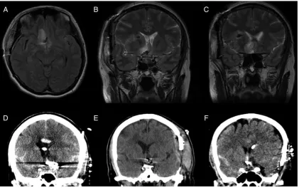
presence of subarachnoid haemorrhage (figure 1.1).
An additional mechanism of delayed ischaemia, cortical spreading depression
has also been demonstrated in a cohort of SAH patients (Woitzik et al, 2012). The
use of nimodipine as a prophylactic treatment and intravascular volume
treatments (triple-H) was established in the 1980’s (Kassell et al, 1982; Allen et al,
1983). A systematic review of eight prospective randomised trials including 1514
patients designed to assess the efficacy of prophylactic nimodipine (Liu et al, 2011),
concluded that nimodipine reduced the rate of delayed cerebral ischaemia by 38%
and the rate of cerebral infarction by 48%. Conversely, the evidence for the use of
triple-H therapy remains sparse despite relatively widespread acceptance. A
systematic review of triple-H therapy components suggested that induced
hypertension was the most effective at improving cerebral blood flow (Dankbaar et
al, 2010).
Figure 1.1 Frontal projection left internal carotid artery angiogram at day 2 post ictus
immediately post coiling (left) and day 6 post ictus (right) performed to investigate an acute right hemiparesis and aphasia. The coiled aneurysm is labelled with an arrowhead. The immediate post coiling appearances demonstrate a normal left M1 MCA calibre and at day 6
show severe left M1 MCA (arrow) and A1 ACA vasospasm.
Endovascular approaches to treating this condition were fist applied in the
1980’s also (Higashida et al, 1989). Transluminal balloon angioplasty (TBA) and
chemical angioplasty using papaverine were the initial methods employed. The use
of the latter agent has now largely been superseded by other vasodilators, most
1.2 Prognosis
1.2.1 Outcome scales
The Glasgow Outcome Scale (GOS) and modified Rankin Scale (mRS) are
the most commonly used systems for measuring the degree of disability or
dependence in activities of daily living in patients who have suffered SAH. The
GOS is a five-point scale (table 1.4) and was originally developed to assess outcome
after traumatic brain injury (Jennett et al, 1981). This has largely been superseded
by the mRS (table 1.5) that was developed to assess outcome, principally following
stroke (van Swieten et al, 1988) and encompasses a seven-point scale. In many
studies, favourable outcomes are dichotomised as GOS scores of 4 and 5 or 5 alone
[image:24.595.107.535.350.556.2]and mRS scores of 0-2 or 0-3.
Table 1.4 The Glasgow Outcome Score.
Score Definition
1 Death.
2 Persistent vegetative state. Patient exhibits no obvious cortical function.
3 Severe disability. Conscious but disabled. Patient depends upon others for
daily support due to mental or physical disability or both.
4 Moderate disability. Disabled but independent. Patient is independent as
far as daily life is concerned. The disabilities found include varying degrees of dysphasia, hemiparesis, or ataxia, as well as intellectual and memory deficits and personality changes.
5 Good recovery. Resumption of normal activities even though there may be
Score Definition
0 No symptoms.
1 No significant disability. Able to carry out all usual activities, despite some
symptoms.
2 Slight disability. Able to look after own affairs without assistance, but
unable to carry out all previous activities.
3 Moderate disability. Requires some help, but able to walk unassisted.
4 Moderately severe disability. Unable to attend to own bodily needs without
assistance, and unable to walk unassisted.
5 Severe disability. Requires constant nursing care and attention, bedridden,
incontinent.
6 Death.
Table 1.5 The modified Rankin Scale.
1.2.2 Factors impacting on patient outcome
Recent studies of in-hospital mortality for SAH suggest that this lies in the
order of 20% and has fallen since the 1980s (Rincon et al, 2013; Lantigua et al,
2015), likely due to advances aneurysm treatment and in critical care. Nevertheless,
up to 30% of the survivors exhibit significant morbidity and will depend on others
for activities of daily living and of those patients who survive the acute phase
non-disabled, many suffer cognitive impairment (Hijdra et al, 1987a). As many as a third
in the International Subarachnoid Aneurysm Trial (ISAT) cohort suffered cognitive
imparirment (Scott et al, 2010).
Established patient risk factors for death or dependency include poor clinical
grade at presentation, older age, aneurysm rebleeding, large aneurysm size, early
ischaemic brain injury, cerebral oedema and intraventricular haemorrhage (Claassen
et al, 2002; Naidech et al, 2005; Rosen et al, 2004; Wartenberg et al, 2011; Lantigua
et al, 2015). Mode of aneurysm treatment and treatment complications,
physiological derangement, complications of invasive imaging with digital
subtraction angiography, cerebral vasospasm and the delayed ischaemic process and
long term imaging follow-up all have implications for early patient outcome and/or
2011; Ferns et al, 2011a; Lantigua et al, 2015). These factors are the subject of this
thesis and will be discussed in turn.
1.3 Aneurysm treatment modality
1.4.1 A brief history of aneurysm treatment
In the 1930’s, the first neurosurgical procedures to protect aneurysms from
rehaemorrhage were performed in Edinburgh, UK and Baltimore, USA with clipping
of aneurysms first successfully performed by Dandy in 1937 (Dandy, 1938) and,
with microsurgery first employed to aid clipping in the 1970’s (Krayenbühl et al,
1972). Endovascular occlusion of aneurysms first employed detachable balloons in
the 1980s (Scialfa et al, 1983) but in the early 1990s, the first use of electrolytically
detachable platinum coils, the mainstay of aneurysm treatment today in most
institutions, was described by Guglielmi (Guglielmi et al, 1991). The first of a
number of land-mark publications as a result of the ISAT trial of 2143 patients,
comparing endovascular coiling with microsurgical clipping was published in 2002
(Molyneux et al, 2002).
Since the development of simple coiling techniques, endovascular treatment
options evolved. Balloon-remodelling and stent-assisted coiling are adjunctive
treatments that allow endovascular occlusion of more complex aneurysms (Pierot et
al, 2009). In the last ten years both luminal and intra-aneurysmal flow diverters have
been developed and although the evidence for their use is still building, they do aid
occlusion of aneurysms with difficult morphology and further add to the
neurointerventionalist’s armamentarium.
1.4.2 Controversies
The mode of aneurysm treatment and complications of treatment are
significant factors which may impact on functional and cognitive outcomes
(Molyneux et al, 2002, Scott et al, 2010, Ayling et al, 2015). Broadly, arguments
for the continued use of clipping in the context of SAH are that there is a perceived
improved ability to fully treat aneurysms of more complex morphology (Johnston et
al, 2008). Conversely, coiling is less invasive, is associated with fewer
peri-operative complications (Ayling et al, 2015) and lower levels of biochemical
markers of brain injury (Shim et al, 2012) but critics of the technique cite incomplete
occlusion as a major flaw, suggesting that in the long term there may be an increased
risk of rebleeding and complications associated with further treatment.
The results of ISAT have gone some way to satisfying this concern. This
trial randomised patients with aneurysms that were deemed suitable for both surgical
clipping and endovascular coiling. At 1 year there was an absolute 6.9% reduction in
the rate of death or dependency for patients treated with coiling rather than clipping
(Molyneux et al, 2002). In longer term follow-up of 1003 patients at 10 years
(Molyneux et al, 2015), 435 (82%) patients treated with endovascular coiling and
370 (78%) patients treated with surgical clipping were independent (mRS 0-2; OR
1.25; 95% CI 0.92-1.71). Patients in the endovascular treatment group were more
likely to be alive and independent at 10 years than were patients in the neurosurgery
group (OR 1.34, 95% CI 1.07-1.67). Although there was a small increased risk of
rebleeding in the coiling group, this did not translate to a significantly worse clinical
outcome when compared with that of the surgically treated group. Patients were
40-times more likely to die from another cause than from the treated aneurysm.
After the initial ISAT results there was a shift in the treatment of aneurysms
in the UK and coiling became the dominant modality. In 2006, an audit of
subarachnoid haemorrhage treatment in the UK demonstrated that 85% of patients
treated for ruptured aneurysms had coiling (Royal College of Surgeons, 2006).
There was much criticism of the trial, particularly from neurosurgeons in the
United States. The study was designed to randomise aneurysms for which the
treating clinicians had equipoise. A principle criticism is that only 20% of eligible
patients with SAH were enrolled into the study and as a result there is considerable
bias in terms of the population included and that there is a significant proportion of
aneurysmal SAH patients to whom the ISAT results do not apply.
As a result, a more recent study from the Berrow institute (the Berrow
of SAH (McDougall et al, 2012). This study was initially constructed as a pilot and
as a result was always underpowered. A total of 725 patients were screened, with
472 assigned to coiling or clipping either by alternating fashion or by lottery for the
last 100 patients. ‘Right of first refusal’ and crossover were allowed but results were
presented in an intent-to-treat analysis. There was a 38% cross-over rate from
clipping to coiling and a large proportion (12%) of patients without an aneurysm
who did not undergo either treatment were also included, making the results
somewhat difficult to interpret. Indeed, whether this can still be interpreted as a
randomised trial is questionable. Initial results at 1 year showed a 10% reduction in
death or disability (mRS >2) in the coiling group. In a post hoc analysis of the
3-year outcome data (Spetzler et al, 2013) in which those patients without an aneurysm
were excluded, the authors concluded that for anterior circulation aneurysms there
was no difference in outcomes for clipping and coiling and that clipping should be
considered a more appropriate treatment due to the risks associated with retreatment,
completeness of obliteration, and subsequent SAH risk.
A subsequent 6-year outcome analysis was interpreted in a similar way
(Spetzler et al, 2015), though independent analysis of the results (Macdonald et al,
2015) highlighted that poor outcomes were observed in 41% of patients (72 of 174)
who underwent clipping versus 35% of patients (57 of 162) who underwent coiling,
with an absolute risk reduction for coiling of 6% and a relative risk reduction of
17%. This is very similar to the results of ISAT. Nevertheless, the authors stand by
their interpretation of the results, and also suggest that scientific data from
well-designed trials is less important than an individual operator’s talent (McDougall &
Spetzler, 2015).
In some quarters, the optimal treatment option for anterior circulation
aneurysms therefore remains a subject of debate and two topics are of particular
importance. Firstly, middle cerebral artery aneurysms lie in an anatomical location
traditionally felt most suitable for surgical clipping and routine use of coiling here
remains contentious. The results of a study assessing a coil-first policy would be
intriguing. Secondly, although functional outcomes (measured using the mRS or
GOS) may not be dissimilar, it has been reported that coiling is associated with
Gilsbach, 1993; Mavaddat et al, 1999; Hadjivassiliou et al, 2001; Scott et al, 2010).
Despite lying within this range, patients may suffer cognitive deficits that prevent
them from working and participating in other activities (Al-Khindi et al, 2010).
Therefore, it is possible that there might be a much larger difference in
outcomes between clipping and coiling but that functional outcome scales are
insensitive to it. A study assessing the anatomical reasons for a difference in
cognition at a defined location would be of value.
1.5 Endovascular treatment of middle cerebral artery aneurysms
Ruptured middle cerebral artery (MCA) aneurysms represented only 303
(14.1%) of the 2143 enrolled patients in ISAT (Molyneux et al, 2002) and of all
aneurysm locations assessed in ISAT there was least difference in functional
outcome between clipping and coiling at the MCA location (Molyneux et al, 2005).
Many specialists consider this anatomical location an indication for surgical
clipping: the location aids surgical access, and in some cases, surgery facilitates
haematoma evacuation. There is also a perceived increased risk for coiling in terms
of thrombo-embolism and infarction at this site because these aneurysms are often
wide-neck and have branches arising from the neck (Ausman et al, 1997, Regli et al,
1999). Recently, several surgical series have been published that demonstrate
excellent clinical results with low rates of morbidity and mortality (Morgan et al,
2010, van Dijk et al, 2011; Choi et al, 2012; Rodrıguez-Hernandez et al, 2013) and this practice has extended to treatment of unruptured aneurysms.
As practice in the United Kingdom has shifted in many institutions to a
coil-first policy, there is a need for assessment of clinical outcomes using this approach to
analyse how this compares to the results in ISAT and also those of recent surgical
series. A retrospective analysis was therefore undertaken of a prospectively acquired
data base including 295 patients with 300 saccular MCA aneurysms treated using
endovascular coiling at a regional neuroscience centre between November 1996 and
June 2010 (Mortimer et al, 2014). This is described in detail in Chapter 2. Of the total, 244 (80.7%) ruptured aneurysms were treated. The technical failure rate was
(91.4%). Complications included rupture in 15 patients (5%), thromboembolism in
17 patients (5.7%), and early rebleeding in 3 patients (1%). Overall permanent
procedural-related morbidity and mortality were seen in 12 patients (7.8%). Of the
ruptured aneurysms, 189 (79.4%) had a favourable clinical outcome (GOS 4–5). A
total of 33 patients (13.6%) died. On initial angiographic follow-up, aneurysm
remnant was seen in 18 aneurysms (8.1%). A total of 13 patients (4.3%) were
retreated.
These results compare very favourably with that of the best published
surgical series (Morgan et al, 2010, van Dijk et al, 2011; Choi et al, 2012; Rodrıguez-Hernandez et al, 2013). They are also in line with the results published in
other smaller endovascular series and ISAT which suggests that they are
reproducible (Molyneux et al, 2002; Iijimaet al, 2005; Quadros et al, 2007; Oishiet
al, 2009; Suzukiet al, 2009; Bracardet al, 2010) and that although the approach was
different to ISAT (treating all-comers as opposed to just those aneurysms with more
favourable morphology for coiling), clinical outcomes, complications, anatomical
results and retreatment rates were in line with published benchmarks for aneurysms
at all locations (Molyneux et al, 2002; Cognardet al, 2011). Therefore, although the conservative view is that surgery should remain the treatment of choice of ruptured
MCA aneurysms, except when there are extenuating comorbidities or overriding
patient preferences, the results suggest that for the range of ruptured MCA
aneurysms, at least equivalent clinical outcomes to the best surgical series can be
obtained by use of conventional endovascular techniques.
The use of adjunctive devices such as stent placement or balloon remodelling
was very infrequent in this series, and intra-aneurysmal flow diverters were not used.
Adjunctive devices have been used in 20.4% of other published MCA series
(Brinjikji et al, 2011a),with the aim of obtaining complete occlusion to minimize the risk for subsequent haemorrhage (Johnston et al, 2008). A critical appraisal of the available literature has suggested that balloon assistance, which increases the range
of lesions that can be treated has a very similar safety profile to coiling without
remodelling (Pierot et al, 2012b). Stent assistance has also been used successfully in large series of MCA aneurysms (Johnson et al, 2013a). Stents allow more complete treatment of more complex lesions and may lower recurrence. However, there are
Intra-aneurysmal flow diverters now represent an additional treatment option
for wide-neck bifurcation aneurysms. Pierot et al (2013) treated 34 ruptured and
unruptured MCA aneurysms with the Woven Endobridge (WEB) device, a first
generation intra-aneurysmal flow diverter. Adequate occlusion (total occlusion or
neck remnant) was observed in 83.3% of aneurysms with an acceptable safety
profile; mortality rate of the treatment was 0.0% and morbidity rate was 3.1%
(intraoperative rupture with an mRS of 3 at 1-month follow-up). It is likely
therefore, that new devices will further increase the range of endovascular options
for aneurysms at this location and are potential avenues for future research into the
optimal strategy for these lesions.
1.6 Brain injury following treatment of anterior communicating artery
aneurysms
Approximately 50% of the patients treated in ISAT suffered SAH as a result
of ruptured anterior cerebral artery (ACA) or anterior communicating artery
(ACOM) aneurysms. Minimal differences in the rate of poor functional outcome or
death assessed using the modified Rankin Scale were demonstrated between clipping
and coiling for aneurysms at this location (27.5% v 24.6%) (Molyneux et al, 2005).
Nevertheless, patients may suffer significant cognitive impairment despite
favourable functional outcome scores (Hutter & Gilsbach, 1993; Mavaddat et al,
1999; Hadjivassiliou et al, 2001, Scott et al, 2010) and it has been recognised for
some time that surgical clipping may be associated with greater rates of cognitive
impairment than coiling for aneurysms treated at this location (Chan et al, 2002;
Fontanella et al, 2003; Proust et al, 2009). Various patterns of neuropsychological
impairment can occur (Böttger et al, 1998).
Injuries to the basal forebrain and the fornix specifically have been
implicated in resulting in memory impairment (Damasio et al, 1985; Phillips et al,
1987; Abe et al, 1998; Wright et al, 1999; Hashimoto et al, 2000; Mugikura et al,
2014; Meila et al, 2015).
The basal forebrain contains a number of important structures including the
nucleus accumbens, substantia innominata, nucleus basalis of Meynert, and medial
case reports and series (Damasio et al, 1985; Phillips et al, 1987; Böttger et al, 1998;
Abe et al, 1998; Goldberg et al, 1999; Wright et al, 1999; Hashimoto et al, 2000)
and a functional imaging study has also suggested that the basal forebrain is likely to
play a role in episodic memory recall (Fujii et al, 2002). The medial septal-diagonal
band of the Brocca complex region is likely to be an important interface within the
so-called septohippocampal system (Böttger et al, 1998). Bilateral basal forebrain
lesions do produce more severe memory deficits (Böttger et al, 1998). The fornix is
a compact fiber bundle connecting the hippocampus with the hypothalamus and a
number of other structures including the septal area of the basal forebrain. It is an
important constituent of the Papez circuit and is involved in the formation and
consolidation of declarative memories (Meila et al, 2015). Diffusion-weighted
MRI-proven infarction limited to the fornices has been reported in conjunction with acute
onset of amnesia, suggesting that an isolated injury to this structure may be critical
(Mugikura et al, 2014). Injuries to other vulnerable structures including the corpus
callosum and caudate nucleus have also been implicated in resulting in
neuro-behavioural sequelae (Mendez et al, 1989; Kasow et al, 2000). Caudate nucleus
lesions may result in depression, agitation, abulia, neglect (right-sided lesions),
memory disturbance (particularly if bilateral), dysarthria, aphasia (left-sided lesions),
movement disorders (ballistic, choreatiform), or motor weakness. (Kumral et al,
1999).
Structures within the basal forebrain, basal ganglia, and limbic system are
vulnerable to injury during neurovascular procedures, probably through occlusion of
ACA or ACOM artery complex perforating branch vessels, notably the recurrent
artery of Heubner (RAH) or subcallosal artery (Mugikura et al, 2014; Meila et al,
2015). Following treatment of ACOM artery aneurysms, a detailed radiological
investigation into the rates and distribution of injury to the above structures is
required to assess whether surgical treatment is associated with greater rates of
injury; a potential cause of inferior cognitive outcomes in the surgical group.
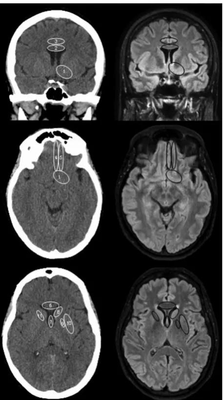
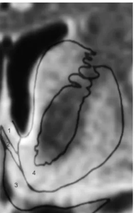
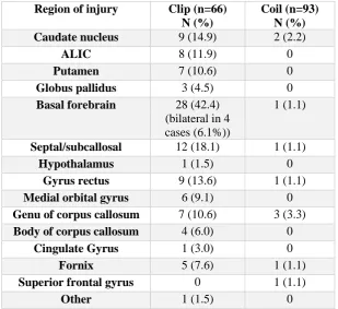
A retrospective dual-centre radiological investigation was undertaken of a
consecutive series of patients with ruptured and unruptured ACOM aneurysms
treated between January 2011 and October 2014 (Mortimer et al, 2016a). This is
described in detail in Chapter 3. Sixty-six patients treated with clipping were
ischemic, 1 haemorrhagic) compared with 4/93 (4.4%) patients in the coiling group
(3 ischemic, 1 haemorrhagic) (p<0.0001). For patients with subarachnoid
haemorrhage, the multivariate OR for infarction for clipping over coiling was 24.42
(95% CI 5.84 to 102.14), p<0.0001. The most common site of infarction was the
basal forebrain (28/66 patients, 42.4%), with bilateral infarction in four patients.
There was injury of the septal/subcallosal region in 12/66 patients (18%).
Clipping of ACOM aneurysms was clearly associated with significantly
higher rates of structural injury than coiling, and this may be a reason for superior
cognitive outcomes in patients treated with coiling in previously published studies.
Chan et al (2002) studied 18 patients who had undergone treatment for ruptured
ACOM aneurysms, half with clipping and half with coiling; 33% of the clipped
patients showed severe impairment of memory and executive function whereas no
coiled patient demonstrated this impairment.
Fontanella et al(2003)assessed 37 consecutive WFNS grade I or II patients
who underwent treatment of ACOM aneurysms within 48 h of rupture; 20 of 37
were treated with clipping and 17 were treated with coiling. Both groups were
compared with 16 angiogram-negative patients with SAH and 18 normal controls.
All patients were neurologically intact at discharge and were independent at 6-month
follow-up after SAH. Surgically treated patients showed a significant worse
performance on the logical memory and on the frontal lobe executive functions
compared with controls, while the endovascular group and the angiogram-negative
group showed a significant decrease only in the literal fluency score. Furthermore,
the surgical group showed a significant impairment in using grammatical and
syntactical rules to produce sentences. Proust et al (2009)studied 36 clipped and 14
coiled patients at 14-month follow-up. They found no difference in executive
dysfunction although there was a significant impairment of verbal memory in the
clipped group.
This raises the question of how to optimally treat these patients and this is
one factor which may sway a decision towards endovascular approaches, and
possibly the use of adjunctive techniques such as stent-assisted coiling for more
complex lesions. A systematic review of stent coiling at all locations in patients with
SAH who were managed with dual antiplatelet therapy demonstrated clinically
haemorrhages. Clinically significant thromboembolic events occurred in 16 (6%) of
288 patients (Bodily et al, 2011). Perhaps a trial comparing more aggressive
endovascular approaches (e.g. using stents or even flow diversion) with clipping for
less favourable aneurysms, particularly for locations such as this, would be
worthwhile. Such a study should include neuropsychological testing as part of the
outcome measures.
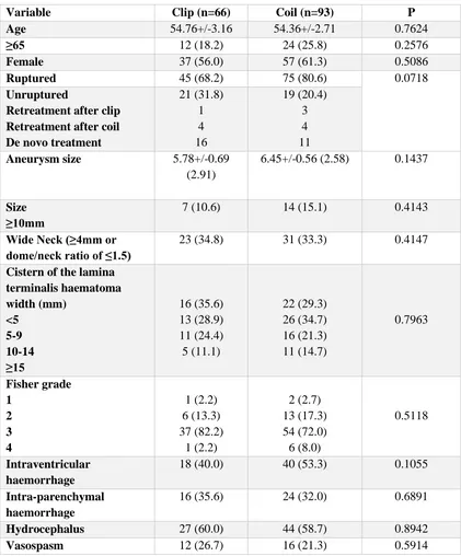
1.7 Physiological derangement and choice of treatment modality
Although there was little reference to physiological derangement or medical
comorbidity or complications in either ISAT or BRAT studies, there is increasing
recognition that these factors are significant contributors to poor outcome after SAH
(Wartenberg et al, 2006; van der Bilt et al, 2009, Lantigua et al, 2015).
Physiological derangement and medical complications are more common in poorer
grade patients (Enblad & Persson, 1997). Only 4.4% of patients recruited into ISAT
were of poor clinical grade (WFNS 4 or 5). The results of ISAT may therefore not
directly apply to poor-grade patients. A recent multicentre prospective observational
study demonstrated that after adjustment for baseline characteristics there was no
significant difference in outcomes for coiling or clipping in this patient group (Zhao
et al, 2015).
Severity of illness scoring systems have shown better correlation with poor
outcomes in SAH patients than acute neurological grading (Gruber et al, 1999) and
there is little evidence regarding the interaction of treatment modality and presenting
physiological derangement rather than neurological grade in isolation. In
comprehensive centres, the decision on whether to clip or coil an aneurysm is often
based on the morphology and location of the aneurysm. An alternative approach is
to subject an acutely unwell patient to the least stressful procedure (namely an
endovascular coiling), if even just to protect the aneurysm dome in the short term in
order that they may recover to undergo a further protective procedure if required
(Waldau et al, 2012).
The Acute Physiology and Chronic Health Evaluation II (APACHE II)
of physiological function (Knaus et al, 1985). This has been validated in the SAH
population (Lantigua et al, 2015). An assessment of how physiologicalderangement
interacts with treatment approach is required to answer whether the physiological
condition of the patient should influence mode of treatment in the sickest patients.
An exploratory analysis of prospectively collected trial data was therefore
undertaken, comparing the outcomes of sixty-nine patients treated with clipping to
sixty-six treated with coiling (Mortimer et al, 2016b). This is described in detail in
Chapter 4. More profound physiological derangement (APACHE II score >15) was
the strongest predictor of poor outcome in the overall cohort (OR 17.80, 95% CI
4.78 to 66.21, p<0.0001). For those with more deranged physiology (APACHE II
score>15; 59 patients), WFNS grade ≥4 (OR 6.74, 1.43 to 31.75) and surgical
clipping (OR 6.33, 1.27 to 31.38) were significant predictors of poor outcome
(p<0.05). Favourable outcome (mRS 0-2) was seen in 11% of surgical patients
compared with 38% of coiled patients in this subgroup. The results indicated an
advantage for coiling in the more profoundly physiologically deranged subgroup.
On multivariate analysis both poor grade and clipping were predictors of poor
outcome. Furthermore, functional outcome differences by treatment modality for the
WFNS grades 1–2 patients with high APACHE II scores tended to significance
(favourable outcomes for clipping were seen in 30% and in 56% for coiling in this
subgroup). Perhaps this did not reach significance as the study was underpowered.
A number of authors have attempted to explain superior outcomes for coiling
based on lower rates of cerebral vasospasm. We did not see a significantly increased
rate of angiographic vasospasm in the clipped patients yet clipped patients did have
significantly increased total norepinephrine dose, ventilated days, and hospital stay
(p<0.05). A large Canadian multicentre study demonstrated that clipped patients
more commonly suffered medical complications, such as urinary tract infection,
pneumonia, cardiorespiratory arrest, and seizures, and that these complications were
linked to poor outcome (Vergouwen et al, 2011c).
It is therefore plausible that patients undergoing a more invasive procedure
may require more inotropic support, more time ventilated, and more time in hospital.
They could therefore take much longer to recover and are therefore more at risk of
question of whether more profoundly sick patients should undergo a coiling as
opposed to a clipping procedure.
1.8 Invasive imaging in patients with perimesencephalic subarachnoid
haemorrhage
Perimesencephalic subarachnoid haemorrhage (PMSAH) accounts for
approximately 5% of SAH and represents a distinct clinic-radiological entity
characterised by haemorrhage centred on the perimesencephalic cisterns, aneurysm
negative angiographic investigation and a more benign clinical course (van Gijn et
al, 1985; Rinkel et al, 1991a; Rinkel et al, 1991b; Brilstra et al, 1997; Flaherty et al,
2005; Greebe et al, 2007). An aneurysm is likely responsible for this pattern of
haemorrhage in less than 1% of cases and CT angiography (CTA) with modern
systems are highly sensitive for aneurysm detection (Westerlaan et al, 2011; Kalra et
al, 2015).
CTA is now commonly performed prior to digital subtraction angiography
(DSA) to aid treatment planning through aneurysm identification and
characterisation. The traditional approach to investigating patients with SAH also
includes mandatory use of DSA, the gold standard angiographic modality. A review
of the literature has demonstrated that the average risk of permanent neurological
complications from DSA in patients with perimesencephalic haemorrhage was
0.74% (95% CI, 0.09% to 2.7%) (Ruigrok et al, 2000). This is a higher rate than
seen in two subequent very large series of all consecutive DSA procedures
performed in 19,826 (Kaufmann et al, 2007) and 2,899 patients (Willinsky et al,
2003). The permenant neurological complication rate was 0.14% and 0.5%
respectively. The relative increased risk in PMSAH patients could relate to age.
Nevertheless, current European and American guidelines are ambiguous as to the
need for invasive imaging with DSA in this setting and the majority of previous
studies are of a relatively low number of patients (Connolly et al, 2012; Steiner et al,
2013). There is a need for an assessment of the diagnostic yield of DSA in the face
of a negative CTA in a large cohort of patients with strictly defined PMSAH to
elucidate the negative predictive value of CTA in this population and to guide
The negative predictive value (NPV) of CTA was assessed in a series of 72
patients with PMSAH treated at a regional neuroscience centre over a 9-year period
(Mortimer et al, 2016c). This is described in detail in chapter 5. A PMSAH pattern
defined as blood centred anterior to the midbrain and/or pons within the pre-pontine
or interpeduncular cistern with possible quadrigeminal or ambient cistern extension;
possible extension into the basal parts of the Sylvain fissures but not the lateral
sylvian fissures; possible extension to the cisterna magna but not centred on the
cisterna magna; and possible extension into the fourth ventricle and occipital horns
of the lateral ventricles.
Of 72 patients, one patient showed a potentially significant finding on DSA
that was not demonstrated on initial CTA (NPV 98.61% (95% CI 92.47% to
99.77%)). However, when cisterna magna extension was excluded from the
definition of PMSAH, no false negative CTAs in 56 patients were encountered (NPV
100% (95% CI 93.56% to 100.00%)). The NPV of normal CTA for an arterial
abnormality in patients with PMSAH is high and our results therefore question the
role of invasive imaging and certainly imply that if a prospective study is required to
fully evaluate this then this would be safe and feasible.
1.9 Delayed cerebral ischaemia, cerebral vasospasm and endovascular
approaches to prevent delayed infarction
Cerebral infarction is strongly associated with poor outcome following SAH
(Furgusson & Macdonald, 2007; Vergouwen et al, 2011b). In the context of SAH,
infarction could be the result of the acute injury, complications of the aneurysm
securing procedure or the delayed ischaemic process. For decades it has been
assumed that the latter results from the development of cerebral vasospasm (Graham
et al, 1983), detected angiographically as luminal narrowing of the proximal cerebral
arteries. The dominant factor determining the development of vasospasm is the
presence of thick subarachnoid clot adjacent to the proximal cerebral arteries
(Inagawa, 2015). Angiographic vasospasm is an independent predictor of poor
outcome following SAH (Vergouwen et al, 2011a). An association between
vasospasm in the development of delayed infarction is supported by the results of
is a correlation between vasospasm severity and the incidence of infarction. The
majority of vasospasm-related infarcts are associated with severe vasospasm,
(Weidauer et al, 2007; Crowley et al, 2011; Inagawa et al, 2014)which results in the
most severe perfusion deficits (Dankbaar et al, 2009; Vatter et al, 2011; Dhar et al,
2012). Of patients with severe vasospasm, 50–100% develop cerebral infarction
compared with 3–5% of patients without significant vasospasm (Weidauer et al,
2007; Crowley et al, 2011; Inagawa et al, 2014). Severe vasospasm is associated
with poor cognition, worse patient-relevant outcomes, and greater inpatient
healthcare resource use (Macdonald et al, 2012).
More recently, the role of vasospasm in delayed ischaemia has been brought
into question largely secondary to the finding that the trial drug clazosentan has been
shown to improve rates of vasospasm but this did not translate into improved
functional outcomes whereas nimodipine has been shown to improve clinical
outcomes without an impact on vasospasm (Petruk et al, 1988; Feigin et al, 1998;
Macdonald et al, 2008; Macdonald et al, 2011).
There are many potential reasons for a failure in correlation between
reductions in the incidence of vasospasm but not in rates of poor outcome in the
clazosentan trials. First, poor grade patients who suffer a more significant acute
neurological injury are those who are more likely to suffer the most severe
vasospasm and therefore detecting differences with current outcome measures would
probably require very large trials or alternative outcome measures. Second, only
severe vasospasm may result in significant reductions in cerebral blood flow so as to
result in infarction and the inclusion of those with moderate vasospasm may dilute
the treatment effect. Third, vasospasm is likely to exert its clinical impact through
infarction: factors such as age (Danière et al, 2014)govern the impact of infarct size
on functional outcome. Furthermore, the eloquence of the infarct area will result in
variable functional impact. Fourth, the systemic side effects of the treatment may
have resulted in poor outcome through alternative mechanisms (medical
complications) in the treatment arm. Lastly and importantly, rescue treatments such
as endovascular intervention and systemic hypertension may have been effective in