A Thesis Submitted for the Degree of PhD at the University of Warwick
Permanent WRAP URL:
http://wrap.warwick.ac.uk/99422/
Copyright and reuse:
This thesis is made available online and is protected by original copyright. Please scroll down to view the document itself.
Please refer to the repository record for this item for information to help you to cite it. Our policy information is available from the repository home page.
Distinct conformations, aggregation
and neuronal internalisation of different
tau strains
by
Thomas Kwaku Karikari
A Thesis Submitted
for the Degree of Doctor of Philosophy in Life Sciences
School of Life Sciences University of Warwick Coventry, United Kingdom
Nyame ne Hene!
(God is King!)
Table of Contents
Table of Contents i
List of Figures iv
List of Tables vii
Acknowledgements viii
Declaration x
Abbreviations xi
Abstract xiii
1| Introduction 1
1.1 Tau protein: structure, isoforms and functional fragments 1
1.2 Physiological and pathophysiological functions of tau 3
1.2.1 Physiological functions of tau 3
1.2.2 Pathophysiological functions of tau 5
1.2.2.1 Pathophysiology arising from loss of physiological functions 6
1.2.2.2 Pathophysiology arising from gain of toxic functions 7
1.3 Tauopathies: disease classification, clinical diagnosis, progression, and differential roles of tau isoforms 13
1.4 Tau mutations as modulators of familial FTD 16
1.5 Theories of tau pathology: aggregation and conformational change 20
1.5.1 Biochemical and biophysical tools for studying tau aggregation and conformation 21
1.6 Proteinopathies and the prion hypothesis 24
1.6.1 Neuronal internalisation and transmission of tau pathology: the transneuronal propagation and the selective vulnerability hypotheses 25
1.7 Drug development against misfolded tau protein 28
1.8 Aims of this thesis 30
2| Materials and methods 32
2.1 Materials 32
2.2 Cloning of tau proteins into pProEx plasmids 33
2.3 SDM 35
2.4 Transformation of E. coli BL21*(DE3) cells with tau-encoding plasmids 37
2.5 Expression of tau proteins 37
2.6 SDS-PAGE and WB 38
2.7 Large-scale expression of tau constructs 39
2.8 Tau protein purification by immobilised metal affinity chromatography (IMAC) 40
2.9 Expression and purification of TEV protease 41
2.10 Protein quantification using the BCA assay 42
2.11 Preparation of Alzheimer-like PHFs 42
2.12 CD spectroscopy 43
2.13 Thioflavin T kinetic assay of tau aggregation 44
2.14 TEM analysis of tau filament formation 44
2.15 Dot blot assay of tau aggregation 44
2.16 AFM analysis of tau aggregation 45
characterisation of labelled proteins 46
2.19 Neuroblastoma cell culture 47
2.20 Differentiated neuroblastoma cell culture 47
2.21 Human induced pluripotent stem cell (hiPSC)-derived cerebral cortical neural stem cells 48
2.22 Immunohistochemistry 48
2.23 Confocal microscopy 49
2.24 Lactate dehydrogenase (LDH) assay of tau oligomer toxicity 49
2.25 Statistical analysis 50
3| A plasmid library for expressing full-length and truncated tau proteins, and their pathologic and cysteine-modified variants 51
3.1 Introduction 51
3.2 Results 52
3.2.1 Creation of a library of plasmids for expressing pathologic variants of tau 52
3.2.2 Introduction of cysteine modifications into the tau proteins 54
3.3 Discussion 68
4| Expression and purification of tau and its FTD variants using a cleavable histidine tag 71
4.1 Introduction 71
4.2 Results 72
4.2.1 Preparation of tau pathogenic mutants 72
4.2.2 Comparison of tau expression in two different E. coli strains 73
4.2.3 Temperature optimisation for tau expression 75
4.2.4 Induction length for optimum tau expression 76
4.2.5 Media supplementation with 0.2 %w/v glucose 77
4.2.6 Purification of tau constructs 78
4.2.7 CD assessment of secondary structure properties of the purified tau 78 4.2.8 Preparation of Alzheimer-like PHF and subsequent imaging using negative-stain TEM 79
4.3 Discussion 84
4.5 Conclusion 88
5| Preparation of stable tau oligomers for cellular and biochemical studies 89
5.1 Introduction 89
5.2 Results 90
5.2.1 Preparation of tau oligomers from monomers 90
5.2.2 Structural characterisation of tau oligomers 91
5.2.3 Oligomer preparation with regards to the aggregation pathway 92
5.2.4 The K18 tau repeat domain construct forms filaments of diverse structural morphology 92
5.2.5 Preparation of fluorescently-labelled tau oligomers 94
5.2.6 Fluorescently-labelled tau oligomers exist principally as trimers 94
5.2.7 Cysteine-specific labelling with maleimide derivatives stabilises tau oligomers 95
5.3 Discussion 100
6| Distinct conformations, aggregation and neuronal internalisation of different tau strains 103
6.1 Introduction 103
6.2.1 FTD mutations alter tau K18 binding to ThT 105
6.2.2 AFM analysis of endpoint ThT assay samples provide insights into the aberrant binding of the tau K18 variants to ThT 108
6.2.3 The FTD familial mutations alter the ultrastructure of tau aggregates 109
6.2.4 Structural features of WT and FTD tau K18 aggregates are unchanged following prolonged incubation 110
6.2.5 FTD mutations alter the dynamics of tau aggregation 111
6.2.6 FTD mutations alter the immunoreactivity of aggregating tau K18 114
6.2.7 AFM structural evidence support the variable immunological reactivity for the tau K18 proteins 115
6.2.8 WT and FTD tau K18 oligomers have distinct conformations 115
6.2.9 Secondary structural features of WT and FTD tau K18 116
6.2.10 Dominant conformers of WT and FTD tau K18 are unaltered in the presence of competing species 117
6.2.11 Preparation and characterisation of stabilised tau K18 oligomers for exogenous application to neurons 121
6.2.12 Optimisation of human neuroblastoma and neuronal cell culture growth conditions 123
6.2.13 Familial FTD mutations increase tau oligomer internalisation in SH-SY5Y cells but not in hiPSC-derived cortical neurons 124
6.2.14 Exogenous tau K18 oligomers are internalised by endocytosis 127 6.2.15 Internalised tau K18 oligomers accumulate in the cytoplasm and nucleus of SH-SY5Y cells and the axons and the neurites and soma of hiPSC-derived neurons 128
6.2.16 Internalised tau K18 oligomers co-localise with the nuclear protein nucleolin 129
6.2.17 Internalised tau K18 oligomers colocalise with endogenous tau 130
6.2.18 Morphological phenotypes of internalised tau K18 132
6.2.19 Internalised tau K18 oligomers do not induce cell death 132
6.3 Discussion 134
6.4 Conclusion 136
7| General Discussion 138
7.1 Rationale 138
7.2 Design and characterisation of a plasmid library for expressing full length and truncated tau proteins 140
7.3 Development of a novel protocol for high-yield expression and purification of tau 141
7.4 Preparation of stabilised tau oligomers 141
7.5 ThT fluorescence is not a universal indicator of proteinopathic aggregation 142
7.6 Internalisation of tau oligomers and the induction of neurotoxicity 143
7.7 Co-localisation of internalised tau with endogenous tau 145
7.8 Co-localisation of internalised tau with nucleolin 147
7.9 Conclusion 148
8| References 150
List of Figures
Figure 1.1. A schematic illustration of tau isoforms and functional
fragments 3
Figure 1.2. Physiological functions of tau 5
Figure 1.3. Pathophysiological functions of tau 8
Figure 1.4. Cysteine-dependent tau aggregation is isoform-specific 11
Figure 1.5. Schematic illustration of the “nucleation-elongation” mechanism of tau aggregation 12
Figure 1.6. Electron micrographs of tau aggregates isolated from AD brains (upper panel; three patients – cases 1 – 3) and in vitro polymerised filaments from recombinant tau K19, and disease variant forms of K18 and htau40 (bottom panel) 13
Figure 1.7. Exonic distribution of disease-associated tau mutations 17
Figure 1.8. Example data from some commonly used biochemical and biophysical assays for evaluating tau aggregation and conformation 24
Figure 1.9 Mechanisms of misfolded tau transmission in the transneuronal propagation model for extracellular tau. 27
Figure 1.10. The selective vulnerability model of tau toxicity 28
Figure 1.11. Dysfunctional tau-targeted drug development targets and approaches 30
Figure 2.1 Bacterial expression plasmids used in this study. 34
Figure 2.2. BCA assay standard plot for estimating protein concentration. 43
Figure 3.1. Clustal Omega sequence alignments confirming the creation of the FTD mutations N279K, V337M, P301L and C291R in the WT K18 tau construct. 53
Figure 3.2. Clustal Omega sequence alignments confirming the creation of the N279K, V337M and P301L pathological mutations in the FLAG-tagged htau40 plasmid. 53
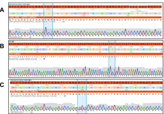
Figure 3.3. Clustal Omega sequence alignments confirming creation of the FTDP-17 mutations N279K and V337M (A and B respectively) in the FLAG-tagged htau40 gene. 54
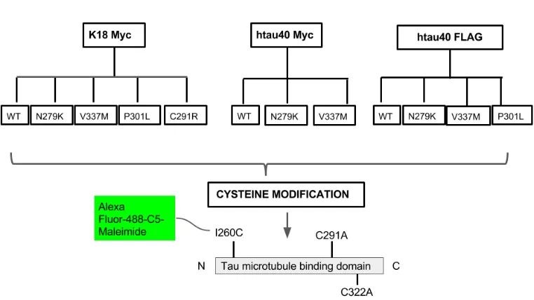
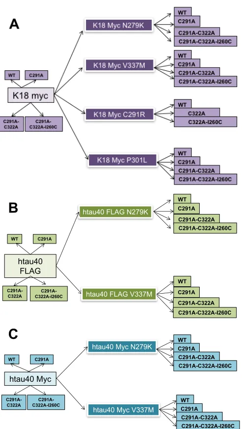
Figure 3.4. Schematic illustration of the creation of four disease- associated exonic mutations in the K18 and htau40 tau proteins. 54
Figure 3.5 Schematic illustration of cysteine modifications induced in the tau protein-expression library hitherto created. 56
Figure 3.6. Cysteine modifications in the WT K18 sequence. 56
Figure 3.7. Evidence of cysteine modifications in the Myc-K18-N279K construct. 57
Figure 3.8. Modification of native cysteine residues in the Myc-K18-V337M construct. 57
Figure 3.9. Evidence of native cysteine modification in the Myc-K18-P301L construct. 58
Myc-htau40 construct. 59 Figure 3.12. Native cysteine residue modification in the
Myc-htau40-N279K construcT 59 Figure 3.13. Modification of the native cysteine residues in the
Myc-htau40-V337M construct. 60 Figure 3.14. Evidence of the creation of amino acid modifications in
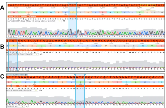
the WT htau40-Flag construct. 60 Figure 3.15. Cysteine modification in the htau40-Flag-N279K construct, showing the C291A, C322A and I260C amino acid modifications in
A, B and C respectively. 61 Figure 3.16. Modification of the native cysteine residues C291 and C322 to alanine (A and B respectively) and the introduction of a new cysteine residue at position 260 (in C) in the htau40-FLAG-V337M construct. 61 Figure 3.17. Modification of the native cysteine residues C291 and C322 to alanine (A and B respectively) and the introduction of a new cysteine residue at position 260 (in C) in the htau40-FLAG-V337M construct 62 Figure 3.18. Description of single nucleotide polymorphisms made in the primary structure of tau, in creating disease-associated and cysteine-
modified variants of the protein. 63 Figure 4.1. The WT K18 expression plasmid used in this study. 73 Figure 4.2. Representative WB analysis of K18 tau expression in the
NEB-5α and BL21(DE3)*pRosetta E. coli strains. 74 Figure 4.3. Representative Western blot analysis showing the influence of induction temperature on expression levels of K18 WT tau. 75 Figure 4.4. Time-course evaluation of K18 WT expression in E. coli
using optical density measurement and WB. 76 Figure 4.5. Media supplementation with 0.2% glucose did not
significantly enhance tau expression. 77 Figure 4.6 Analysis of IMAC-based tau purification using SDS-PAGE followed by WB. 80 Figure 4.7. Confirmation of cleavage of the polyhistidine tag. 81 Figure 4.8. Characterisation of the purified tau proteins using CD
spectroscopy and the preparation of Alzheimer-like PHFs. 82 Figure 4.9. Representative electron micrographs showing negative-
stained Alzheimer-like filaments of distinct morphologies prepared
from the purified tau proteins. 83 Figure 5.1. Characterisation of oligomer formation by WT and pathologic tau K18 using an oligomer-specific antibody. 91 Figure 5.2. Structural characterisation of tau K18 oligomers using AFM. 92 Figure 5.3. The tau monomer preparations from which the oligomers were produced were capable of recapitulating the tau aggregation pathway in vitro. 93 Figure 5.4. WT tau K18 forms mature filaments of diverse morphology. 93 Figure 5.5. Comparison of two methods of fluorophore labelling of tau oligomers. 95 Figure 5.6. Preparation of tau K18 oligomers stabilised by crosslinking
with maleimide derivatives. 97
Figure 5.7. Crosslinking with AF-maleimide stabilises tau K18 WT
granular conformation. 99 Figure 5.9 Extracellularly-applied stabilised tau K18 oligomers are
internalised by human neuroblastoma cells and cortical neurons. 100 Figure 5.10. Schematic illustration of the tau oligomer stabilisation
method described in this study 101
Figure 5.11. Schematic illustration of the chemical reaction involved in labelling of tau protein with maleimide derivatives. 102 Figure 6.1. Schematic illustration of the molecular arrangement of the
N279K and V337M tau mutations. 106
Figure 6.2. FTD mutations alter tau K18 binding to ThT 108 Figure 6.3. The V337M and N279K FTD mutations induce changes
in the structural properties of tau K18 filaments. 109 Figure 6.4. The structural distinctions between WT and FTD tau K18
aggregates are maintained following extended aggregation reactions. 111 Figure 6.5. AFM imaging provides insights into the stages of tau K18
aggregation in the presence of FTD mutations 113 Figure 6.6. The V337M and N279K familial FTD mutations alter the
immunoreactivity of tau K18 112
Figure 6.7. Characterisation of the conformation, shapes and size
distribution of WT and FTD tau K18 oligomers 116 Figure 6.8. Cross-seeding of tau K18 variants with filamentous WT
aggregates does not alter the morphological characteristics of
aggregates formed 118 Figure 6.9. Dominant conformers of tau K18 WT, V337M and N279K are unaltered in the presence of competing conformers 119 Figure 6.10. Characterisation of the size distribution of fluorescently
labelled tau K18 and its V337M and N279K pathologic variants
using non-denaturing SDS-PAGE and TEM 122 Figure 6.11. DLS assessment of the sizes of AF-maleimide–labelled tau
K18 WT oligomers 123
Figure 6.12. Optimisation of parameters for cell culture studies 125 Figure 6.13. Uptake of exogenous tau K18 oligomers by SH-SY5Y
cells 126 Figure 6.14. The V337M and N279K mutations significantly enhance the cellular uptake of tau K18 oligomers 126 Figure 6.15. Internalisation of extracellular tau K18 oligomers in hiPSC neurons occurs by endocytosis, and the internalised oligomers localise to the cell soma and the neurites 127 Figure 6.16. Extracellular tau K18 oligomers are taken up by
SH-SY5Y cells through endocytosis 128 Figure 6.17. Internalised tau K18 oligomers localise to the cytoplasm
and the nuclei in SH-SY5Y cells 129 Figure 6.18. Exogenous tau K18 oligomers that are internalised in
the nuclei of SH-SY5Y cells co-localise with the nucleolar protein
nucleolin 130 Figure 6.19. The nuclear protein nucleolin is an interacting partner of tau K18 oligomers internalised in hiPSC neurons 131 Figure 6.20. Internalised tau oligomers in hiPSC neurons co-localise
oligomers 133 Figure 6.22. Internalisation of tau K18 oligomers does not lead to cell death 133 Figure 7.1 Internalised WT and FTD tau K18 likely seed the aggregation of endogenous tau and may have critical effects on ribosomal biosynthesis. 146
List of Tables
Table 1.1. MT binding effects and clinical phenotypes of some MAPT mutations associated with FTDP-17 18 Table 2.1. Primers used in SDM to generate FTD variants of tau 35 Table 2.2. Details of PCR settings used in SDM to create FTD tau
variants 36 Table 2.3. Primers used in SDM to introduce cysteine modifications in the WT tau construct 37 Table 2.4. DNA sequencing primers designed and used in this study 37 Table 2.5. Preparation of hand-cast gels and buffers for SDS-PAGE
and WB 39 Table 2.6. Primary and secondary antibodies used in this study 40 Table 3.1. Details of pathological mutations created in the htau40
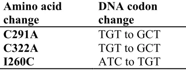
isoform and the K18 fragment of tau 52 Table 3.2. Codon changes made in the cysteine modifications induced in the tau proteins to enable chemical labelling with maleimide 55 Table 3.3. A comprehensive collection of plasmids created in this study for the recombinant expression of human tau proteins, described according to the encoded genetic construct. 64 Table 4.1. Summary of studies on expression and purification of
recombinant tau 81 Table 6.1. Previously-reported pathological consequences of the
V337M and N279K familial mutations and their impacts on aggregation and filament morphology of tau protein. 107 Table 6.2. Summary of biochemical and biophysical characterisation of the influence of the V337M and N279K familial FTD mutations on the
Acknowledgements
I would like to express my sincere gratitude to my supervisor, Prof. Kevin G. Moffat, for his unflinching support, guidance and mentoring throughout my PhD. Kevin, thank you for the opportunity to explore this exciting new project. I will miss our humour-filled conversations!
I would also like to appreciate the members of my Advisory Panel, Prof. Bruno G. Frenguelli and Prof. David I. Roper, for their constructive feedback, which kept me on track towards building a coherent scientific argument.
The Warwick experience would not be possible without the generous studentship from the University of Warwick’s Chancellor’s Scholarship and the Biotechnology and Biological Sciences Research Council. You turned a dream into reality!
Special appreciation to Dr. Eric J. Hill of Aston University, Birmingham, and his lab members for bringing me up to speed with stem cell culture techniques. I would also like to thank Prof. Alison Rodger and her group at the Department of Chemistry, for expert advice on, and access to, analytical biophysics equipment. My further thanks go to Dr. Neil Wilson, Department of Physics, and Mr. Ian Hands-Portman for training and the use of atomic force microscopy and transmission electron microscopy respectively. I am thankful to Dr. Mussa Quareshy for useful tips on protein purification, and Greg Walkowiak for assisting with microplate reader use. To the several MBio and BSc students I worked with during my PhD, including Alexandra Turner, Rachel Thomas and Sophie Keeling, thank you for offering me the opportunity to share the skills gained.
Declaration
I hereby declare that the material contained in this thesis is, to the best of my knowledge, my own original work unless otherwise cited, indicated, or is commonly known. This thesis has not been previously submitted for any degree at this or any other institution.
Abbreviations
AD Alzheimer’s disease
AFM Atomic force microscope
AF-maleimide Alexa Fluor® 488 C5-maleimide AGD Argyrophilic grain disease APP Amyloid precursor protein
BCA Bicinchoninic acid
BSA Bovine serum albumin
CBD Corticobasal degeneration
cDNA complementary deoxyribonucleic acid
CD Circular dichroism
CREB cAMP Response Element Binding CSF Cerebrospinal fluid
DYRK1A Dual-specificity tyrosine phosphorylation-regulating kinase 1A ddH2O Double distilled water
DLS Dynamic light scattering DMSO Dimethyl sulphoxide
DNA Deoxyribonucleic acid
DPBS Dulbecco’s phosphate buffered saline
DTT Dithiothreitol
ECL Electrochemiluminiscent
EDTA Ethylenediaminetetraacetic acid
EGTA ethylene glycol-bis(β-aminoethyl ether)-N,N,N',N'-tetraacetic acid FTD Frontotemporal dementia
FTLD-tau Frontotemporal lobar degeneration with tau pathology
FTDP-17 Frontotemporal dementia with Parkinsonism on chromosome 17 GSK3 Glycogen synthase kinase 3
IMAC Immobilised metal affinity chromatography
hiPSC human induced pluripotent stem cell IPTG Isopropyl β-D-1 thiogalactopyranoside
LDH Lactate dehydrogenase
LMW Low molecular weight
MAPT Microtubule associated protein tau gene MEM Minimal essential medium
NEM N-ethyl maleimide
NFT Neurofibrillary tangles NMR Nuclear magnetic resonance PBS Phosphate buffered saline PCR Polymerase chain reaction PHF Paired helical filament
PiD Pick’s disease
PIPES Piperazine-N,N′-bis(2-ethanesulfonic acid)
PSEN1 Presenilin 1
PSEN2 Presenilin 2
PSP Progressive supranuclear palsy
RT Room temperature
SDS Sodium dodecyl sulphate
SDS-PAGE Sodium dodecyl sulphate-polyacrylamide gel electrophoresis SDM Site directed mutagenesis
SF Straight filament
RT Room temperature
TCEP Tris(2-carboxyethyl)phosphine hydrochloride TEM Transmission electron microscopy
TEV Tobacco Etch Virus
ThS Thioflavine S
ThT Thioflavin T
UK United Kingdom
WB Western blot(ting)
Abstract
A shared property of several neurodegenerative diseases is the neuronal accumulation of aggregated tau protein. These include Alzheimer’s disease (AD) and frontotemporal dementia (FTD). Many studies have suggested that aggregated tau accumulation in AD brains involves: (i) internalisation of extracellular tau (aggregated or not) into neurons; (ii) induction of endogenous tau aggregation by the internalised tau; and (iii) secretion of part or whole of this aggregated tau complex. This complex then initiates a new cycle of internalisation, aggregation and secretion. While this AD mechanism has strong evidential support, it is unclear if it applies to FTD. It was therefore investigated if and how two FTD-associated tau mutations, V337M and N279K, affect in vitro wild type (WT) tau aggregation and conformation, and studied the cell biological effects of their respective extracellular oligomers.
A library of 43 plasmids for expressing full-length and truncated tau and their FTD variants were created, in conjunction with the establishment of a new high-yield tau purification method. Consequently, in vitro biochemical assays showed that the FTD variants distinctively altered the immunological reactivity, the stages of aggregation, and the structural phenotypes of aggregated WT tau four-repeat domain, K18. Internalisation of WT and FTD tau K18 extracellular oligomers was significantly different in human neuroblastoma cells and human stem-cell derived neurons. Internalisation seemed to occur by endocytosis, and the internalised oligomers localised to the nucleus and cytoplasm of human neuroblastoma cells and the soma and neurites of stem cell-derived neurons. Moreover, internalised oligomers co-localised with endogenous tau and the nuclear protein nucleolin, without inducing cell death.
1|
Introduction
A major function of tau protein is to support the intact neuronal cytoskeleton, enabling efficient trafficking between sub-neuronal compartments. The dysfunction of tau is, however, implicated in the pathogenesis of many neurodegenerative diseases including the focus of this thesis, frontotemporal dementia (FTD). Whilst there is strong evidence supporting the mechanisms of disease, it is currently unclear if and how single nucleotide polymorphisms in tau associated with different forms of FTD may influence disease onset and progression. This introduction covers the structural biochemistry of tau and its functions in physiological and pathophysiological conditions. Furthermore, the mechanisms by which abnormal tau protein may lead to distinct forms of disease and the probable therapeutic targets involved are discussed. The chapter concludes with the aims of this study.
1.1 Tau protein: structure, isoforms and functional fragments
4R-tau) due to exon 10 inclusion (Goedert and Jakes, 1990). Several tauopathies are associated with this irregular splicing of exon 10, by influencing the 3R:4R ratio (section 1.3).
Tau monomers in physiological conditions have little or no propensity to aggregate owing to their random, intrinsically disordered conformation. Using X-ray scattering and solution nuclear magnetic resonance (NMR), it has been demonstrated that tau lacks significant amounts of secondary structure (Mukrasch et al., 2009). Tau is a highly dynamic protein in terms of solubility and adaptability to different solution conditions: this property may have been acquired due to the protein’s flexible disordered character. Tau has an overall hydrophilic nature and is therefore highly soluble in water (Jeganathan et al., 2008; Mukrasch et al., 2009). The protein can also adapt to high temperatures and high acidity, which adds to its flexibility (Jeganathan et al., 2008). Nonetheless, the amino acid charge distribution varies between the different regions: the N terminus, the C terminus and the MT binding region are mainly acidic, neutral and basic respectively (Kolarova et al., 2012). These differences are believed to contribute to the protein’s varied functions in physiology and pathophysiology. Moreover, the tau isoforms have an anomalous behaviour with sodium dodecyl sulphate (SDS) which leads to a decrease in their mobility on SDS-polyacrylamide gel electrophoresis (SDS-PAGE) (Guo et al., 2017).
1.2 Physiological and pathophysiological functions of tau
Tau is widely known for its physiological function in stabilising MTs. Nonetheless, the protein has other physiological functions. Moreover, disease-causing properties of the protein can be manifested through multiple pathways.
N1 N2 P1 P2 R1 R2 R3 R4
N Proline-rich region Repeat domain C
N1 P1 P2 R1 R2 R3 R4
2N4R(htau40; 441 amino acids)
1N4R(htau34; 412 amino acids) P1 P2 R1 R2 R3 R4 0N4R(htau24; 383 amino acids)
R4 R3 R1 P2 P1 N2
N1 2N3R(htau39; 410 amino acids)
R4 R3 R1 P2 P1
N1 1N3R(htau37; 381 amino acids)
R4 R3 R1 P2
P1 0N3R(htau23; 352 amino acids)
R4 R3
R1 K18(129 amino acids) Tau repeat domain fragments
Tau isoforms in the adult human brain
R1
R2 R3 R4
K19(98 amino acids)
Microtubule-binding domain
Figure 1.1. A schematic illustration of tau isoforms and functional fragments. Alternative splicing of exons 2, 3 and 10 (colour-coded) produces six isoforms with different combinations of N terminus inserts and the C-terminus repeat domains (namely 0N3R, 1N3R, 2N3R, 0N4R, 1N4R and 2N4R). Due to the significance of the repeat domains in binding to and stabilising MTs, tau fragment constructs consisting of the two possible repeat domain combinations (repeats R1 – R4 referred to as K18 and R1-R3-R4 known as K19) are often used as functional truncation forms of tau for biochemical and cellular studies.
1.2.1 Physiological functions of tau
et al., 2015). These two hexapeptide motifs are critical for tau aggregation, and therefore suggests that MT binding and self-aggregation are conflicting functions of tau (section 1.2.2.2). Due to its extra repeat region, 4R tau binds MTs more efficiently than 3R tau (Goedert and Jakes, 1990; Zhong et al., 2012). The projection domain binds to the neuronal plasma membrane and may contribute to neurite development (Brandt et al., 1995). Moreover, the length of the projection domain is a determining factor of axon diameter and MT filament spacing length (Chen et al., 1992). Deleting or reducing MAPT expression leads to impaired MT density and morphology, indicating that intact tau may be essential for cellular function (Bolkan and Kretzschmar, 2014).
Tau can also regulate axonal transport, by modulating the actions of the motor proteins dynein and kinesin, which function to ensure efficient protein trafficking from the axon to the soma (retrograde) and from the soma to the axon (anterograde) respectively (Dixit et al., 2008; Stamer et al., 2002). This is achieved through competition of tau with dynein and kinesin for MT binding, moderating retrograde and anterograde transport and altering the accumulation of cargo in the soma (Dixit et al., 2008; Stamer et al., 2002). As a result, tau can influence the number of kinesin molecules bound to MTs by occupying binding sites on MTs or by the interaction of its N-terminus with specific enzymes that regulate MT-kinesin binding (Kanaan et al., 2011). Furthermore, tau can modulate axonal transport by binding to the dynein interacting partner, dynactin (Magnani et al., 2007). As the functions of dynein and kinesin oppose each other (Belyy et al., 2016), tau is able to contribute to the control of both anterograde and retrograde transport.
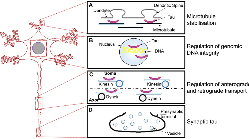
Tau has also been found in eukaryotic nuclei, both in neurons and non-neuronal cells (Loomis et al., 1990; Sultan et al., 2011). Nuclear tau in human and monkey kidney cell lines co-localise with Tau-1, a pan-isoform tau antibody (Loomis et al., 1990). In these cells, Tau-1 co-localisation occurs in the nucleolar organiser region of the chromosome, suggesting that tau may have a role in regulating genomic deoxyribonucleic acid (DNA) (Loomis et al., 1990). Notably, tau homologues were not found in non-primate cells, possibly due to a conserved function of nuclear tau in primates (Loomis et al., 1990). Fig. 1.2 provides a schematic illustration of the neuronal location and physiological functions of tau.
Introduction: Normal Tau Function
1.
Dendrite Dendritic Spine Tau Microtubule Nucleus B C D Microtubule stabilisation
Regulation of genomic DNA integrity
[image:21.595.144.555.283.513.2]Regulation of anterograde and retrograde transport Tau DNA Axon A Presynaptic terminal Vesicle Synaptic tau Soma Dynein Kinesin Kinesin Dynein
Figure 1.2. Physiological functions of tau.
(A) The most widely characterised role of tau is the stabilisation of MTs and the promotion of MT assembly. (B) In the nucleus, tau binds to specific chromosomal regions and possibly regulates the integrity and activity of genomic DNA. (C) Another function of axonal tau is the modulation of axon-soma shuttling of cellular material through the anterograde and the retrograde transport mechanisms, by regulating the interaction between MTs and the motor proteins kinesin and dynein required for these processes. (D) Synaptic tau is believed to be important for synaptic plasticity, although little is known about this property. Figure design concept taken from Wang and Mandelkow (2016).
1.2.2 Pathophysiological functions of tau
functions. Loss of function effects of tau include: (i) decrease in tau expression levels, which reduces the amount of tau required for efficient MT stabilisation; and (ii) loss of the MT stabilisation function due to abnormal phosphorylation. Examples of the gain of function effects include: (i) self-polymerisation of soluble, free tau to form disease-associated aggregates; (ii) disease-associated mutations that change disease-causing pathways; and (iii) extracellular release and trans-synaptic propagation of misfolded tau.
1.2.2.1 Pathophysiology arising from loss of physiological functions
Elimination or reduction of tau levels
Loss of the MT stabilisation function
Adjacent to the MT-binding region are proline-rich regions (P1 and P2 in Fig. 1.1) that contain many phosphorylation sites, although many other sites are distributed across the entire tau sequence (Hanger et al., 2007). Excessive phosphorylation of tau reduces its ability to bind to MTs, hence disrupting the protein’s main physiological function (Wang et al., 2013). Increasing levels of phosphorylation may lead to a complete loss of the protein’s affinity to MTs, resulting in soluble tau accumulation in neurons, impairing neuronal homeostasis either by the sheer space occupied or by interfering with specific processes such as the protein degradation system (Stoothoff and Johnson, 2005). Moreover, the loss of tau’s MT binding affinity can lead to MT disassembly, disrupting cargo transport. Because the extent of tau phosphorylation directly corresponds to reduction in MT binding, which may have disease-causing implications (Stoothoff and Johnson, 2005), the amount of phosphorylated tau is used as a biomarker in staging AD and other tau-positive diseases (Hampel et al., 2010). Importantly for this thesis, free forms of MT-detached tau can self-aggregate into large structures that may define disease progression (section 1.2.2.2).
1.2.2.2 Pathophysiology arising from gain of new toxic functions
Self-polymerisation of free tau to form aggregates
In its hyperphosphorylated state, the MT domain of tau cannot effectively bind MTs: this results in free floating forms of soluble tau (Fig. 1.3). These MT-unbound forms of tau can lead to disease through self-aggregation. The two hexapeptide motifs 275VQIINK280 and 306VQIVYK311 at the beginning of repeats R2 and R3 respectively are critical for the ability of monomeric tau molecules to aggregate. Removal of one or both motifs reduces or abolishes tau’s aggregation competence (Bergen et al., 2000; Li and Lee, 2006). These motifs modulate the structural conversion of natively unfolded monomeric tau to b-sheet-rich
contacts leading to tau aggregation (Xiang et al., 2017), suggesting that this motif regulates tau aggregation. It is therefore unsurprising that this structural peptide is capable of forming tau aggregates independently of the surrounding tau sequences (Stöhr et al., 2017), and that tau protein lacking this structural peptide is unable to aggregate (Li and Lee, 2006).
Introduction: Pathological Tau
A
D C B
Dendritic spine loss
No entry into the nucleus
Loss of affinity to microtubules Microtubule disassembly Tau aggregation
Post synaptic dysfunction Dysfunctional
Tau
Tau direct and indirect pathway of neuronal dysfunction
Aggregation and seeding Phosphorylation
Tau aggregation
Seeds Monomers
Induced aggregation
Figure 1.3. Pathophysiological functions of tau.
Dysfunctional tau, generated due to overproduction or abnormal phosphorylation, can lead to disease through different routes, such as: (A) presynaptic terminal mislocalisation, causing spine loss through a cascade of events; (B) lost affinity to MTs resulting in MT disassembly, impaired axonal transport and tau aggregation; (C) aggregated tau acting as seeds or templates to induce further aggregation; and (D) impaired neurotransmission by affecting the production and/or activity of specific post-synaptic proteins. Figure design concept taken from Wang and Mandelkow (2016).
pro-aggregant hexapeptide structural element 275VQIINK280 in 4R isoforms also likely contribute to their higher aggregation rates.
Cysteine Binding Possibilities
Four-repeat Tau (two native cysteines)
S S
2. Oxidised Monomer 1. Reduced Monomer
HS
SH
3. Partially Oxidised Dimer
S HS S SH 5. Trimer S HS S S SH S Etc.
4. Fully Oxidised Dimer
S S S S 6. Tetramer S HS S S S S S SH N….. …..N OR A
1. Reduced Monomer SH
2. Fully Oxidised Dimer S S
Cysteine Binding Possibilities
B Three-repeat Tau (one native cysteine)
Cysteine Binding Possibilities
C Four-repeat and Three-repeat Tau bonds1. Dimer S S SH 3R 4R 2. Trimer S S S S
Figure 1.4. Cysteine-dependent tau aggregation is isoform-specific.
Tau-bound microtubules
Free tau dissociated
from MTs
Soluble tau
Aggregation-competent monomers due to conformational change
Monomers
Neurofibrillary tangles
Dimerisation
Dimers
Filamentous aggregation
Protomers
Nucleation
Oligomers
Edit into Nucleation-Elongation mechanism
Neurotoxicity
Figure 1.6. Electron micrographs of tau aggregates isolated from AD brains (upper panel; three patients – cases 1 – 3) and in vitro polymerised filaments from recombinant tau K19, and disease variant forms of K18 and htau40 (bottom panel).
The recombinant PHFs, assembled in the presence of the polyanionic cofactor heparin, share close features (such as a paired helical characteristic, 10-25 nm width and a crossover repeat of ∼80 nm) with the brain-derived PHFs. Figure taken from Barghorn et al., (2004).
1.3 Tauopathies: disease classification, clinical diagnosis, progression, and differential roles of tau isoforms
A major clinical feature of AD is intraneuronal NFT accumulation consisting of PHFs and SFs polymerising from both 4R and 3R tau (Wischik et al., 1988). Tau filaments in AD are mostly PHFs, with crossover distances of ~80 nm and width of 10–20 nm, and SFs 15 nm wide (Wischik et al., 1988). A strong evidence
supporting a causative role for tau in AD is obtained from amyloid-b processing.
Amyloid-b is a proteolytic product of the transmembrane protein amyloid precursor protein (APP) whose processing occurs by two broad pathways: amyloidogenic and non-amyloidogenic, with the former leading to amyloid-b
production through g-secretase-mediated proteolysis of APP (O’Brien and Wong, 2011). As a multiprotein complex consisting of, among other proteins, presenilin 1 (PSEN1) and presenilin 2 (PSEN2), g-secretase activity can influence APP
proteolysis and hence amyloid-b generation (Bergmans and De Strooper, 2010). For example, substituting the aspartyl residues D257 and D385 in PSEN1 to Alanine reduces APP cleavage and hence amyloid-b production (De Strooper et
al., 1998; Wolfe et al., 1999). Since amyloid-b is primarily an extracellular protein, its intracellular effects are triggered by activating specific channels and receptors (e.g., tyrosine kinase receptors and Ca2+ channels). Extensive interactions between candidate protein kinases (phosphatases, serine/threonine kinases, and tyrosine kinases) lead to increased tau phosphorylation, which then induces NFT formation and subsequent neurodegeneration and neuronal loss (Hanger et al., 2009). Based on this pathway, tau toxicity manifests downstream of abnormal amyloid-b production. Pathological mutations in APP, PSEN1 and PSEN2 would then induce and/or require tau dysfunction to cause disease. For example, 87% of a cohort of early-onset AD patients carrying APP or PSEN1, or PSEN2 mutations had abnormal cerebrospinal fluid (CSF) levels of total tau, phosphorylated tau and amyloid-b (Lanoiselée et al., 2017). Moreover, the V717I
APP mutation causes increased amounts of amyloid-b by enhancing g-secretase cleavage of APP in stem cell-derived neurons, which then leads to increased total and phosphorylated tau levels (Muratore et al., 2014). Importantly, total tau overproduction was rescued by amyloid-b-specific antibody treatment, suggesting
a causal link between altered amyloid-b production and tau levels (Muratore et al., 2014). Furthermore, altered APP metabolism increases tau production and
levels (Moore, et al., 2015). This connected pathway of events, referred to as the amyloid cascade hypothesis, provides evidence demonstrating that dysfunctional tau has a central role in AD pathogenesis, dependent on altered APP metabolism
and/or amyloid-b levels (Hardy and Allsop, 1991). Another piece of evidence supporting this hypothesis is that tau knock-out mice do not develop NFTs, suggesting that upstream dysfunctional APP and/or amyloid-b activity is required for tau toxicity (Chin et al., 2004, 2005). Indeed, reduction in tau levels is neuroprotective due to its obstruction of amyloid-b-induced toxicity (Vossel et al., 2015).
NFT accumulation in AD first occurs in the transentorhinal cortex, and tends to extend to other brain regions including the neocortex and the hippocampus. This gradual development of tau pathology in distinct anatomical brain regions is used in staging AD, as first described over two decades ago (Braak and Braak, 1991). NFT development in AD often occurs alongside memory and cognitive decline (Guillozet et al., 2003; Nelson et al., 2012).
sometimes present (Josephs et al., 2008). CBD and PSP both form predominantly SFs with few twisted filaments which are often 15-30 nm wide (Ksiezak-Reding et al., 1996; Takauchi et al., 1983). However, the crossover periodicity of SFs in
the two diseases tends to differ, being ∼100 nm and 160 nm for PSP and CBD
respectively (Ksiezak-Reding et al., 1996; Takauchi et al., 1983). Tau aggregation in PiD involves mainly 3R isoforms, and leads primarily to SFs 160 nm in periodicity and 15 nm wide (Kato and Nakamura, 1990). As a rare form of frontal lobe dementia, PiD mainly affects the cortex and limbic lobe (Barker et al., 2002). PiD can be due to sporadic or familial causes, with the latter due to specific tau mutations (Hogg et al., 2003; Murrell et al., 1999).
FTDP-17 has several shared clinical features with the other FTDs – PiD, CBD, and PSP – including the affected anatomical regions (cortex, brainstem, and basal ganglia), clinical features (dementia, psychosis, and focal cortical syndrome), and tau isoforms involved (either 4R only or 4R and 3R jointly) (Dickson et al., 2011). The majority of familial FTDP-17 are caused by tau mutations (section 1.4), with the rest often occurring due to mutations in the GRN gene on chromosome 17q21 that codes for progranulin (Cruts et al., 2006), and C9orf72 on chromosome 9p21 (DeJesus-Hernandez et al., 2011; Renton et al., 2011). FTD patients with progranulin mutations have TDP-43 pathology, which explains the absence of tau-positive inclusions (Cenik et al., 2012; Perry et al., 2013; Rademakers et al., 2012).
1.4 Tau mutations as modulators of familial FTD
repeat region emphasises the importance of this region not only in tau physiology but also pathophysiology. Moreover, because many tau mutations cluster around exon 10, they can only be produced through alternative splicing which may alter the isoform balance (Fig. 1.7).
Many mutations in MAPT are associated with familial forms of FTD, including FTDP-17, CBD, PiD, AGD, and PSP (Goedert and Jakes, 2005) The pathological MAPT mutations underlying these tauopathies may confer disease in a number of ways: (i) at the mRNA level (through alternative splicing) or (ii) at the protein level (by altering MT-binding efficiency or by enhancing/reducing protein aggregation). As shown in Table 1.1, familial FTDP-17 exhibits extensive heterogeneity in disease characteristics which overlaps with features of other FTDs.
Figure 1.7. Exonic distribution of disease-associated tau mutations.
Over 100 mutations in the MAPT gene are associated with different brain disorders involving dysfunctional tau. Majority of these mutations are single point changes or deletions that change the protein’s amino acid composition. These alterations can impact physiological functions such as MT stabilisation and can lead to disease through reduced MT binding, changes in aggregation kinetics, and isoform imbalance. Most mutations cluster around the MT repeat region, which spans part of exon 9 through to exon 12. Mutations in exon 10 are only evidence in 4R tau isoforms. Figure from Ghetti et al., (2015).
MT binding whilst V337M and R406W decreased tau’s ability to promote MT stabilisation (Hong et al., 1998). Other mutations that reduce MT binding in vitro include N279K, G272V and ΔK280 (Barghorn et al., 2000). The ability to alter MT binding is not limited to mutations in the MT binding region, as A152T, R5L and R5H in the N terminus also cause reduction in MT binding of tau (Coppola et al., 2012; Magnani et al., 2007). Since truncation at glutamine 124 in AD brains alters tau MT-binding abilities (Derisbourg et al., 2015), the N-terminus mutations may have direct functions in tau-MT association
Table 1.1. MT binding effects and clinical phenotypes of some MAPT
mutations associated with FTDP-17
Table adapted from Liu and Gong, (2008)
Exon Mutation Effect on MT binding
Tau isoforms involved Clinical phenotype
1 R5H N/A Mostly 4R PSP
1 R5L N/A 1N3, 4R AD
9 K257T Reduced 3R>4R PiD
9 I260V N/A Mostly 4R N/A
9 L266V N/A 3R PiD
9 G272V N/A 3R PiD
10 N279K Variable* 4R PSP
10 DK280 Reduced 3R>4R FTDP-17
10 L284L No change 4R maybe AD
10 N296N No change Mostly 4R CBD
10 N296H N/A Mostly 4R FTDP-17
10 DN296 Reduced N/A PSP
10 P301L Increased 4R FTDP-17
10 P301S N/A Mostly 4R FTDP-17 and CBD
10 G303V N/A Mostly 4R PSP
10 S305N No change Mostly 4R CBD
10 S305S N/A Mostly 4R PSP
11 L315L No change N/A N/A
11 S320F Reduced N/A PiD
11 S320Y N/A N/A PiD
12 Q336R Increased N/A PiD
12 V337M Reduced 3R, 4R FTDP-17
12 E342V N/A Mostly 4R FTDP-17, PiD
12 S352V N/A 3R, 4R PiD
13 G389R Reduced 4R>3R PiD
13 R406W N/A 3R, 4R PSP
N/A = not available Inhibit exon 10 inclusion Enable exon 10 inclusion
Other MAPT mutations have additional suggested mechanisms. Mutations located in exon 10 have additional effects in the form of increased 4R isoform production which can influence the overall isoform distribution and lead to dysfunction through the overproduction of tau. As shown by Hong et al., (1998), insoluble human brain-derived tau aggregates from patients with N279K or P301L have exclusive involvement of 4R tau contrary to those from V337M and R406W brains that separated with both 3R and 4R tau in SDS-PAGE assays. How exon 10 mutations such as N279K increase 4R production and selectively induce 4R tau aggregation is unclear as this appears independent of MT binding effects. This could occur by promoting exon 10 splicing at the mRNA level, possibly by altering a splicing enhancer element that regulates exon 10 synthesis (Dawson et al., 2007; Hong et al., 1998). Although P301L is located in exon 10, it has opposite effects on MTs compared to N279K, indicating the two may be acting independently. Contrary to N279K, mutations such as G272V ΔK280, and L266V all inhibit exon 10 inclusion, thus reducing 4R tau production (Liu and Gong, 2008).
The aggregation of tau containing specific mutations, irrespective of the isoforms involved, can also lead to neurodegeneration via differential aggregation kinetics. P301L and DK280 increase thioflavine S-based tau aggregation, independent of
1.5 Theories of tau pathology: aggregation and conformational change
As discussed (section 1.2.2), a well-known route to tau dysfunction is hyperphosphorylation, which disrupts the protein’s MT stabilisation function. This therefore suggests that gain-of-function effects of tau that occur downstream of MT destabilisation, such as aggregation, will be dependent on the protein’s phosphorylation status. Motivated by this hypothesis, the tau research field has conducted intensive research aimed at identifying critical tau phosphorylation sites and kinases that may regulate aggregation (Šimić et al., 2016). It has however been found that tau aggregation can occur independently of phosphorylation, indicating that hyperphosphorylation may not be the sole route to disease-related aggregation (Tepper et al., 2014). Although there are disagreements as to which stage of tau aggregation (e.g., oligomers and PHFs; section 1.7) best correlates with disease, it is generally accepted that tau aggregation can lead to disease (Goedert, 2016; Goedert and Spillantini, 2017). If WT tau aggregation can cause disease, how then do FTD mutations affect this mechanism and therefore clinical outcomes? As specific mutations can alter tau’s MT-stabilisation and aggregation functions (Table 1.1; section 1.2), it can be hypothesised that they do so by modifying tau’s mechanism of disease. This could occur through multiple means, including: (i) by inducing conformational changes that make the mutated protein a more preferred substrate for aggregation (Jicha et al., 1999); (ii) by influencing the aggregation rate, as illustrated by significant lag time changes in kinetic aggregation assays (Combs and Gamblin, 2012); and (iii) by reducing the protein’s affinity for MTs (which may also occur due to conformational change) (Table 1.1).
rate of nucleation and/or elongation, as shown previously (Barghorn et al., 2000; Combs and Gamblin, 2012) . However, these induced effects are likely to act downstream of the initial conformational change that tends to enable or enhance aggregation competence. For example, a “correct turn” conformational change in the third repeat region is essential to induce aggregation even in the presence of the 306VQIVYK311 hexapeptide motif, as tracked by time-dependent NMR assays (Jiji et al., 2016). Such a conformational change which influences initiation of aggregation should perhaps occur before the nucleation stage (Fig. 1.5). Another form of conformational change could occur at the elongation stage of tau aggregation. In early stage AD (Braak stages I and II) brains: the monoclonal antibody MC1 recognises soluble forms of aggregated tau that appear before PHF and NFT formation in vulnerable brain areas, indicating that the MC1 epitope is an early pathological signal (Weaver et al., 2000). The conformational significance of the MC1 epitope may be the reason why the S320F FTD mutation causes reduced MT binding (Rosso et al., 2002).
1.5.1 Biochemical and biophysical tools for studying tau aggregation and conformation
Much of the insights into tau conformation and aggregation have been obtained from in vitro experiments on recombinant or brain-derived proteins using biochemical and biophysical tools. Those used in this thesis are introduced here.
Kinetic assays – using fluorescent dyes
2010). Nonetheless, this property is dependent on the forms of tau used, buffer conditions, spectrophotometer and the tau-ThT/S ratio (Xue et al., 2017). The principle of ThT/S binding to tau filaments has been applied in clinical histopathology to diagnose tauopathies (Bussière et al., 2004; Rajamohamedsait and Sigurdsson, 2012). The mechanism of ThT/S binding to tau is not fully understood: one model suggests that ThT binds to amino acid side surface chains that are arranged in parallel to the b-sheet axis of tau filaments (Biancalana and Koide, 2010). Binding to proteinopathic aggregates can drastically increase the fluorescence intensities of both ThT and ThS, but the main difference is that a corresponding forward shift in emission spectrum is observed for ThT but not ThS for which no change in the excitation or the emission spectra is recorded (Groenning, 2010; LeVine, 1999). This results in a consistently high background fluorescence in the case of ThS, making it unsuitable for quantitative analysis (LeVine, 1999).
Secondary structure determination: circular dichroism (CD)
CD spectroscopy is routinely used to ascertain the structural transition of tau from its random coiled conformation to b-sheet formation. A CD spectrum is obtained due to a sample’s differential absorption of left and right circularly polarised light. The extent to which a test molecule absorbs the two light waves which are arranged 90 O out of phase leads to the generation of an electrical field signal. The resultant differences in the electric field generated by the clockwise- and anti-clockwise-facing lights is calculated to give the CD readout at each wavelength. Signature CD spectra measured in the far UV range differ for given proteins depending on their predominant secondary structure content (Fig. 1.8). Unfolded proteins, such as monomeric tau, have negative peaks at 198 – 200 nm, b
-sheet-enriched proteins like PHFs have negative peaks at ~220 nm, whilst soluble aggregated tau (e.g., oligomers) have an intermediate negative peak between 200 and 220 nm. Other proteins with majority a-helix content have two negative
Ultrastructure of aggregated species: atomic force microscopy (AFM) and transmission electron microscopy (TEM)
AFM and TEM are similar techniques used to probe the ultrastructural properties of aggregated tau. AFM involves three-dimensional scanning of a sample surface with a cantilever-suspended flexible probe. Deflection of the cantilever occurs in response to probe-sample surface contacts on a piezoelectric scanner, leading to parallel images formed along the probe tracks. These images are then projected by laser signals onto photodiode detectors and processed with specific algorithms. There are three main AFM modes, depending on probe-sample interactions: contact, non-contact and tapping. Tapping mode imaging is often preferred as it eliminates the disadvantages of the other methods regarding poor resolution and frictional forces that can damage sample surfaces (Carvalho and Santos, 2012; Dufrêne, 2002).
In TEM, an electron beam passes through the sample and the micrometer-scale image formed based on electron-sample interactions is magnified to enhance resolution. As complementary techniques, TEM and AFM compensate for their shortcomings. For example, heavy metal staining in TEM can disrupt some nanoscale details. This challenge can be addressed by using AFM which does not use sample staining and therefore retains samples in their native state. Moreover, small aggregated proteins (e.g. oligomers made of <10-20 monomers) whose imaging can be problematic with TEM can be easily done with AFM. Nonetheless, AFM imaging is extremely slow and gives poor image resolutions. On the contrary, TEM images have better resolution and are achievable within shorter times (Tinker-Mill et al., 2014).
Dynamic light scattering (DLS)
tau proteins), using the hydrodynamic radius which relates to the intensity of molecule fluctuations (Stetefeld et al., 2016).
B A
Figure 1.8. Example data from some commonly used biochemical and biophysical assays for evaluating tau aggregation and conformation.
(A) example standard curve from ThT or ThS kinetic assays (Abedini et al., 2016). (B) Standard CD spectra for proteins which have predominantly a-helices (black), b sheets (red), and unfolded/random coiled (green) conformations. Figure taken from http://www.ap-lab.com/circular_dichroism.htm, accessed on 10th September, 2017.
1.6 Proteinopathies and the prion hypothesis
Proteinopathies is a catch-all term for neurodegenerative diseases characterised by specific post translational modifications, involving the disease-related aggregation and neuronal accumulation of otherwise unstructured proteins, leading to neurodegeneration and eventual neuron loss. The term proteinopathies looks at the similarities between the molecular pathogenesis of neurodegenerative diseases such as AD, Parkinson’s disease (PD) and Huntington’s disease: the respective proteins underlying these diseases are tau/amyloid-b, a-synuclein, huntingtin and mutated forms of tau (in familial cases). Several other diseases have also been classified as proteinopathies (Golde et al., 2013).
protein are gradually distributed throughout the brain, leading to synaptic dysfunction and neuron death (Frost and Diamond, 2010; Golde et al., 2013).
1.6.1 Neuronal internalisation and transmission of tau pathology: the transneuronal propagation and the selective vulnerability
hypotheses
Although mounting evidence suggests that tau dysfunction causes neurodegeneration, the mechanistic basis for this association is not fully understood. Two main mechanisms have been proposed, referred to as the trans-neuronal propagation and the selective vulnerability spread hypotheses.
Transneuronal propagation
Papanikolopoulou and Skoulakis, 2011), neuronal and neuron-like tissues (Michel et al., 2014; Usenovic et al., 2015; Wauters et al., 2016), ex vivo brain tissues (Fá et al., 2016) and post-mortem brains (Lasagna-Reeves et al., 2012a). The minimal tau peptide sequence required for this propagation behavioural is thought to be a 31-residue peptide that includes the hexapeptide motif in R3 (Stöhr et al., 2017). Neuronal accumulation and propagation of dysfunctional tau causes toxicity by impairing neurotransmission through neurite retraction and soma loss (Stancu et al., 2015; Usenovic et al., 2015), damaging electrical communication (Fá et al., 2016; Lasagna-Reeves et al., 2012a), and triggering memory and cognitive changes (Fá et al., 2016; Stancu et al., 2015).
Selective vulnerability model
A critique of the transneuronal propagation model argues that the physical internalisation, release and subsequent propagation proposed may be unrealistic because: (i) the model is incapable of explaining cell autonomy in degeneration and death; (ii) the Braak staging (Braak and Braak, 1991) used as a premise does not support the proposed transmission hypothesis; and (iii) the model does not address the patient heterogeneity and the brain regional vulnerability observed in proteinopathic protein accumulation. On this basis, an alternative model, referred to as the “selective vulnerability” hypothesis has been proposed. This model suggests that neuronal vulnerability to toxic insults (e.g., from misfolded tau) is selectively dependent on its viability: neurons stressed from the extracellular build-up of tau (or due to any other reason) would be easier targets compared to healthy ones. Healthy cells may become susceptible over time as the disease progresses and/or the levels of the stressor increase (Walsh and Selkoe, 2016). There are various mechanisms of transmission reported for the trans-neuronal propagation (Fig. 1.9), whilst Fig. 1.10 describes the counter selective vulnerability model.
Tau seeds 1
Fibrils
Ectosomes
Donor neuron Receiving
neuron Vesicular
bodies
Macropinocytosis 2
3 4
Nanotubules
Exosomes
5 Extracellular Space
Free Tau
6 Through Membrane
Endosome 7
Receptor-mediated
Figure 1.9. Mechanisms of misfolded tau transmission in the transneuronal propagation model for extracellular tau.
Figure 1.10. The selective vulnerability model of tau toxicity.
(A) The transneuronal spread model proposes physical transfer of misfolded tau between synaptically connected neurons, and thus brain regions, without accounting for cell autonomy. (B) The selective vulnerability model suggests that neurons of compromised viability will be first affected by possible insult from misfolded tau. Vulnerability can be transferred trans-synaptically through diffusible metabolic factors, instead of protein internalisation and release (Walsh and Selkoe, 2016).
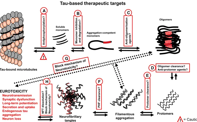
1.7 Drug development against misfolded tau protein
phosphorylates tau serine 202 leading to a pathology-associated conformational change (Martin et al., 2013; Seifert et al., 2008). Nonetheless, DYRK1A also phosphorylates the cAMP Response Element Binding (CREB) transcription factor, which regulates learning and memory (Yang et al., 2001). Hence, CREB inhibition is likely to lead to non-specific effects. Glycogen synthase kinase 3 (GSK3) is a tau protein kinase which has a central role in amyloid b-induced tau
pathology: exposure to amyloid b increases GSK3 activity, tau phosphorylation,
memory defects, apoptotic cell death and increased amyloid b expression (Hoshi et al., 1996; Takashima et al., 1996). GSK3 phosphorylates tau at multiple sites in human AD brains (Hanger et al., 2009; Martin et al., 2013), and its overexpression in NFT-forming transgenic mice causes hyperphosphorylation and neurodegeneration (Lucas et al., 2001). Lithium treatment rescues both GSK3-induced hyperphosphorylation and downstream neurodegeneration effects in transgenic mice (Klein and Melton, 1996). Other GSK3 inhibitors include specific compounds from the anilinomaleimide (Smith et al., 2001), paullone (Leost et al., 2000), indirubin (Leclerc et al., 2001), and thiadiazolidinone (Martinez et al., 2002) families. Some of these compounds are non-specific for GSK3 because they also inhibit tau kinases from the cyclin-dependent kinase family, including CDK2 and CDK5 (Bhat et al., 2004; Leclerc et al., 2001). Since CDK5 inhibition activates GSK3, such compounds may not be ideal drug candidates (Hanger et al., 2009). Whilst tau kinase inhibition may be a promising target, there is a need to be cautious in this approach because not all forms of phosphorylation may be harmful (Ittner et al., 2016).
respectively). Formed filamentous aggregates can be otherwise targeted by degradation (option F), as explored for amyloid b (Yan et al., 2006). Should PHF degradation not prove successful, an alternative strategy will be to inhibit NFT accumulation with small molecules or immunotherapeutic agents (option H). Importantly, all the approaches proposed here should be feasible in both intra- and extra-cellular contexts.
While there are clearly several intervention points being investigated, the targeting of the cellular entry and exit of tau, which appears to be the central hub of many neurotoxic properties, may be productive in diseases involving tau and indeed other proteins (Fig. 1.10).
Tau-bound microtubules Soluble monomers Aggregation-competent monomers Filamentous aggregation Protomers Oligomers
Tau-based therapeutic targets
NEUROTOXICITY
• Neurotransmission
• Synaptic dysfunction
• Long-term potentiation
• Secretion and uptake
• Endogenous tau aggregation
• Neuron loss
An ti p h o sp h o ry la ti o n ? 1 A 1 D !
! = Caution
Mo n o m er i n h ib iti o n an d d eg rad at io n ? An ti -oligom er is at ion ag en ts? 1
B 1C
[image:46.595.153.484.323.528.2]Oligomer clearance? Anti-protomer agents? Pr o to m er cl earan ce? PH F c le ar an ce ? 1 E Neurofibrillary tangles NF T d is so lu ti o n ? Bl o ck me ch an is m o f Ne u ro to xi ci ty ? 1 H 1 G 1 F
Figure 1.11. Dysfunctional tau-targeted drug development targets and approaches.
Description of each numbered approach has been provided in the text in section 1.7.
1.8 Aims of this thesis
i. To develop genetic resources and a simple method of expressing and purifying tau protein and a selection of its FTD variants;
ii.To develop a new method of preparing low molecular weight (LMW) tau K18 oligomers of enhanced stability for use in biochemical and cell biology assays;
iii. To study the in vitro conformation and aggregation of tau K18 WT, V337M and N279K; and
2| Materials and methods
2.1 Materials
dehydrogenase (LDH) cytotoxicity assay kit (88954) was obtained from Pierce Biotechnology, Rockford, Illinois, USA). The cOmplete protease inhibitor cocktail tablets (11836145001) and DNAse I (11284932001) were obtained from Roche Diagnostics GmbH (Mannheim, Germany). Isopropyl β-D-1 thiogalactopyranoside (IPTG; MB1008) was purchased from Melford Laboratories Limited (Ipswich, Suffolk, UK). Tris(2-carboxyethyl)phosphine (TCEP; A2233,0001) was procured from Applichem GmbH, Damstadt, Germany. SynaptoRedTM C2 (FM4-64; 70021) was obtained from Biotium (Hayward, California, USA). The QIAprep Spin Miniprep kit (27104) was obtained from QIAGEN GmbH, Hilden, Germany. Marvel dried skimmed milk was obtained from Premier Foods Company, UK.
2.2 Cloning of tau proteins into pProEx plasmids
Figure 2.1. Bacterial expression plasmids used in this study.