University of Warwick institutional repository: http://go.warwick.ac.uk/wrap
A Thesis Submitted for the Degree of PhD at the University of Warwick
http://go.warwick.ac.uk/wrap/56924
This thesis is made available online and is protected by original copyright. Please scroll down to view the document itself.
The Role of NSP1 in Rotavirus
Pathogenesis
Fan Zhang
Submitted for the degree of Doctor of Philosophy
School of Life Sciences
University of Warwick
United Kingdom
Table of contents...i
List of Figures...v
List of Tables ...vii
Acknowledgements...viii
Declaration...ix
Abbreviations ...x
Summary...xii
Chapter 1: Introduction ...1
1.1 Rotavirus...2
1.1.1 The discovery of rotavirus………...2
1.1.2 Classification of rotaviruses……….3
1.1.3 Structure of Rotavirus particle…...6
1.1.4 Rotavirus genome and gene-protein coding assignments...7
1.1.5 Viral protein structure and function...10
1.1.5.1 Structural proteins…...11
1.1.5.2 Non-structural proteins...13
1.1.6 Rotavirus replication cycle…...15
1.1.6.1 Virus attachment and entry...18
1.1.6.2 Transcription and translation...20
1.1.6.3 Genome replication...22
1.1.6.4 Virus assembly and release...23
1.1.7 Rotavirus pathogenesis...24
1.1.8 Rotavirus vaccines...25
1.2 Innate immune system…...26
1.2.1 Overview………...26
1.2.2 Interferon regulatory factors regulated pathway...31
1.2.3 NFκB regulated pathway...33
1.3 Non-structural protein NSP1...36
1.3.1 NSP1 shows high levels of primary sequence variability………..36
1.3.2 NSP1 can interact with Interferon Regulatory Factors (IRFs)...39
1.3.3 NSP1 can induce a proteasome-dependent degradation of β-TrCP …..43
1.3.4 Other NSP1 targets...45
2.2.1 Bacteriological techniques...59
2.2.1.1 Growth of bacteria………...59
2.2.1.2 Preparation of competent cells...59
2.2.1.3 Transformation of competent bacterial cells by heat shock....60
2.2.1.4 Transformation of competent bacterial cells by electroporation………60
2.2.1.5 Extraction of plasmid DNA (Mini Prep, Midi Prep and Maxi Prep)………61
2.2.1.6 Quantification of nucleic acid……….61
2.2.1.7 Blue/white selection of bacterial colonies………..61
2.2.2 Manipulation of DNA...62
2.2.2.1 Standard Polymerase Chain Reaction (PCR) ...62
2.2.2.2 Site-Direct Mutagenesis PCR...62
2.2.2.3 Restriction Enzyme Digest...63
2.2.2.4 De-phosphorylation of vector DNA...63
2.2.2.5 Conversion of 5’ overhangs restriction enzyme ends to blunt ends……….64
2.2.2.6 Phenol/Chloroform extraction………64
2.2.2.7 Ethanol precipitation of DNA………...64
2.2.2.8 Agrose gel electrophoresis of DNA………65
2.2.2.9 Agarose Gel Extraction of DNA……….65
2.2.2.10 DNA Ligation...65
2.2.2.11 DNA Sequencing...66
2.2.2.12 Transfection of plasmid DNA...66
2.2.3 Manipulation of RNA...67
2.2.3.1 Extraction of RNA from cultured 293 cells...67
2.2.3.2 Reverse-Transcription (RT) PCR...68
2.2.3.3 Transfection of cells by poly (I:C) ...68
2.2.4 Mammalian cell culture techniques...69
2.2.4.1 Maintenance of tissue culture cells...69
2.2.4.2 Growth of rotavirus...69
2.2.4.3 Titration of virus stocks...70
2.2.4.4 MG132 treatment...70
2.2.4.5 Mammalian two-hybrid assay...70
2.2.5 Protein expression and analysis...71
2.2.5.1 Coupled Transcription/Translation system...71
2.2.5.2 Determination of protein concentration...71
2.2.5.3 Harvesting of total cellular protein...71
2.2.5.4 SDS-Polyacrylamide Gel Electrophoresis (SDS-PAGE) ...72
2.2.6.1 Preparation of cell lysates...75
2.2.6.2 β-Galactosidase Assay...75
2.2.6.3 Bright-Glo Luciferase Assay...76
2.2.6.4 Luciferase Assay Data Analysis...76
Chapter 3: Generation of chimeric rotavirus NSP1 hybrids...77
3.1 Introduction...78
3.2 Cloning vector preparation for generating chimeric NSP1 hybrid cDNAs……..85
3.3 Cloning of the UKtcNSP1 and OSUNSP1 genes into the mpCR2.1 vector...87
3.4 Sequential mutagenesis PCR of UKtcNSP1 and mpCR2.1-OSUNSP1………..95
3.5 Generating chimeric NSP1 hybrid constructs using mutated UKtcNSP1 and OSUNSP1 cDNAs………..……….100
3.6 Discussion ...104
Chapter 4: Analysis of protein interactions between rotavirus NSP1 and host cellular proteins………..…106
4.1 Introduction...107
4.2 Molecular cloning of protein coding sequences for interaction studies in the mammalian two-hybrid assay system..………..………..111
4.3 Protein-protein interaction studies using the mammalian two-hybrid assay system………...124
4.4 NSP1 expression in transfected cells………..129
4.5 Expression of NSP1 proteins in pCDNA3.1 vector………131
4.6 Protein interaction studies via co-immunoprecipitation……….134
4.7 Discussion………...140
Chapter 5:Functional analysis of NSP1 hybrid proteins…………...142
5.1 Introduction...143
5.2 pcDNA3.1-NSP1 constructs do not stimulate the activation of the IFNβ promoter...144
5.3 pCI-neo-NSP1 constructs can cause deduction of IFNβ promoter activity...150
5.4 Analysis of protein expression of all NSP1 hybrid genes in pCI-neo vector...152
5.5 Mapping of the interaction sites with different pCI-neo-NSP1 hybrids analysed in the reporter assay...157
5.6 The effect of NSP1 hybrid genes on NFκB activity………...163
5.7 NSP1 induced degradation of IRF3 and/or β-TrCP………...165
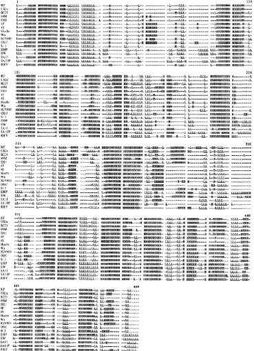
Figure 1.1 Structure of mature rotavirus particle at 9.5-Å resolution…………..8 Figure 1.2 The rotavirus replication cycle………..17 Figure 1.3 Three classes of PRRs for RNA virus recognition………...29 Figure 1.4 Signalling pathways triggered by viral infection……….35 Figure 1.5 Conservation of predicted secondary structure across NSP1………40 Figure 3.1 Sequence comparision between NSP1s of different rotavirus
strains……….80 Figure 3.2 Schematic diagrams illustrating the selected restriction enzyme (RE) sites to be generated in both UKtc and OSU NSP1 sequences………..82 Figure 3.3 Illustration of the cloning vector pCR2.1 indicating the selected RE sites already exist in the vector sequence……….………...87 Figure 3.4 Modification of the pCR2.1 cloning vector for use in generating NSP1 Hybrids………...89 Figure 3.5 Cloning of UKtcNSP1 into mpCR2.1 vector………91 Figure 3.6 Cloning of OSUNSP1 into mpCR2.1 vector……….93 Figure 3.7 The cloning strategy employed for constructing NSP1 genes
containing six selected RE sites………96 Figure 3.8 Analysis of RE sites generated in UKtcNSP1 and OSUNSP1 cDNA clones………..98 Figure 3.9 Analysis of the chimeric NSP1 hybrid pA………..101 Figure 3.10 Schematic illustrations of the chimeric NSP1 hybrid sequences generated between UKtcNSP1 and OSUNSP1………...………..103 Figure 4.1 Schematic representation of the mammalian two-hybrid assay…..110 Figure 4.2 IRF3 obtained from vector pEFplink2………...112 Figure 4.3 Experimental strategies for re-amplifying IRF3 coding sequence into the mammalian two-hybrid vectors………..112 Figure 4.4 Schematic diagrams of the mammalian two-hybrid system cloning vectors pM and pVP16, showing the multi-cloning site sequence and unique restriction enzyme sites………...113 Figure 4.5 Confirmation of the constructs pM-IRF3 and pVP16-IRF3………115 Figure 4.6 Confirmation of the constructs pM-β-TrCP and pVP16-β-TrCP…118 Figure 4.7 Cloning of UKtcNSP1 into mammalian two-hybrid vectors pM and pVP16………...121 Figure 4.8 Cloning of OSUNSP1 into mammalian two-hybrid vectors pM and pVP16………...123 Figure 4.9 Reporter gene plasmid used in the mammalian two-hybrid
Figure 4.14 pCDNA3.1-FLAG-NSP1s cannot be detected in 293 cells………..133 Figure 4.15 Map of cloning vector pCI-neo………..135 Figure 4.16 Analysis of protein interaction between NSP1 and IRF3 via Co-Immunoprecipitation………..137 Figure 5.1 Studies on IFNβ promoter activities in 293HEK cells………...146 Figure 5.2 Studies of IFNβ reporter activity in 293 cells infected with selected rotaviruses..……….149 Figure 5.3 Studies of IFNβ reporter activities in 293 cells transfected with
parental pCI-neo-NSP1 plasmids……….……….151 Figure 5.4 Expression of NSP1 hybrid gene constructs labelled with 35S
methionine analysed in in-vitro transcription translation system ……….…....153 Figure 5.5 mRNA expressions analysis of all NSP1 constructs by RT-PCR …155 Figure 5.6 Schematic illustration of INFβ promoter sequence including four PRDs……….………157 Figure 5.7 Studies of PRDI/III promoter activities in 293 cells transfected with NSP1 hybrid gene constructs ………..………..160 Figure 5.8 NFκB ConA promoter activities of NSP1 hybrid constructs ...……164 Figure 5.9 Construction of HA-tagged pcDNA3-IRF3 plasmid ………166 Figure 5.10 Testing of parental UKtc and OSU NSP1 genes and hybrid
constructs between these two genes in proteasomal based degradation assays of IRF3 and β-TrCP ………...169 Figure 6.1 Schematic of proposed structural and functional domains in
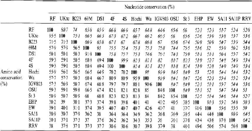
Table 1.1 General characteristics of Group A rotaviruses……….4
Table 1.2 Gene-protein coding assignments of rotavirus………..10
Table 1.3 Levels of nucleotide and amino acid conservation across gene 5 and NSP1 sequences…………...37
Table 2.1.1 List of suppliers used in this study………..48
Table 2.1.2 List of cell lines used in this study………...49
Table 2.1.3 List of Rotavirus strains used in this study………50
Table 2.1.4 List of bacterial strain used in this study………50
Table 2.1.5 List of primary antibodies used in this study……….50
Table 2.1.6 List of secondary antibodies used in this study………..51
Table 2.1.7 List of primers used in this study……….52
Table 2.1.8 List of plasmids used in this study………...56
Firstly, I would like to thank my supervisor Dr Keith Leppard and my previous
supervisor Professor Malcolm McCrae for all their invaluable guidance and support
in every aspect of my work as well as life. Special thanks must also go to all the
present and past members of the rota/adenovirus research group for their assistance
and help in the laboratory over the last few years. It is a pleasure to thank all
members in Warwick Virology Research Group for asking great questions and giving
important feedback throughout the last few years.
I would like to thank all my family, especially my parents for their understanding,
support and love during the entire preparation of this project and thesis. I would also
like to thank all my friends for their patient and support over the last few years,
especially those who have always been supportive under any circumstances. I could
not have done this without them.
I would like to dedicate this thesis to my grandmother, who I lost during the course
I hereby declare that, all the work described in this thesis was undertaken by the
author alone under the supervision of Dr Keith Leppard and Professor Malcolm
McCrae unless explicitly stated in the text. This thesis has not previously been
aa Amino acids
AD Activating domain
Amp Ampicillin
APS Ammonium persulphate
ATP Adenosine triphosphate
bps Base pairs
BSA Bovine serum albumin
C- Carboxyl-
cDNA Complementary DNA
cpe Cytopathic effect
cryoEM Cryo-electron microscopy
DBD DNA binding domain
dH2O Distilled water
DMEM Dulbecco’s modification of Eagle’s minimal essential medium
DMF Dimethylformamide
DNA Deoxyribonucleic acid
dNTP Deoxynucleotide triphosphate
ds Double-stranded
DTT Dithiothreitol
E.coli Escherichia coli
EDTA Ethylenediamine tetracetic acid
ELISA Enzyme-linked immunosorbent assay
EM Electron microscopy
ER Endoplasmic reticulum
FCS Foetal calf serum
GFP Green fluorescent protein
GMEM Glasgow modification of Eagle’s minimum essential medium
IEM Immunoelectron-microscopy
IF Immunofluorescence
IP Immunoprecipitation
IPTG Isopropyl β-D-1-thiogalactopyranoside
m.o.i. Multiplicity of infection
MOPS 3-(N-morpholino) propanesulphonic acid
mRNA Messenger RNA
N- Amino-
NLS Nuclear localisation signal
NSP Non-structural protein
NP40 Nonidet P40
OD Optical density
ONPG O-nitrophenyl β-D-galactopyranoside
ORF Open reading frame
PABP Poly A binding protein
PAGE Poly-acrylamide gel electrophoresis
PBS Phosphate buffered saline
PCR Polymerase chain reaction
Pfu Plaque forming unit
RNA Ribonucleic acid
rpm Revolutions per minute
SAP Shrimp alkaline phosphatase
SD Standard deviation
SDS Sodium dodecyl sulphate
ss Single-stranded
SV40 Simian virus 40
TBE Tris borate EDTA
TBS Tris-buffered saline
TEMED N,N,N’N’-tetramethylethylenediamine
Tris Tris-(hydroxymethyl)-methylamine
UTR Untranslated region
VP Viral protein
v/v Volume to volume
WT Wild type
NSP1, a non-structural protein encoded by rotavirus gene segment 5, has been suggested as a virulence determinant for rotavirus and to function as an antagonist of the interferon signalling pathway. Although non-essential for rotavirus replication in cell culture, and is the least conserved in all rotavirus proteins, NSP1 from different rotavirus strains of different species has been demonstrated to interact with several cellular proteins involved in the IFNβ induction pathway. NSP1 from a bovine rotavirus strain (UKtc) has been shown to interact with and to degrade IRF3 in a proteasome dependent manner whereas NSP1 from a porcine rotavirus strain (OSU) fails to target IRF3 but is able to interfere with IFNβ production via similar targeting of β-TrCP.
The research presented in this thesis sought to gain a better understanding of the molecular determinants of NSP1 specificity for targeting the IFNβ pathway by mapping the regions in NSP1 sequences responsible for targeting specific cellular proteins.
NSP1 hybrid constructs with sequences from both UKtcNSP1 and OSUNSP1 were generated and their interactions with both IRF3 and β-TrCP were tested in a series of assays. The initial attempts to map interaction sites using the mammalian two-hybrid assay were not successful. No reporter plasmid signal was generated indicating the expected interaction. The failure of this assay might be due to the insufficient
expression of the NSP1 proteins as subsequent modification of the expression vector was shown to improve the expression level of NSP1 proteins in subsequent reporter assay analysis.
Using IFNβ promoter reporter assays to demonstrate the functional consequence of NSP1 action in IRF3, it was found that the constructs containing the entire C-terminal part of UKtcNSP1 were able to reduce IRF3-induced IFNβ promoter activity. Such constructs also caused IRF3 degradation in a proteasome dependent manner in agreement with previous studies. However, the sequence containing the last 135 amino acids from UKtcNSP1 was not sufficient for these activities.
Collectively, these data suggested that the sequence between amino acid position 165 and 135 from the C-terminus are required for this interaction and subsequent
degradation of IRF3.
Similar experiments focused on determining the interaction site for β-TrCP on NSP1 were more difficult to interpret according the data presented. Unexpectedly in the light of published data, not only OSUNSP1 was able to degrade β-TrCP but UKtcNSP1 appeared to have the similar effect, as well as two reciprocal pairs of NSP1 hybrid constructs.
In summary, it appears that sequences from the C-terminal part of UKtcNSP1 can function in a heterogeneous NSP1 context to target IRF3 from human cells. Further analysis is clearly required to fulfil the understanding of the role of NSP1 in rotavirus pathogenesis, including its interaction with β-TrCP.
Chapter 1
1.1 Rotavirus
1.1.1 The discovery of rotavirus
Acute diarrheal diseases have been one of the major causes of death in children, and
the young in a wide range of other mammalian and avian species worldwide.
Diarrheal disease is one of the most important causes of death in children,
particularly in those less than five years old in developing countries; in developed
countries, the mortality rate is considerably lower however, it still represents a
common infirmity, especially among the youth (Monto et al., 1983; Estes and
Kapikian, 2007; Martella et al., 2010).
Until the early 1970s, no important causative viral factors related to infectious
diarrheal diseases had been discovered (Yow et al, 1970). The discovery of the
Norwalk virus associated with viral gastroenteritis in older children and adults was a
big breakthrough, revealing the important role of specific viruses in causing
infectious diarrheal diseases (Kapikian et al., 1972). In 1973, another viral particle of
about 70nm in diameter that was associated with severe diarrhea in infants and young
children was directly visualized in duodenal mucosa by thin-section electron
microscopy (EM), and due to the wheel-like structure observed it was subsequently
designated rotavirus (rota: Latin for wheel) (Bishop et al., 1973; Flewett et al., 1974;
Bishop et al., 1974). Human rotaviruses were then linked to previous discoveries of
viral particles causing severe diarrhea in infant mice (EDIM), calves (NCDV) and
monkeys (SA11) (Adams et al., 1963; Mebus et al., 1971; Malherbe and
Investigators from many countries also reported the detection of rotavirus and it was
soon found that rotaviruses were the most important etiologic agents of acute viral
gastroenteritis in infants and young children, causing more than half a million deaths
annually (Parashar et al., 2006). Globally almost every child has experienced
rotavirus gastroenteritis by the age of 5, and children in the poorest areas of the
world, such as South-East Asia and sub-Saharan Africa account for more than 80%
of rotavirus related deaths (Ramig, 2004; Bishop, 2009).
1.1.2 Classification of rotaviruses
Rotavirus is one of the 16 recognized genera of the family Reoviridae, together with
Aquareovirus, Cardoreovirus, Coltivirus, Crabreovirus, Cypovirus, Dinovernavirus,
Fijivirus, Idnoreovirus, Mimoreovirus, Mycoreovirus, Orbivirus, Orthoreovirus,
Oryzavirus, Phytoreovirus and Seadornavirus (Attoui et al, 2006; Estes and
Kapikian, 2007; Deng et al, 2012). Members of the family Reoviridae are the largest
and most diverse group of double-stranded RNA (dsRNA) viruses in terms of host
range. All members of the Reoviridae family have dsRNA genomes consisting of
10-12 segments and all of them have RNA-dependent RNA polymerase activity
associated with the core of the virion, as cells do not have enzymes capable of
transcribing dsRNA templates. The transcription process of the Reoviridae family is
conserved, such that it uses template dsRNA contained within the viral particles
rather than free RNA (Patton, 1986; Estes and Kapikian, 2007).
tests such like immunofluorescence (IF), enzyme-linked immunosorbent assay
(ELISA) and immunoelectron-microscopy (IEM) (Pedley et al., 1986; Mattion et al.,
1994; Estes and Kapikian, 2007). Rotaviruses group A, B and C are found in humans
and other animals; human rotavirus infections are predominantly caused by group A
rotaviruses (Parashar et al., 2003), group B rotaviruses associated with epidemics of
severe diarrhea was primarily reported in adults in China (Chen et al,. 1985; Hung et
al, 1983) and group C rotaviruses have been reported to cause sporadic cases of acute
diarrhea in children older than 3 years of age (Von Bonsdorff and Svensson, 1988).
Rotaviruses of groups D, E, F, and G have been found only in other animals to date
(Saif et al,. 1994). The genome of an avian group D rotavirus strain has been
reported but much less data has yet been published on rotavirus groups E-G (Trojnar
et al., 2010; Wakuda et al., 2011). The focus of this thesis is the Group A
rotaviruses, which have been studied in more details than any other group. The
[image:18.595.115.521.558.766.2]general characteristics of Group A rotaviruses are summarised in Table 1.1.
Table 1.1 General characteristics of Group A rotaviruses (Adapted and summarised from Estes and Kapikian, 2007)
Structure
1. Mature virions are approximately 70nm (100nm including VP4 spikes) in diameter and possess a triple layered icosahedral protein capsid
2. 60 protein spikes extend from the outer layer of the particle 3. Capsid contains all enzymes for mRNA production
Genome
1. 11 segments of double-stranded RNA and capable of genetic reassortment with co-infecting members of the same group
2. Each RNA segment codes for at least one protein
Replication
1. Cytoplasmic replication in infected cells
2. Viral cultivation in vitro is facilitated by treatment of mature virus with proteolytic enzymes
Group A rotaviruses are classified further into two independent serotypes, i.e:
G-serotype and P-G-serotype (Hoshino and Kapikian, 1996). Serotypes are defined by
reactivity of viruses in plaque reduction or fluorescent foci reduction neutralization
assays in antibody-negative animals (Estes and Kapikian, 2007). These assays
measure the reactivity of antibody against the two neutralizing antigens VP7 and
VP4 (Hoshino and Kapikian, 1996). At least 16 VP7 (G) serotypes have been
identified and strains of animal and human origin may fall within the same serotype.
Among these G serotypes, G1-4 and G9 are the most common human strains
worldwide (Kobayashi et al., 2007; Angel et al., 2007). When studies on the
protease-sensitive protein VP4 revealed the importance of anti-VP4 neutralization
antibody in protection from rotavirus infection in vivo, a serotyping system based on
VP4, named P-serotype, was proposed (Gorziglia et al., 1990). Nevertheless,
discrimination of P-serotypes is more difficult to characterize. Although human
rotavirus P-serotypes can be determined by ELISA with specific monoclonal
antibodies, this method has not become common. So far at least 14 different
P-serotypes have been discriminated (Kobayashi et al., 2007).
Sequence analysis and hybridization assays based on VP7 and VP4 genes are used to
define the genetic typing of rotavirus, i.e: G (VP7)-genotype and P (VP4)-genotype
(Kobayashi et al., 2007). G-genotype has shown a clear correlation with the
serotypes whereas the sero-genotypic correlation has not been clearly defined in
P-typing (Estes and Kapikian, 2007). At least 28 different P-genotypes have been
reported (Kobayashi et al., 2007). To integrate the P-serotype and genotype
Arabic number in brackets is used to indicate genotype; for instance, the human Wa
strain is represented as P1A[8] (Estes and Kapikian, 2007).
1.1.3 Structure of Rotavirus particle
Mature infectious rotavirus particles are approximately 100nm in diameter including
the spikes and possess a triple-layered icosahedral protein capsid, hence these are
called triple-layered particles (TLPs). The TLP is composed of an outer layer with
protein spikes extending from the surface, an intermediate layer and an inner core
layer surrounding the dsRNA genome (Flewett et al., 1973). The smooth outer layer
forming the majority of the outer capsid consists of 780 molecules of the
neutralization antigen VP7 arranged as trimers. 60 spikes composed of dimers of
haemagglutinin VP4 protrude to a length of around 120Å from this smooth surface
(Prasad et al., 1990). This outer layer is lost during the uncoating process to leave
double-layered particles (DLPs), which are described as rough particles and are
transcriptionally active but not infectious (González et al., 1995). The intermediate
layer is composed of 780 molecules of VP6 arranged as trimers (Bican et al., 1982).
This VP6 layer may provide structural integrity to the rotavirus capsid by enhancing
the morphologic homogeneity and long-term stability of the particle (Zeng et al.,
1996). Single-layered particles (SLPs or cores) are seen infrequently; they are
non-infectious and usually consist of the enzymes VP1 and VP3 together with the dsRNA
genome, surrounded by an outer surface layer formed by 120 copies of VP2 (Labbé
et al., 1991; Lawton et al., 1997). A distinctive feature of the viral particle is the 132
aqueous channels that penetrate the virion to link the inner core with the outer
transcription and export nascent RNA transcripts during viral replication (Lawton et
al., 1997; Estes and Kapikian, 2007).
Three-dimensional reconstruction studies of the viral particle using cryo-electron
microscopy (cryoEM) and image processing techniques have provided a detailed
description of the structures of triple-and double-layered rotavirus particles (Li et al.,
2009). Current understanding of virions has been enhanced reaching a resolution of
about 9.5-Å and all three layers establish an icosahedral organization (Figure 1.1).
1.1.4 Rotavirus genome and gene-protein coding assignments
There are 11 segments of dsRNA with sizes ranged from 3302 bps to 667 bps in the
rotavirus genome, with around 25% of it forming a dodecahedral structure (Estes and
Kapikian, 2007). The nucleotide sequence for all eleven RNA segments has been
elucidated for a number of strains and general features have been described.
The open reading frame (ORF) of each segment is surrounded by conserved
untranslated regions (UTRs) of varying length dependent upon the gene segment.
Although some of the genes possess additional in-phase (such as gene 7, 9, and 10),
or out-of-phase (gene 11) ORFs, evidence up to date indicates that all the segments
are monocistronic except gene 11, which encodes two non-structural proteins NSP5
and NSP6 (Mattion et al,. 1991; Rainsford and McCrae, 2007). In general, the gene
sequences of the dsRNA segments are A+U rich (58% to 67%) (Estes and Kapikian,
Figure 1.1 Structure of mature rotavirus particle at 9.5-Å resolution.
VP4 spikes are indicated in red, VP7 outer layer in yellow, VP6 intermediate layer in
blue, VP2 inner layer in green and the orange colour indicates the internal density
consisting of RNA genome and polymerase complex (figure taken from Li et al.,
The positive RNA strand has a conserved 5’ cap sequence m7GpppG(m)Gpy but lacks a 3’ poly-A tail (Imai et al., 1983; McCrae and McCorquodale, 1983); however, the
3’ end of the positive sense strand has the conserved sequence 5’-UGUGACC-3’ as
the minimal essential promoter for the negative RNA strand synthesis (Wentz et al.,
1996). Similar features of the RNA termini are found in the primary structures of the
genomes of other viruses in the family Reoviridae such as orbivirus and in other
virus families with segmented RNA genomes including Orthomyxoviridae and
Arenaviridae (Estes and Kapikian, 2007). The use of polyacrylamide gel
electrophoresis (PAGE) in rotavirus genome analysis is now popular for detecting
viruses and monitoring viral outbreaks and transmissions. The electrophoretic pattern
of group A rotavirus genomes is made up by four high-molecular-weight segments
indicating gene 1-4, two middle-sized segments indicating gene 5 and 6, a distinctive
triplet of segments indicating gene 7-9 and the two smallest segments indicating gene
10 and 11(Estes and Kapikian, 2007). However, in viruses with genome
rearrangements, these typical RNA segments are decreased in concentration or
missing in the electrophoretic profile and are replaced by additional more slowly, or
rarely more rapidly, migrating bands of dsRNA (Desselberger, 1996). Virus isolates
with rearrangements in segments 5, 6, 8, 10 and 11 have been characterized; of these,
segments 5 and 11 appear to be rearranged most frequently (Estes and Kapikian,
2007).
The rotavirus genome segments code for six structural proteins VP1-VP4, VP6 and
VP7 that are found in virus particles and six non-structural proteins NSP1-NSP6,
and McCorquodale, 1983); (b) analysis of reassortant viruses (Kantharidis et al.,
1983; Mason et al., 1983; Liu et al., 1988); and (c) immunological studies with
specific antibodies (Both et al., 1983; Greenberg et al., 1983). The eleven gene
segments and the corresponding viral proteins are summarised in Table 1.2 (Estes
[image:24.595.152.487.320.610.2]and Kapikian, 2007).
Table 1.2 Gene-protein coding assignments of rotavirus. (Adapted from Estes and Kapikian, 2007)
Genome segment Protein product
Location in virus particles
1 VP1 Core
2 VP2 Core
3 VP3 Core
4 VP4 Outer capsid
5 NSP1 Non-structural
6 VP6 Inner capsid
7 NSP3 Non-structural
8 NSP2 Non-structural
9 VP7 Outer capsid
10 NSP4 Non-structural
11 NSP5
NSP6 Non-structural
1.1.5 Viral protein structure and function
A characteristic feature of rotavirus-infected cells is the presence of highly
viroplasms (Petrie et al., 1982; Petrie et al., 1984). Viroplasm formation is typical for
infections by several members of the Reoviridae family and these structures are sites
of rotavirus protein accumulation, particle assembly, RNA packaging, and genome
replication (Patton et al., 2006; Contin et al., 2010).
1.1.5.1 Structural proteins
VP1 has been found to be a minor (~2% of the virion mass) but consistent
component of rotavirus complexes that has polymerase activity (Gallegos and Patton,
1989; Zeng et al., 1996). VP1 is a nucleotide-binding protein that specifically binds
the 3’ end of viral mRNA in the absence of any other proteins. In addition, VP1
appears to be the RNA-dependent RNA polymerase and functions as both the viral
replicase and transcriptase (Patton, 1996).
VP2 forms the inner core layer of the viral particle and constitutes ~12% of the
virion protein (Kumar et al., 1989). It is known to be essential for replicase activity
and is also involved in transcriptase activity (Mansell and Patton, 1990; Estes et al.,
1983). VP2 has been shown to have sequence-independent RNA binding activity and
to associate with both ssRNA and dsRNA, but ssRNA preferentially, which is
thought to facilitate its role in viral replication and encapsidation (Boyle and Holmes,
1986; Labbé et al., 1994).
VP3 is found in early replication intermediates in the viroplasm and associates with
viral mRNA at early stages in virus replication cycle (Gallegos and Patton, 1989). It
al., 1998; Sandino et al., 1994). VP3 is a guanylyltransferase and has a function in
mRNA capping by transferring GMP to the 5’ phosphate end of nascent RNAs
(Pizarro et al., 1991; Liu et al., 1992). VP3 has also been demonstrated to have a
sequence-independent affinity for ssRNA, but not for dsRNA (Patton and Chen,
1999).
VP4 proteins make up the spikes of the outer layer of the viral particle; it is the viral
haemagglutinin and determines the host range and cell tropism (Offit et al., 1986;
Estes and Kapikian, 2007). Proteolytic cleavage of VP4 to produce VP5* and VP8*
potentiates infectivity by greatly enhancing penetration of the virus into cells, but
does not affect absorption on to the cell surface (Kalijot et al., 1988; Fukuhara et al.,
1988).
VP6, which forms the intermediate layer of the virion, is the major structural
component of the virion, forming ~51% of the total protein content (Estes and
Kapikian, 2007). VP6 spontaneously forms trimers which are particularly stable and
contains several highly immunogenic epitopes (Estes et al., 1987).
VP7 forms the smooth external layer of the outer capsid and constitutes ~30% of the
virion protein (Estes and Kapikian, 2007). It is known to be the major neutralisation
antigen and is largely responsible for serotype classification of rotaviruses (Sabara et
al., 1985). VP7 is a glycoprotein with three potential sites for N-linked glycosylation
although only two of these sites appear to be used in different rotavirus strains
(Kouvelos et al., 1984). It is co-translationally glycosylated as it is inserted into the
1.1.5.2 Non-structural proteins
NSP1, the least conserved of all rotavirus proteins, is approximately 55kDa in size.
Immunofluorescent staining of virus infected cells has shown that NSP1 is localized
throughout the cytoplasm and also associated with the cytoskeleton (Hua and Patton,
1994a). By contrast, all other rotavirus proteins are mostly concentrated in the
viroplasms. NSP1 does not appear to be essential for rotavirus replication in cell
culture since viable virus mutants only able to encode a highly truncated form of the
protein can still replicate, although they give smaller virus plaques (Taniguchi et al.,
1996). NSP1 has been suggested as a virulence determinant for rotavirus. This was
initially reported from studies in a mouse model using rotavirus reassortants (Broome
et al., 1993). Moreover, NSP1 functions as an antagonist of the interferon (IFN)
signalling pathway (Arnold and Patton, 2009). Since this thesis is focused on NSP1,
a more detailed discussion of this protein will be presented later in this chapter.
NSP2 accumulates predominantly in the viroplasm in rotavirus-infected cells (Petrie
et al., 1984). It has been shown to interact with NSP5 and to form the viroplasm
structure without the presence of other viral proteins (Fabbretti et al., 1999).
Furthermore, NSP2 is able to interact with the viral polymerase VP1 and it possesses
nucleoside triphosphatase (NTPase), RNA triphosphatase (RTPase) and
helix-destabilizing activity (Kattoura et al., 1994; Taraporewala et al., 1999; Aponte et al.,
1996; Vasquez-Del Carpió et al., 2006).
rotavirus mRNA binding activity (Poncet et al., 1993). NSP3 has also been shown to
interact with the scaffold translation initiation factor eIF4G (Piron et al., 1998).
eIF4G can bring the initiation factor 4E (eIF4E) and the poly A binding protein
(PABP) together to induce circularisation of cellular mRNA for efficient initiation of
translation (Preiss and Hentze, 1998). Due to the interaction of NSP3 with eIF4G, the
PABP and its associated mRNA are evicted from the cellular translation complex
causing in the block of cellular protein synthesis, and suggesting that NSP3 has a role
in ensuring efficient translation of viral mRNA (Piron et al., 1998).
NSP4 is a transmembrane glycoprotein with three hydrophobic amino-terminal
regions maintained in the ER membrane and a hydrophilic carboxy-terminus
extended into the cytoplasm (Ericson et al., 1982; Chan et al., 1988). NSP4 is known
to be cytotoxic and functions as a viral enterotoxin in rotavirus-induced diarrhoea by
inducing calcium-dependent chloride secretion across the mammalian small
intestinal mucosa (Tian et al., 1995; Ball et al., 1996; Zhang et al., 2000). In
addition, NSP4 plays a crucial role in viral assembly and facilitates the budding of
viral DLPs into the ER lumen to form infectious TLPs (Estes and Kapikian, 2007).
NSP5 is the product of the 5’ proximal ORF in gene 11 and is 26 kDa in size. It is a
phosphoprotein that exists in several different phosphorylated forms (Afrikanova et
al., 1996). NSP5 forms homo-dimers and is known to interact with NSP2 to form the
viroplasm (Poncet et al., 1997). Recent studies demonstrated that the interactions of
NSP5-NSP2 and NSP5-VP2 are able to recruit all other viral proteins found in the
viroplasms, suggesting that NSP5 plays an important role in the assembly of
NSP5 has also been shown to be involved in viral replication (Campagna et al.,
2005).
NSP6 is encoded by a second out-of-phase ORF in gene 11 with a protein size of 12
kDa. A relatively low level expression of NSP6 is observed in infected cells and the
protein co-localizes with NSP5 in viroplasms (Mattion et al., 1991; Torres-Vega et
al., 2000). It has been suggested that NSP6 may play non-essential roles in regulation
of the viral replication cycle based on the findings that two viable rotaviruses, the
lapine Alabama strain and the porcine OSU strain, encode truncated NSP6 (Gorziglia
et al., 1989; González and Burrone, 1989).
1.1.6 Rotavirus replication cycle
Rotavirus has a rapid replication cycle in permissive cells with a maximum yield of
virus after 10-12 hours (McCrae and Faulkner-Valle, 1981). Although the natural cell
tropism for rotavirus is the differentiated enterocyte in the small intestine, suggesting
that specific receptors for rotavirus are expressed in these cells, recent investigations
have also recognised extraintestinal spread of rotavirus (Azevedo et al., 2005; Blutt
et al., 2006; Kim et al., 2011). Studies of the initial stages of rotavirus replication
have been primarily conducted in continuous cell cultures derived from MA104
(monkey kidney epithelial cell line) and Caco-2 (polarized human colon carcinoma
cell line). Both of the cell lines are highly permissive for most of the mammalian
rotavirus infection and Caco-2 cells in particular are thought to reflect the in vivo
The general features of rotavirus replication based on studies in cultures of MA104
are summarised as follows (Estes and Kapikian, 2007), and a schematic
representation of the virus replication cycle (Figure 1.2) demonstrates the process known so far (Trask et al., 2012).
(a) Cultivation of most rotavirus strains requires the addition of exogenous
proteases to the culture medium to ensure the activation of viral infectivity by
cleaving the outer capsid protein VP4.
(b) Replication is entirely cytoplasmic and occurs within cytoplasmic viroplasms.
(c) Host cells do not contain enzymes to replicate dsRNA; thus rotavirus supplies
the necessary enzymes.
(d) Viral mRNA transcripts are used both to produce proteins, and as a template
for negative stranded RNA synthesis.
(e) Replication occurs in nascent subviral particles and during the replication,
both parental dsRNA remain associated with the partially uncoated viral
particle, free dsRNA or free negative stranded RNA is never found in
infected cells.
(f) Subviral particles are produced in association with viroplasms and the
progeny virus particle matures by budding through the membrane of the ER
and is then released by cell lysis.
(g) Intracellular calcium levels are important for controlling virus assembly and
Figure 1.2 The rotavirus replication cycle.
The rotavirus virion first attaches to the target cell followed by virion delivery to the
early endosome. The incoming TLP is then uncoated by losing the VP7 outer layer
partially and subsequently releases the DLP into the cytosol. This activates the
internal polymerase VP1 and VP3 complex to transcribe capped positive-sense
RNAs, which then serve either as mRNAs or as templates for synthesis of
negative-sense RNAs. When VP1 and VP3 bind the viral positive-negative-sense RNAs, the genome
packaging is initiated. Condensation of VP2 triggers dsRNA synthesis. VP6 then
assembles onto the nascent core to form the progeny DLP, followed by the assembly
of the outer capsid. Through an undefined mechanism, the DLP-VP4-NSP4 complex
buds into the ER. Subsequent removal of the ER membrane and NSP4 permits
assembly of the ER-resident outer capsid protein VP7, thus the formation of the TLP.
Release from the infected cell exposes the virion to trypsin-like proteases, resulting
in the specific cleavage of VP4 to produce the fully infectious virion (figure taken
1.1.6.1 Virus attachment and entry
Viral proteins and host cell surface molecules are essential for rotavirus infections. A
broad range of cells can be infected by rotaviruses with different efficiencies
(Crawford et al., 2006). The current opinion on rotavirus cell entry is that it is a
coordinated, multistep process involving sequential interactions with several ligands
and a series of conformational changes in the capsid proteins VP4 and VP7 (Estes
and Kapikian, 2007). It has been demonstrated that the proteolytic cleave of VP4 and
the glycosylation of VP7 are not essential for the initial binding process (Fukuhara et
al., 1988). A variety of cellular receptors have been reported to be involved in the
binding process: N-acetylneuraminic acid (sialic acid), which functions as an
attachment receptor for some rotavirus strains; the heat shock cognate protein hsc70,
and four integrins namely α2β1, ανβ3, α4β1 and αxβ2 (Lόpez and Arias, 2006).
Observations on cells pre-treated with neuraminidase (NA), or pre-incubation of
virus with sialic acid-containing compounds, showed reduced infectivity of some
viral strains, indicating that sialic acid is able to mediate the initial viral attachment
to susceptible cells (Lόpez and Arias, 2006). However, most human rotavirus isolates
are not affected by prior NA treatment of cells; they can still use sialic acid located in
the internal regions of oligosaccharide structure such like ganglioside GM1 for viral
recognition (Lόpez and Arias, 2006; Haselhorst et al., 2009). To date four integrins
including α2β1, ανβ3, α4β1 and αxβ2, have been identified to function at the
post-attachment level (Lόpez and Arias, 2006). Studies have shown that VP4 from a
number of rotavirus strains, including those from humans, monkeys and calves, was
viral protein responsible for ανβ3 integrin binding was identified to be VP7 (Graham
et al., 2003; Zárate et al., 2004). In addition, integrins α4β1 and αxβ2 have also been
reported to promote post-attachment steps in infections by interactions with both
VP4 and VP7, and VP7 alone respectively (Graham et al., 2005; Fleming et al.,
2007; Graham et al., 2003). Furthermore, studies on Hsc70 have shown that it
promotes viral entry at a post-attachment step by interacting with VP4 and VP6
(Gualtero et al., 2007).
The entry of the viral particles into host cells is driven by the interaction between the
virus and its receptors on the cell surface. Observations made with EM of
rotavirus-infected cells suggested that the virus internalization was achieved via endocytosis
(Ludert et al., 1987). It has been reported that an inhibitor of the vacuolar
proton-ATPase pump, bafilomycin A, is able to block rotavirus infectivity, supporting the
endocytotic mechanism for virus entry (Chemello et al., 2002). However, studies
have also revealed that rotavirus infectivity is not inhibited by either preventing the
acidification of endosomes or by treatment with drugs that block the intracellular
traffic of the endocytic vesicles, these results suggested that the endocytotic pathway
might not to be universal for cell entry of all rotaviruses (Lόpez and Arias, 2006).
Instead, another possible mechanism by direct penetrating of cell membrane was
proposed for rotavirus internalization. This was based on the observation that
rotavirus infection induced a rapid permeabilization of the cell membrane (Estes and
Kapikian, 2007). In summary, it is possible that different viral strains may utilize
different mechanisms for their internalization and the precise mechanisms for each
The uncoating process of rotaviruses includes the removal of the outermost layer
made from VP4 and VP7. It has been proposed that these proteins are solubilized
within an endocytic vesicle because of the low calcium concentration, and this was
supported by the inhibition of uncoating of the porcine rotavirus strain OSU in
MA104 cells by the calcium ionophore A23187 (Ludert et al., 1987). However,
different observation have been made by Cuadras et al, who showed that the
uncoating of rhesus rotavirus strain RRV and monkey rotavirus strain SA11 were not
affected by the use of compounds which increased the intracellular calcium level
(Cuadras et al., 1997). Therefore, the importance of calcium levels for viral entry
might also vary between the different viral strains used in different studies.
1.1.6.2 Transcription and translation
The virus-associated transcriptase in TLPs can be activated in vitro by treatment with
a chelating agent or by heat shock treatment (Cohen and Dobos, 1979; Spencer and
Arias, 1981). Such treatments result in the conversion from TLPs to DLPs by losing
the outer capsid proteins and subsequently permit transcription to proceed (Estes and
Kapikian, 2007).
Synthesis of viral transcripts is initiated by the endogenous viral RNA-dependent
RNA polymerase VP1 which utilizes viral dsRNA as template to generate positive
sense ssRNA (Lawton et al., 1997). The nascent RNA transcripts are capped at their
5’ end by the virion-associated guanylyl and methyltransferase activities present in
through channels in the VP2 and VP6 layers; multiple mRNA transcripts can be
released simultaneously from an actively transcribing particle (Lawton et al., 2001).
Each genome segment is transcribed by a specific polymerase complex, and the
resulting transcripts exit through the channel system at the axis adjacent to their site
of synthesis. The positive-stranded RNA transcripts encode the rotavirus proteins and
also serve as templates for the negative stranded RNAs synthesis to make progeny
dsRNAs (Estes and Kapikian, 2007).
Viral mRNAs released from DLPs are not polyadenylated but capped, and the
cellular translation machinery is used to translate viral proteins, with cellular
ribosomes being recruited for synthesis of all 12 viral proteins (Patton et al., 2006).
Viral protein translation is facilitated by the action of NSP3, which has been shown
to have an important role in shutting off cellular protein synthesis and enhancing
viral protein synthesis by interacting with eIF4G and with RoXaN (rotavirus X
protein associated with NSP3). RoXaN is a novel cellular protein containing at least
five zinc binding domains, a paxillin leucine-aspartate repeat (LD) motif facilitating
protein-protein interactions, and another protein-protein interaction domain called the
tetratricopeptide repeat region (TRR) (Vitour et al., 2004). Complex consists of
NSP3, RoXaN and elF4G can be detected in rotavirus-infected cells, indicating that
RoXaN is involved in translation regulation (Patton et al., 2006; Harb et al., 2008). It
has been shown that viral proteins are neither synthesized in equal amounts nor are
the amounts in each case similar to those of their corresponding gene transcripts
(Johnson and McCrae, 1989). VP6 and NSP4 were found to be synthesized in the
were the least actively produced (Johnson and McCrae, 1989; Rainsford and
McCrae, 2007).
1.1.6.3 Genome replication
The viroplasm structures provide the sites for synthesis of viral RNAs and formation
of DLPs (Petrie et al., 1984; Fabbretti et al., 1999). The kinetics of RNAs synthesis
in infected cells showed that positive- and negative- stranded RNAs were initially
detected 3 hours after infection (Stacy-Phipps and Patton, 1987). It is assumed that
rotavirus RNA replication takes place in a conservative manner; both strands of
parental dsRNA remain within partially uncoated particles, after synthesis, dsRNA
remains associated with subviral particles, and free dsRNA is not found in cells
(Patton et al., 2006). Studies in SA11 infected cells revealed that the replicase
complexes consist of the core proteins (VP1 and VP3), small amounts of VP6, large
amounts of NSP3, and relatively less amounts of NSP1 and NSP2
(Helmberger-Jonew and Patton, 1986) however, the role of individual proteins in RNA genome
replication will be understood better if they are studied in vivo with pure species of
native rotavirus proteins (Estes and Kapikian, 2007). siRNA knock down
experiments demonstrated that NSP4 plays a role in regulating mRNA synthesis and
possibly genome encapsidation although the mechanism remains to be determined
(Silvestri et al., 2005). In addition, a recent study showed that the replication
efficiency of rotavirus RRV in MA104 cells is dependent on the
1.1.6.4 Virus assembly and release
Rapid assembling of DLPs serves as an intermediate stage in the formation of TLPs
(Estes and Kapikian, 2007). Viral cores including VP1, VP2 and VP3, are
accumulated in viroplasms (Korolev et al., 1981). The assembly of VP1 and VP2 is
dependent on both their high affinity for ssRNA and their interaction with NSP2 and
NSP5 (Patton et al., 2006; Arnoldi et al., 2007). VP2 forms the structural basis of the
viral core and its simultaneous association with VP1 and VP3 directs the formation
of a functional viral core (Zeng et al., 1998). During DLP formation in infected
mammalian cells, VP6 has been observed in the exterior region of viroplasms, which
allows VP6 to associate with newly assembled viral cores to form DLPs as they
move toward the periphery of viroplasms (López et al., 2005). The glycoproteins
VP7 and NSP4 are synthesised on ribosomes associated with the membrane of the
ER. NSP4 acts as an intracellular receptor on the ER membrane; it binds newly made
DLPs released from viroplasms and mediates the budding of DLPs into the ER
lumen. A receptor role for NSP4 is supported by the observation that DLPs bind to
ER membranes containing only NSP4 (Meyer et al., 1989; Au et al., 1989),
suggesting that NSP4 alone was sufficient to drive DLPs into the ER lumen.
It has been observed that rotavirus maturation is calcium-dependent. Studies revealed
decreased rotavirus production in cells maintained in calcium-depleted medium, and
these viruses do not bud into the ER (Shahrabadi and Lee, 1987; Poruchynsky et al.,
1991). In undifferentiated cells such as MA104, EM studies have shown that the
cells like Caco-2, progeny virions are selectively released through the apical
membrane involving actin and lipid rafts (Jourdan et al., 1997; Chwetzoff and
Trugnan, 2006; Gardet et al., 2007).
1.1.7 Rotavirus pathogenesis
Rotavirus replicates in non-dividing, mature enterocytes near the tips of the villi.
Pathologic changes of infections caused by rotaviruses are mainly limited to the
small intestine; nevertheless, the severity of infection varies among animal species
across different studies (Burke and Desselberger, 1996). The current understanding
of rotavirus pathogenesis is based primarily on working with different animal
models, in which rotavirus infection is associated with several of disease including
no visible lesions, slight lesions such as enterocyte vacuolization and larger lesions
such as crypt hyperplasia and villus blunting (Ramig, 2004).
Pathogenesis of rotavirus infection is multifactorial, both host and viral factors can
affect the consequences of infection. There are some major host factors recognised to
affect the severity of rotavirus-associated disease: (i) the age of the animals at
infection can result in different clinical symptoms including biliary atresia, diarrhea
and some extraintestinal replication of virus (Estes and Kapikian, 2007); (ii)
increased severity of rotavirus diarrhea can be caused by malnutrition as this delays
the recovery of the small intestines (Ramig, 2004); and (iii) the expression of
intestinal mucins and the rate of epithelial cell replacement can affect rotavirus
infection in the host (Ramig, 2004). Furthermore, the enteric nervous system (ENS)
diarrhoea (Lundgren et al., 2000). A recent study analysed the total gene expression
profile of HT29 cells infected with either simian (SA11), bovine (A5-13) or human
(Wa) rotavirus strains using microarrays. 131 genes involved in innate immune
responses, stress responses, apoptosis and protein metabolism were similarly induced
by all three strains (Bagchi et al., 2012). In terms of the viral factors, studies on
rotavirus-infected intestinal epithelial cells showed that the infection is different
depending on whether or not the virus requires sialic acid for initial binding (Estes et
al., 2001).
1.1.8 Rotavirus vaccines
Three live rotavirus vaccines have been developed, Rotashield (Wyeth), RotaTeq
(Merck) and Rotarix (GSK). Both Rotashield and RotaTeq are reassortant viruses
based on simian and bovine rotaviruses respectively with gene segments encoding
VP4 and VP7 derived from human rotaviruses (Estes and Kapikian, 2007). By
contrast, Rotarix is a human rotavirus that was attenuated by passaging in cell
culture. RotaShield was the first of these vaccines to be licensed in the United States
in 1998 although was withdrawn subsequently because the detection of a temporal
association between vaccine administration and gut intussusception (Simonsen et al.,
2005). The other two rotavirus vaccines were released in 2006 and no similar link
with intussusception has been observed (Angel et al., 2007). Ongoing vaccination
programs in general show great reductions in clinical burden by both of the vaccines.
However, despite the success of vaccination programs in the US, Australia and some
from emerging manufacturers may be on the market for the next decade, providing
competition and potentially reducing the cost of the program (Weycker et al., 2009;
Lopman et al., 2012).
1.2 Innate immune system
1.2.1 Overview
The body is protected from infection by a variety of effector cells and molecules
involved in the immune system. An adaptive immune response has its specificity as
the production of antibodies is against a definite pathogen or its products; and an
adaptation to infection with that pathogen is developed through the lifetime of an
individual. On the other hand, the innate immune response can be immediately
activated against a wider range of pathogens but has no specificity for any individual
pathogen and does not lead to lasting immunity (Biron et al., 2002). The studies
included in this thesis are mainly focused on innate immunity.
The first line of defence against an invading pathogen is the innate immune response.
Synthesis and secretion of type I interferons (IFN) can exhibit antiviral,
anti-proliferative and immunomodulatory functions and provide a vital aspect of the
antiviral innate immune response (Honda et al., 2005). Innate immunity is based on
an intricate system of host pattern recognition receptors (PRRs) that specifically
recognize pathogen-associated molecular patterns (PAMPs) (Janeway, 1989; Akira et
al., 2006; Unterholzner and Bowie 2008). PRRs are germline-encoded, non-clonal,
leading to different anti-pathogen responses. Four different classes of PRRs found to
date include Toll-like receptors (TLRs), retinoic acid-induced gene-1 (RIG-I)-like
receptors (RLRs), nucleotide oligomerization domain (NOD)-like receptors (NLRs)
and C-type lectin receptors (CLRs). The first three of these PRRs are able to
recognise viral nucleic acids (Geijtenbeek et al., 2009a; Ting et al., 2005; Eisenächer
and Krug, 2012) and among them, TLRs and RLRs are important for producing
various cytokines and type I interferns (IFNs), while NLRs are able to regulate
interleukin-1b (IL-1b) maturation by activating caspase-1 (Pétrilli et al., 2007a).
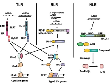
Figure 1.3 illustrates these three PRRs for RNA virus recognition (Takeuchi and
Akira, 2009).
Toll-like receptors (TLRs) are transmembrane proteins and they are the mammalian
homologs of Toll, which was discovered to mediate recognition of pathogens by the
innate immune system in Drosophila species (Lemaitre et al., 1996). There are at
least 13 identified mammalian TLRs that share similar extracellular and intracellular
domains (Brikos and O’Neil, 2008). The molecular basis of TLR signalling is
dependent on the conserved part of their intracellular domain called the
Toll/interleukin-1 receptor (TIR) domain, which functions as a linker to connect to
the membrane. The glycosylated extracellular domain consists of leucine-rich repeats
(LRRs) and they are connected to the cytoplasm by a hydrophobic transmembrane
sequence (Gay and Gangloff, 2008). Although historically the role of TLRs in the
host immune response to bacteria and fungi was initially more apparent, the more
significant role of TLRs to virus infections has been established as several viral
Based on the similarities on the sequence, structure and function, a subgroup of
TLRs including TLR3, TLR7, TLR8 and TLR9 has been identified to recognize
nucleic acids (Hornung et al., 2008). ssRNA, anti-viral drugs and short dsRNA
containing certain sequence motifs can be sensed by both TLR7 and TLR8 (Heil et
al., 2004; Hornung et al., 2005; Diebold et al., 2004; Uematsu and Akira, 2008).
dsRNA viruses and synthetic analogues of dsRNA (Poly(I:C)) are recognised by
TLR3 (Alexopoulou et al., 2001; Beutler et al., 2007), and the ligand for TLR9 is the
DNA-containing CpG motifs (Hemmi et al., 2000). In addition, a number of specific
viral structural proteins have been shown to activate signal transduction through
other TLRs, for instance, envelope proteins of Measles virus can activate the signal
transduction through TLR2 and F protein of respiratory syncytial virus can trigger
Figure 1.3 Three classes of PRRs for RNA virus recognition.
ssRNA from viruses is recognized by TLR7 in plasmacytoid dendritic cells (pDCs),
whereas dsRNA is detected by TLR3 in conventional dendritic cells (cDCs). TLR7
and TLR3 trigger signaling cascades via the myeloid differentiation factor 88
(MyD88) and Toll/IL-1 receptor (IL-1R) homology domain-containing adapter
inducing IFN- β (TRIF), respectively. RIG-I and MDA5 recruit another adapter
protein, IFN-β promoter stimulator-1 (IPS-1). TRIF and IPS-1 share signaling
molecules for phosphorylation of IRF3 and IRF-7 by TBK1/IKK-i.
MyD88-dependent signaling directly activates IRF-7 in pDCs. Phosphorylated IRF-3 and
IRF-7 activate the expression of type I IFN genes. Simultaneously, TLRs and RLRs
induce the translocation of NF-κB to induce the expression of cytokine genes.
NALP3, one of the NLR proteins, detects the presence of dsRNA and induces the
catalytic activity of caspase-1 via an adapter, apoptosis-associated speck-like protein
In 2005, a new pathway was identified when the retinoic acid-induced gene-1
(RIG-I)-like receptors (RLRs) was discovered. This pathway recognizes viral nucleic acids
exclusively in the cytoplasm of infected cells and it has been shown to be
independent of the TLR system (Rothenfusser et al., 2005). The RLRs consist of
three cytosolic RNA helicases namely RIG-I, MDA5 (melanoma
differentiation-associated gene 5) and LGP2 (laboratory of genetics and physiology-2), which are all
capable of unwinding dsRNA molecules (Andrejeva et al., 2004; Tanner and Linder,
2001). RIG-I can bind both Poly (I:C) and viral dsRNA and is activated only in the
presence of dsRNA although over-expression of the N-terminal region of RIG-I
containing two tandem caspase recruitment domains (CARDs) is sufficient to
activate IRF3 and NF-κB without viral infection (Seth et al., 2006). In contrast, the
nature of RNAs for MDA5 activation is less understood although it shares the
N-terminal CARDs structure with RIG-I and can also activate the IFNβ promoter
(Meylan et al., 2005). LGP2 doesn’t contain the N-terminal CARDs, but has a
repressor domain at the C-terminus. This domain is able to not only facilitate the
binding of dsRNA but also to inhibit multimerization and the signalling of RIG-I, but
not MDA5, thus to act as a negative regulator of the RIG-I pathway (Yoneyama et
al., 2005; Saito et al., 2007; Pippig et al., 2009).
The transcriptional activation of type I interferon (IFN) genes is a key step in the
mammalian antiviral response. IFN genes include multiple IFNα isoforms, IFNβ and
other members of the type I interferon family such like IFN-ω, ε, and κ (Der et al.,
1998). In humans there are at least 13 subtypes of IFNα (Randall and Goodbourn,
2008), though only a single gene exists for IFNβ. Both IFNα and IFNβ are produced
cells and epithelial cells. The activation of IFN regulatory factors (IRFs) and/or
NFκB is triggered by the engagement of PRRs by PAMPs, which subsequently
induce the production and secretion of type I IFN proteins. The binding of these type
I IFN proteins to their receptors on the surface of both infected and uninfected cells
prompts a cascade of signalling events involving signal transducers and activators of
transcription (STAT) and Janus kinases (JAK) molecules, which can translocate to
the nucleus after phosphorylation. They then induce the expression of many
IFN-stimulated genes (ISG) that encode proteins with antiviral properties to establish an
antiviral state. Importantly, this occurs in both infected and uninfected cells and so is
responsible for helping to control the spread of infection (Takeuchi and Akira, 2007;
Randall and Goodbourn, 2008).
1.2.2 Interferon regulatory factors regulated pathway
Nine interferon regulatory factors (IRF1-9) have been established and are
characterized by a conserved DNA-binding domain near the N-terminus of the
protein (Taniguchi et al., 2001). IRF3-9 have an IRF-associated domain (IAD)
within the carboxy terminus which is important for protein-protein interactions upon
phosphorylation-mediated activation (Taniguchi et al., 2001). The IAD typically
promotes either homodimer or heterodimer formation among IRF family members,
but they can also interact with other protein families such as members of the STAT
family (Taniguchi et al., 2001). Several IRFs have been implicated in type I IFN
Early studies identified that the main steps in the activation of IRF3 and IRF7 are
similar (Servant et al., 2002a). IRF3 is localized to the cytoplasm in a monomeric
and inactivated form in uninfected cells (Mercurio et al., 1997). Subsequent to viral
infection, IRF3 becomes phosphorylated and dimerized (Honda et al., 2006). The
dimers accumulate in the nucleus, bind consensus DNA sequences, recruit histone
acetyltransferases (HATs) to promote gene induction, and then are degraded in a
proteasome-dependent manner (Sato et al., 1998; Yoneyama et al., 1998).
Hyperphosphorylation of serine and threonine residues near the C-terminus of IRF3
by TBK1 and IKKi is necessary for IRF3 activation (Hemmi et al., 2004).
Hyperphosphorylated IRF3 dimers form a complex with histone acetyltransferases
such as CREB-binding protein (CBP) or p300 (Yoneyama et al., 1998; Lin et al.,
1998). Binding both DNA and CBP or p300 results in nuclear retention of IRF3
dimers (Kumar et al., 2000); while in the nucleus, IRF3 is targeted for
proteasome-mediated degradation dependented on a peptidyl-prolylisomerase, PIN1 (Saitoh et
al., 2006). The E3 ubiquitin ligase that directs IRF3 polyubiquitination to signal its
degradation is likely to be a Skp1-Cull1-F-box multiprotein complex, based on the
requirement of cullin 1 for IRF3 degradation (Bibeau-Poirier et al., 2006).
IRF7 has a short half-life, and is only present in high concentrations when a viral
infection is in progress (Taniguchi and Takaoka, 2002). Sequence alignments show
that IRF7 has serine and threonine residue spacing near its C-terminus similar to that
found in IRF3 (Servant et al., 2002b). IRF7 is also activated by
hyperphosphorylation mediated by TBK1 and IKKi similar to IRF3 (Sharma et al.,
2003). The expression of IRF7 in most cells is low and the expression is upregulated
IRF7, was essential for the early phase of type I IFN induction since IRF3-/- mice have an impaired type I IFN response (Sato et al., 1998). However, it has also been
demonstrated that IRF7-/- mice completely lost the ability to express type I IFN (Honda et al., 2005). Therefore, a revised model suggests that IRF3/IRF7
heterodimers or IRF7 homodimers are important for the initial induction of type I
IFN (Honda et al., 2005). Following induced expression of IRF7 by IFN signalling,
IRF7 is the major driver of a positive feedback mechanism that upregulates the
expression of a wide range of type I IFN genes and it is considered the ‘master
regulator’ of the IFN response, as it also induces the expression of IFNα (Honda et
al., 2005).
1.2.3 NFκB regulated pathway
NFκB is a dimeric transcription factor made up with members of the Rel family
including NFκB1 (p105/p50), NFκB2 (p100/p52), Rel (c-Rel), RelA (p65) and RelB.
NFκB is required for the optimal expression of IFN-β (Takeuchi and Akira, 2009).
The activity of NFκB is tightly regulated by the inhibitor IκB. Phosphorylation of
IκB by IκB kinases (IKK) leads to its rapid ubiquitination by the E3 ligase
Skp1/Cull/F-box complex named SCFβ-TrCP and subsequent degradation by the 26S proteasome. This in turn leads to the translocation of NFκB subunits to the nucleus
and promotes binding and transcription of NFκB target genes (Holloway et al., 2009;
Kroll et al, 1999).
Karin, 2006). As a negative feedback loop, activated IKK also phosphorylates the
IKKγ regulatory subunit; this leads to dissociation of IKKγ and conformational
changes in IKKα and IKKβ, which allows for the recognition and dephosphorylation
of the kinase domain serine residues to inactivate the IKK complex (Kray et al.,
2005; Palkowitsch et al., 2008). Once activated, the IKK complex can phosphorylate
three NFκB inhibitor (IκB) proteins, IκBα, IκBβ, and IκBε (Didonato et al., 1997).
Phosphorylated IκB is recognized by the SCFβ-TrCP E3 ubiquitin ligase complex leading to polyubiquitination of IκB, which is subsequently degraded by the
proteasome (Kroll et al., 1999).
The most common NFκB dimer is composed of the p65 and p50 subunits and is
usually inhibited by association with IκBα (Hayden et al., 2008). IκBα in complex
with p65:p50 normally shuttles between the cytoplasm and the nucleus, but is
predominantly found in the cytoplasm (Baeuerle and Baltimore, 1988). The
inhibitory activity of IκBα was initially recognised as a function of maintaining the
cytoplasmic localization of p65:p50, however it was then found that the p65:p50 still
remains mostly cytoplasmic in cells deficient in all three IκB proteins (Tergaonkar,
2006). Robust nuclear accumulation of p65:p50 free from IκB inhibition occurs
when the DNA binding of this dimer is enhanced by phosphorylation of the p65
subunit on serine 276 (Vermeulen et al., 2003). This phosphorylation has been
shown to be essential for p65:p50 interaction with the histone acetyltransferases,
CBP and p300 (Chen et al., 2005). CBP/p300 then acetylates p65 to maximize
transcriptional activity of this complex (Chen et al., 2005). Figure 1.4 summarises
Figure1.4 Signalling pathways triggered by viral infection.
Antiviral signalling is initiated by the recognition of dsRNA in the cytoplasm,
followed by the activation of IRF3 and IRF7 via TBK1-and IKKi-dependent
phosphorylation, and the activation of NFκB via stimulation of phosphorylated IκB.
After translocation of these proteins to the nucleus, the type I interferon promoters