Original Article
Effects of cathepsin B on proliferation, activation, and
melanin synthesis of human hair follicle melanocytes
Yuan Si1, Junbin Zhang2, Jingzhu Bai3, Qun Li3, You Mo3, Jiang Wu3, Ruihua Fang3
1Department of Dermatology, The Third Affiliated Hospital of Sun Yat-sen University, Guangzhou, Guangdong Province, China; 2Department of Orthopedics, The Third Affiliated Hospital of Sun Yat-sen University-Lingnan Hos-pital, Guangzhou, Guangdong Province, China; 3Department of Dermatology, Guangzhou First People’s Hospital, Guangzhou, Guangdong Province, China
Received May 18, 2018; Accepted June 14, 2018; Epub August 15, 2018; Published August 30, 2018
Abstract: Objective: The aim of this study was to investigate the effects of cathepsin B on proliferation, activation, and melanin synthesis of human hair follicle melanocytes (HFM). Methods: HFM from normal people were cultured,
in vitro, and its morphology, proliferation, tyrosinase activity, and melanin synthesis were observed in different con-centrations of cathepsin B (0.3-1.2 ng/mL). Expression of pan-actin protein, after treatment, was detected by cell immunofluorescence assay while proliferation, tyrosinase activity, and melanin synthesis were detected via methyl thiazolyl tetrazolium method. Results: After treatment with cathepsin B for 7 days, the dendrites of HFM proliferated more in number and length compared to the control group. Expression of pan-actin and melanin was significantly higher than those in control group (both P<0.01), accompanied with increased tyrosinase activity, especially at a concentration of 0.6 ng/mL. Conclusion: Cathepsin B can induce proliferation of human HFM while increasing ty-rosinase activity and melanin synthesis.
Keywords: Cathepsin B, human hair follicle melanocyte, proliferation, tyrosinase activity, melanin synthesis
Introduction
Reduction of pigmented melanocytes in hair follicles is the main cause of graying hair. Recovery of hair follicle melanocytes (HFM) is due to the migration of melanocytes with undamaged outer root sheaths from the middle and lower part of the hair follicle to the upper part, where cathepsins are located [1-4]. Cathepsin B, one of the cysteine proteases, is 80% homology of cathepsin Land and is found in hair follicles from outer root sheaths to inner root sheaths, as well as in HFM [4]. In addition, cathepsin B acts as a proapoptotic mediator in human epidermal melanocytes exposed to Ultraviolet A/B when it is released from lyso-somes [5].
Whether cathepsin B plays a role in human HFM remains unknown. In this study, the effects of cathepsin B’s overexpression or knock-down on morphology, proliferation, ty- rosinase activity, and melanin content of HFM were observed.
Materials and methods
Main reagents and instruments
the immunofluorescence microscope was ob- tained from Beijing Liuyi, China.
Cell culture
Human HFMs were separated from a healthy donor’s scalp via the separate method. They
was washed by PBS 3 times, 10 minutes each time. The primary antibody mouse anti-pan actin immunoglobulin G (diluted with 1% FBS PBS, 1:500) was added and incubated at room temperature for 16 hours. It was cleaned with 0.1% tween 20/PBS 4 times, 10 minutes each time. Goat anti-mouse secondary antibody con-Figure 1. Morphologic changes of hair follicle melanocytes (400×); A. control
[image:2.612.90.376.72.339.2]group; B. 0.3 ng/mL cathepsin B group; C. 0.6 ng/mL cathepsin B group; D. 1.2 ng/mL cathepsin B group.
Figure 2. The number of hair follicle melanocytes dendrites in each group compared with the control group, HFM dendrites in the cathepsin B groups increased and the number of dendrites was highest in the 0.6 ng/mL ca-thepsin B group.
were cultured with MEM with penicillin 100 IU/mL, strepto-mycin 100 IU/mL, sodium bicarbonate 2 g/L, 4-(2-hy- droxyethyl)-1-piperazinee-thanesulfonic acid 2.98 g/L, sodium pyruvate 0.24 g/L, and 10% FBS. Cells were taken out from liquid nitrogen and warmed in a water ba- th at 37°C, immediately [6]. Next, cell suspension was transferred into the culture dish with RPMI1640 medium and cultured in an incubator containing 5% carbon dioxide. Afterward, the medium was changed every 2 days and regular subculture was car-ried out.
Morphology observation
Final concentrations of ca- thepsin B were set at 0.3 ng/ mL, 0.6 ng/mL, and 1.2 ng/ mL, respectively. In the con-trol group, only phosphate buffer solution (PBS) was ad- ded in the culture medium. After culturing for a week, the morphology of HFM was observed by a high-powered microscope.
Effects of cathepsin B on ex-pression of pan-actin protein
[image:2.612.91.372.405.580.2]jugated fluorescein isothiocyanate (diluted with 1% FBS PBS, 1:250) was added and incubated for 2 hours at room temperature, then washed 3 times with TPBS, 10 minutes each time. Finally, it was cleaned 3 times with 0.9% sodi-um chloride solution, 10 minutes each time. Fluorescence intensity was analyzed by image J.
Detection of proliferation, activation, and melanin synthesis
HFM proliferation measurement: proliferation rate was calculated by MTT assay with the fol-lowing formula [6]. Proliferation rate = (A490 with methoxsalen - A490 without methoxsa- len)/(A490 without methoxsalen - A490 with medium) *100%.
Measuring method of tyrosinase activity re- ferred to the report by Ando et al. [7]. Tyrosinase activity = A475 in cathepsin B group/A475 in control group *100%.
Measuring method of melanin content referred to reports by Ando et al. [7, 8]. Melanin content
HFM morphologic changes
After 1 week of culturing, the number of den-drites of HFM at each concentration of cathep-sin B group was significantly increased. Cell bodies were also increased. Pigment granules were observed in the cytoplasm under a high-powered microscope, especially in the 0.6 ng/ mL cathepsin B group. See Figures 1, 2.
Pan-actin protein expression
Expression of pan-actin protein was detected by immunofluorescence and mainly expressed in the cell membrane. Compared with the con-trol group, expression of pan-actin in cathepsin B groups increased, it was the highest at 0.6 ng/mL cathepsin B. See Figures 3, 4.
HFM proliferation
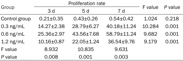
[image:3.612.87.524.72.165.2]Obvious proliferation of HFM could be observ- ed at 3 days, 5 days, and 7 days (all P<0.01). Compared with the other three groups, effects reached the maximum in the 0.6 ng/mL cathepsin B group (compared with 0.3 ng/mL Figure 3. Expression of pan-actin protein detected by immunofluorescence (400×); A. Control group (0 ng/mL ca-thepsin B); B. 0.3 ng/mL caca-thepsin B group; C. 0.6 ng/mL caca-thepsin B group; D. 1.2 ng/mL caca-thepsin B group. The red immunofluorescence indicated pan-actin protein.
Figure 4. Optical density analysis of immunofluorescence in each group compared with control group (0 ng/mL cathepsin B), expression of protein in cathepsin B groups all increased and was the highest in 0.6 ng/mL cathepsin B group.
= A400 in cathepsin B group/A400 in control group * 100%.
Statistical processing
Observation results were statistically ana-lyzed by SPSS23.0 software. All measure-ment data are expressed as mean ± stan-dard deviation. One-way analysis of va- riance and Bonferroni’s post hoc tests were used for comparison among groups. Enumeration data are expressed as num-ber and rate and comparison among groups was tested by χ2. Differences were statistically significant when P<0.05.
[image:3.612.92.323.226.368.2]group, P = 0.003, compared with 1.2 ng/mL group, P = 0.001, and compared with the con-trol group, P = 0.001). Proliferation rates in the 3 groups all evidently increased with prolong-tion of time (all P = 0.001). See Table 1.
Tyrosinase activity
After treatment with cathepsin B for 3 days, 5 days, and 7 days, HFM showed significant increases in tyrosinase activity. Activity was the highest in the 0.6 ng/mL cathepsin B group (compared with 0.3 ng/mL group, P = 0.025; compared with 1.2 ng/mL group, P = 0.011; compared with the control group, P = 0.004). Effects of cathepsin B on promoting tyrosinase
[image:4.612.91.404.99.204.2]hair follicles. In recent years, one study found that, due to HFM being different from epider-mal melanocytes in morphological distribution, antigen expression, and functional characteris-tics, as well its role in the pathogenesis and treatment of the graying of hair, it may become a source of melanocytes for treatment of gray-ing hair. Its regulation factors, however, are not clear yet [9]. Cathepsin B is a cysteine protein-ase in lysosomes, playing a very extensive role and participating in many physiological and pathological processes [10]. One study has shown that expression of cathepsin B in photo-aging skin and photo-aging fibroblasts is decreased and time-dependent. This relates to the decline Table 1. Proliferation of HFM treated by different concentrations of
ca-thepsin B
Group 3 d Proliferation rate5 d 7 d F value P value
Control group 0.21±0.35 0.43±0.26 0.54±0.42 1.024 0.218
0.3 ng/mL 14.27±2.38 28.79±6.27 40.18±11.24 10.284 0.001
0.6 ng/mL 25.36±2.97 43.56±7.68 58.79±11.24 9.682 0.001
1.2 ng/mL 10.16±0.87 22.05±1.24 36.54±9.76 9.179 0.001
F value 8.932 10.835 9.631
[image:4.612.92.403.253.357.2]P value 0.008 0.001 0.003
Table 2. Effects of different concentrations of cathepsin B on tyrosinase activity of hair follicle melanocytes
Group Tyrosinase activity F value P value
3 d 5 d 7 d
Control group 101.64±1.26 106.74±3.94 111.28±4.57 1.237 0.304 0.3 ng/mL 122.53±10.36 166.85±20.32 182.94±20.35 6.273 0.029 0.6 ng/mL 144.38±19.06 227.53±45.72 271.86±39.05 9.305 0.003 1.2 ng/mL 112.05±15.83 133.43±14.65 148.09±22.34 7.316 0.011
F value 6.059 8.294 9.351
P value 0.025 0.011 0.004
Table 3. Effects of different concentrations of cathepsin B on melanin synthesis of hair follicle melanocytes
Group Melanin synthesis F value P value
3 d 5 d 7 d
Control group 101.42±2.09 108.75±5.36 115.09±6.05 1.089 0.218 0.3 ng/mL 142.36±23.47 209.78±40.32 228.79±46.58 9.307 0.012 0.6 ng/mL 172.36±28.57 304.56±48.95 332.32±54.08 5.203 0.028 1.2 ng/mL 131.86±18.06 166.85±31.24 174.56±28.95 4.109 0.032
F value 7.834 8.018 9.657
P value 0.018 0.012 0.002
activity in the 3 groups all evidently increased with prolongation of time (P = 0.029, P = 0.003 and P = 0.011). See Table 2.
Melanin synthesis
After treatment with ca- thepsin B for 3 days, 5 days, and 7 days, HFM showed significant incr- eases in melanin syn-thesis. Melanin content was the highest in the 0.6 ng/mL cathepsin B group (compared with the 0.3 ng/mL group, P = 0.018; compared with the 1.2 ng/mL gr- oup, P = 0.012; com-pared with the control group, P = 0.002). Ef- fects of cathepsin B on melanin synthesis in the 3 groups all sig-nificantly increased wi- th prolongation of time (P = 0.012, P=0.028 and P = 0.032). See Table 3.
Discussion
of self-repair ability of photoaging skin [11]. The effects of cathepsin B on proliferation and acti-vation of HFM requires further study.
This study indicated that cathepsin B showed obvious effects of promoting proliferation at 0.3 ng/mL cathepsin B, reaching the maximum at 0.6 ng/mL. Effects were decreased at 1.2 ng/mL compared with that at 0.3 ng/mL. Proliferation rates in the 3 cathepsin B groups increased in a time-dependent manner. Com-pared with the control group, tyrosinase activi-ty and melanin synthesis increased significant-ly on the 5th day in the 0.3 ng/mL cathepsin B group, reaching the maximum in the 0.6 ng/mL group. Effects decreased in the 1.2 ng/mL group compared with the 0.3 ng/mL group but were still better than the control group. Therefore, cathepsin B can induce proliferation of human HFM, while increasing tyrosinase activity and melanin synthesis at suitable concentrations.
Additionally, after treatment with cathepsin B, HFM dendrites lengthened and increased and visible pigment granules appeared in the cyto-plasm. Dopa staining turned to positive from negative and both cell bodies and dendrites were stained, suggesting that HFM was acti-vated. This suggested that cathepsin B recep-tors might exist in HFM. Therefore, cathepsin B could directly stimulate proliferation and acti-vation of HFM and participate in the pigment recovery of graying hair.
Cathepsin B can degrade myosin, troponin, myogenic protein, and actin. It plays an impor-tant role in extracellular matrix remodeling through the degradation of matrix, regulation of interstitial vascular proliferation, and assis-tance of cytokine regulation [12-14]. This study detected expression of pan-actin protein in melanocytes treated with cathepsin B by immu-nofluorescence. The results showed that pan-protein expression increased in 3 cathepsin B groups. It was the highest in 0.6 ng/mL ca- thepsin B group, indicating that cathepsin B could promote pan-actin protein expression in melanocytes. Melanosome transport includes two processes, the transport of melanosomes within melanocytes and transport of melano-somes from melanocytes to keratinocytes. Proteins involved in transport of melanosomes within melanocytes mainly including kinesin, dynein, and myosin. A large number of studies
have shown that the above three proteins play an important role in the synthesis of melanin [15-19]. This study found that cathepsin B pro-moted the synthesis of pan-actin proteins as well as the synthesis of melanin. The mecha-nism might be that cathepsin B continuously cuts dipeptides from the desmin C-terminal via dipeptidase and its cleavage conditions do not depend on the existence of hydrophobic free amino acids at cleavage position [20]. The spe-cific mechanisms, however, still require further research.
Previously, a study by Gopinathan et al. found that cathepsin B can promote proliferation and differentiation of pancreatic ductal carcinoma cells through mitogen-activated protein kinase pathways [21]. A study by Rajah et al. has sug-gested that cathepsin B inhibitor z-FAFMK can decrease levels of caspase-8, glutathione, and reactive oxygen species, thus inhibiting prolif-eration of T-cells [22]. In short, the mechanism by which cathepsin B activates and proliferates cells may involve a variety of channel proteins but specific mechanisms are not yet clear. In conclusion, cathepsin B can induce prolifera-tion of human HFM, while increasing tyrosina- se activity and melanin synthesis. The specific mechanisms still require further study.
Disclosure of conflict of interest
None.
Address correspondence to: Ruihua Fang, Depart- ment of Dermatology, Guangzhou First People’s Hospital, No.1 Panfu Road, Yuexiu District, Guang- zhou 510180, Guangdong Province, China. Tel: +86-020-81048064; E-mail: fangruihua165@163.com
References
[1] Tobin DJ. Age-related hair pigment loss. Curr Probl Dermatol 2015; 47: 128-38.
[2] Sarin KY, Artandi SE. Aging, graying and loss of melanocyte stem cells. Stem Cell Rev 2007; 3: 212-7.
[3] Nishimura EK, Granter SR, Fisher DE. Mecha-nisms of hair graying: incomplete melanocyte stem cell maintenance in the niche. Science 2005; 307: 720-4.
dif-ferentiation of human hair follicle and nail. J Invest Dermatol 2009; 129: 1232-42.
[5] Bivik CA, Larsson PK, Kågedal KM, Rosdahl IK, Ollinger KM. UVA/B-induced apoptosis in hu-man melanocytes involves translocation of ca-thepsins and Bcl-2 family members. J Invest Dermatol 2006; 126: 1119-27.
[6] Machan S, El Shabrawi-Caelen L, Nikolay E, Kerl H, Requena L and Cerroni L. Follicular ma-lignant melanoma: primary follicular or follicu-lotropic? Am J Dermatopathol 2015; 37: 15-19.
[7] Ando H, Itoh A, Mishima Y and Ichihashi M. Correlation between the number of melano-somes, tyrosinase mRNA levels, and tyrosi-nase activity in cultured murine melanoma cells in response to various melanogenesis regulatory agents. J Cell Physiol 1995; 163: 608-614.
[8] Alhadidi N, Griffith JL, Aljamal MS and Hamzavi I. Role of recipient-site preparation techniques and post-operative wound dressing in the sur-gical management of vitiligo. J Cutan Aesthet Surg 2015; 8: 79-87.
[9] Kumar A, Mohanty S, Nandy SB, Gupta S, Khai-tan BK, Sharma S, Bhargava B and Airan B. Hair & skin derived progenitor cells: in search of a candidate cell for regenerative medicine. Indian J Med Res 2016; 143: 175-183. [10] Shah AN, Marfatia RK and Saikia SS. A study
of noncultured extracted hair follicle outer root sheath cell suspension for transplantation in vitiligo. Int J Trichology 2016; 8: 67-72. [11] Zheng Y, Lai W, Su XY, Wan MJ, Xie XY and Ye
ZZ. Expression and significance of cathepsin B in photoaging skin. Chinese Journal of Derma-tology 2010; 43: 32-35.
[12] Ciescinska C, Pawlak-Osinska K, Marzec M, Kazmierczak K, Malukiewicz G, Drewa G and Czajkowski R. Prevalence of impaired hearing and vision in patients with vitiligo. Acta Derma-tovenerol Croat 2016; 24: 20-24.
[13] Takeo M, Lee W, Rabbani P, Sun Q, Hu H and Lim CH. Ednrb governs regenerative response of melanocyte stem cells by crosstalk with wnt signaling. Cell Reports 2016; 15: 1291.
[14] Gan EY, Cario-Andre M, Pain C, Goussot JF, Taieb A, Seneschal J and Ezzedine K. Follicular vitiligo: a report of 8 cases. J Am Acad Derma-tol 2016; 74: 1178-1184.
[15] Alapati K, Kesanakurti D, Rao JS and Dasari VR. uPAR and cathepsin B-mediated compart-mentalization of JNK regulates the migration of glioma-initiating cells. Stem Cell Res 2014; 12: 716-729.
[16] Ma W, Ma L, Zhe H, Bao C, Wang N, Yang S, Wang K, Cao F, Cheng Y and Cheng Y. Detec-tion of esophageal squamous cell carcinoma by cathepsin B activity in nude mice. PLoS One 2014; 9: e92351.
[17] Gupta R, Nalla AK, Gogineni VR, Chetty C, Bhoopathi P, Klopfenstein JD, Tsung AJ, Moha-nam S and Rao JS. uPAR/cathepsin B overex-pression reverse angiogenesis by rescuing FAK phosphorylation in uPAR/cathepsin B down regulated meningioma. PLoS One 2011; 6: e17123.
[18] Sun Y, Li BH and Wu GP. Effect of cathepsin B on proliferation and apoptosis of human um-bilical vein endothelial cells. Journal of Clinical Cardiology 2015; 213-215.
[19] Bao W, Fan Q, Luo X, Cheng WW, Wang YD, Li ZN, Chen XL and Wu D. Silencing of cathepsin B suppresses the proliferation and invasion of endometrial cancer. Oncol Rep 2013; 30: 723-730.
[20] Baron CP, Jacobsen S and Purslow PP. Cleav-age of desmin by cysteine proteases: calpains and cathepsin B. Meat Science 2004; 68: 447-456.
[21] Gopinathan A, Denicola GM, Frese KK, Cook N, Karreth FA, Mayerle J, Lerch MM, Reinheckel T and Tuveson DA. Cathepsin B promotes the progression of pancreatic ductal adenocarci-noma in mice. Gut 2012; 61: 877-884. [22] Rajah T and Chow SC. Suppression of human T