Original Article
The influence of left atrial appendage closure on
the structure and function of the left atrium
Huakang Li1, Qing Yao1, Bing Shen2, Zhihui Zhang1, Zhiyuan Song1
Departments of 1Cardiology, 2Ultrasound, Southwest Hospital, Third Military Medical University, Chongqing, China Received September 19, 2016; Accepted April 28, 2017; Epub April 15, 2018; Published April 30, 2018
Abstract: Objectives: We aimed to investigate the influence of left atrial appendage closure (LAAC) on the structure and function of the left atrium (LA). Methods: The study included 67 consecutive patients with non-valvular atrial fibrillation (NVAF) who had undergone percutaneous LAAC using the Watchman device. The size, end-diastolic vol-ume, end-systolic volvol-ume, end-diastolic area, and end-systolic area of the LA were determined before LAAC, 48 h after LAAC, and at the first follow-up (45-60 days) using echocardiography. Additionally, the N-terminal pro-brain natriuretic peptide (NT-proBNP) levels were measured before LAAC; 6 h, 24 h, and 48 h after LAAC; and at the first follow-up (45-60 days). Results: LAAC was successfully performed in the 67 patients. The end-diastolic volume, end-systolic volume, end-diastolic area, and end-systolic area of the LA were significantly lower at 48 h after LAAC and at the first follow-up (45-60 days) than before LAAC. Additionally, the size of the LA was significantly lower at 48 h after LAAC than before LAAC. Moreover, the NT-proBNP levels were significantly higher at 6 h and 24 h after LAAC than before LAAC (P < 0.01 and P = 0.013, respectively). However, the NT-proBNP levels before and 48 h after LAAC did not differ significantly (P = 0.26), and the NT-proBNP levels were significantly lower at the first follow-up (45-60 days) than before LAAC (P < 0.01). Conclusion: In conclusion, LAAC might cause reductions in the size, end-diastolic volume, end-systolic volume, end-diastolic area, and end-systolic area of the LA, and variations in plasma BNP levels at different time points.
Keywords: Brain natriuretic peptide, left atrial appendage, atrial fibrillation, left atrium, watchman device
Introduction
Atrial fibrillation (AF) is the most common
arrhythmia in clinical practice [1]. It has been
shown to increase the risk of ischemic stroke
[2] and has been reported to be an underlying
factor in up to 20% of stroke cases among elderly patients [3]. Left atrial appendage clo -sure (LAAC) has been investigated as an alter-native treatment for stroke prevention in patients with non-valvular atrial fibrillation (NVAF) [4-8], and its safety and efficacy have
been shown previously [5]. However, it is
unknown whether LAAC has any significant pathophysiological implications. The left atrial appendage (LAA) plays a role in the release of
brain natriuretic peptide (BNP). In response to pressure overload and ventricular volume expansion, BNP is secreted by cardiomyocytes [9]. Previous studies have demonstrated that
BNP levels were higher in patients with AF than health people, and that the levels significantly
decreased after recovering to sinus rhythm [10]. The N-terminal pro-brain natriuretic pep
-tide (NT-proBNP) is an N-terminal fragment without activity that is produced after the release of BNP. The half-life of NT-proBNP is lon
-ger than that of BNP, and the former one is more stable. What’s more, its concentration
can reflect the synthesis of BNP in a short time. Limited information is available on the influ-ence of LAAC on NT-proBNP levels. Additionally, the influence of LAAC on the structure of the left atrium (LA) is unclear. Therefore, in the present study, we aimed to investigate the influ
-ence of LAAC on the structure and function of
the LA.
Materials and methods
Study population
LAAC at our center between September 2014 and January 2016. All patients underwent
transthoracic echocardiography (TTE) and transesophageal echocardiography (TEE) to evaluate the presence of an intra-cardiac thrombus and determine the structure of the heart. This study was approved by the ethics committee of Southwest Hospital, and all patients were provided written informed con
-sent prior to LAAC. The inclusion criteria were as follows: (1) presence of NVAF; (2) high risk of
bleeding as assessed with the HAS-BLED score; (3) CHA2DS2 score > 2; and (4)
contrain-dication for oral warfarin or unwilling to use warfarin over a long period. The exclusion crite
-ria were as follows: (1) preoperative TEE show
-ing suspicious or clear thrombosis; (2) AF with cardiac valve disease; (3) serious heart failure (NYHA class IV); (4) acute myocardial infarction; and (5) liver and kidney dysfunction.
NT-proBNP measurement
Plasma samples were collected using
dispos-able tubes, and immediately centrifuged at 41°C. The plasma was then frozen and stored at 4°C. Plasma NT-proBNP levels were mea -sured using the Roche BNP Immunoassay kit
(Roche Diagnostics, USA). Plasma NT-proBNP levels were measured before LAAC; 6 h, 24 h, and 48 h after LAAC; and at the first follow-up after discharge (45-60 days).
TTE and TEE
All patients underwent TTE and TEE. TEE was performed using a 3-dimensional TTE system.
According to previously published methods,
TEE was performed to determine whether there
was any LAA thrombosis during the application
of different angles (0°, 45°, 90°, and 135°) to
[image:2.612.89.525.69.418.2]measure the maximum orifice size and depth of
the LAA [11] (Figure 1). Prior to TEE, all patients signed an informed consent form. Lidocaine
hydrochloride was administered at the throat to
anesthetize the posterior pharynx and tongue. The size, end-diastolic volume, end-systolic vol -ume, end-diastolic area, and end-systolic area
of the LA were detected using TTE before LAAC, 48 h after LAAC, and at the first follow-up after
discharge (45-60 days). Additionally, the LAA
was checked for thrombi and its 3-dimensional structure was assessed using TEE.
Catheterization and closure procedure
All patients underwent cardiac catheterization and LAAC via the right femoral vein. After gen
-eral anesthesia, the pressures of the pulmo -nary artery, right ventricle, right atrium, and LA
were assessed during catheterization. LAA angiography was used to assess the shape of the LAA. The Watchman device (Atritech, Plymouth, MN, USA) was used for closure. According to the depth and width of the LAA, we selected the device size. The diameter of the device was approximately 20% larger than the diameter of the LAA to prevent device displace
-ment and migration. On TEE and LAA angiogra
-phy, we identified the position of the device, and ensured that it was in a fixed position. We
then completely released the device. Statistical analysis
All data are presented as means ± standard
errors. The repeated measures analysis of vari
-ance was used to evaluate the size, end-dia -stolic volume, end-sy-stolic volume,
end-diastol-ic area, and end-systolend-diastol-ic area of the LA; size of the right atrium; and NT-proBNP levels at the different time points. All statistical analyses were performed using SPSS software (version 13.0; IBM Corp., Armonk, NY, USA). Differences
were considered statistically significant when
the P-value was < 0.05.
Results
Patient and procedural characteristics
The patient characteristics are summarized in
Table 1. LAAC was successfully performed in
the 67 study patients, without any operative
complications. The sizes of the devices used in the patients ranged from 21 to 33 mm. A minor residual shunt (blood flow velocity < 4 mm on
color Doppler) was noted in 8 patients, and the
shunt disappeared at 48 h after LAAC in 6 of these patients and at 45-60 days after LAAC in
the other 2 patients. All patients underwent
TTE at 48 h after LAAC.
Follow-up
All the patients completed the first follow-up (45-60 days). TTE, TEE, and NT-proBNP mea
-surements were performed. No complications were noted at the follow-up.
TTE and cardiac catheterization
The size, end-diastolic volume, end-systolic vol -ume, end-diastolic area, and end-systolic area
of the LA, and the size of the right atrium before LAAC, 48 h after LAAC, and at the first follow up
(45-60 days) are presented in Table 2. The
end-diastolic volume, end-systolic volume,
end-dia-stolic area, and end-syend-dia-stolic area of the LA were significantly lower at 48 h after LAAC and at the first follow-up (45-60 days) than before LAAC. Additionally, the size of the LA was significantly lower at 48 h after LAAC than before LAAC; however, the size of the LA before LAAC and at the first follow-up (45-60 days) did not differ significantly. The mean pressures of the pulmo -nary artery, right ventricle, right atrium, and LA are presented in Table 3.
NT-proBNP levels
The NT-proBNP levels before LAAC; 6 h, 24 h, and 48 h after LAAC; and at the first follow-up
(45-60 days) are presented in Figure 2. The NT-proBNP levels were significantly higher at 6 h and 24 h after LAAC than before LAAC (1464.6 pg/mL vs. 850.6 pg/mL, P < 0.01 and 1075.2 pg/mL vs. 850.6 pg/mL, P = 0.013, respectively). However, the NT-proBNP levels before and 48 h after LAAC did not differ signifi
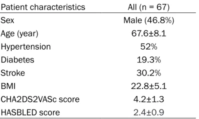
-Table 1. Patient characteristics
Patient characteristics All (n = 67)
Sex Male (46.8%)
Age (year) 67.6±8.1 Hypertension 52%
Diabetes 19.3%
Stroke 30.2%
BMI 22.8±5.1
[image:3.612.92.291.85.204.2]Table 2. Echocardiographic data of the size, end-diastolic volume, end-systolic volume, end-diastolic area, and end-systolic area of the left atrium; size of the right atrium
Echocardiograph 0 48 h 45-60 d
LA size (mm) 45.8±5.6 44.1±5.8 (P < 0.01) 45.5±5.9 (P = 0.25) RA size (mm) 44.7±6.6 44.2±5.9 (P = 0.20) 44.4±5.5 (P = 0.32) LA end-diastolicvolume (ml) 63.5±22.9 60.9±19.6 (P = 0.004) 61.9±19.7 (P = 0.02) LA end-systolic volume (ml) 93.6±28.7 86.1±28.7 (P < 0.01) 88.9±28.5 (P < 0.01) LA end-diastolic area (cm2) 23.0±3.8 21.8±3.6 (P = 0.02) 22.0±3.7 (P = 0.04)
[image:4.612.91.306.235.301.2]LA end-systolic area (cm2) 27.7±5.1 26.2±4.7 (P < 0.01) 26.4±4.9 (P = 0.002) LA: left atrium; RA = right atrium.
Table 3. Results of cardiac catheterization Mean pressure Pre-operation (mmHg) Post-operation LA 15.8±1.8 15.9±1.9
RA 10.1±3.2
RV 18.1±4.7
PA 22.3±5.7
LA = left atrium; RA = right atrium; RV = right ventricle; PA = pulmonary artery.
cantly (850.6 pg/mL vs. 904.5 pg/mL, P = 0.26), and the NT-proBNP levels were signifi
-cantly lower at the first follow-up (45-60 days) than before LAAC (705.1 pg/mL vs. 850.6 pg/ mL, P < 0.01).
Discussion
The present study found that the NT-proBNP levels were significantly higher at 6 h and 24 h after LAAC than before LAAC and were signifi
-cantly lower at the first follow-up (45-60 days) than before LAAC, and that the NT-proBNP lev
-els before and 48 h after LAAC were not differ
-ent. Additionally, the study found that the
diastolic volume, systolic volume,
end-diastolic area, and end-systolic area of the LA were significantly lower at 48 h after LAAC and at the first follow-up (45-60 days) than before LAAC. Furthermore, the study found that the size of the LA was significantly lower at 48 h after LAAC than before LAAC, and that the size of the LA before LAAC and at the first follow-up (45-60 days) was not different.
The LAA originates from the main body of the LA [12]. It is the most common site of cardiac thrombus formation in patients with NVAF, and more than 90% of all thrombi in the LA have
been reported to originate in the LAA [13]. During sinus rhythm, the LAA has normal
con-traction function and adequate blood flow, pre
-venting thrombus formation inside its cavity. However, during AF there is a decrease in LAA contractility and function with Doppler veloci
-ties and dilation of the LAA [14, 15], which increase the risk of thrombus formation inside
its cavity. Percutaneous LAAC is an alternative therapy to prevent thromboembolic events in
patients with NVAF [16, 17], and its safety and efficacy have been shown previously [4]. To the best of our knowledge, the present study is the first to evaluate variations in the structure of the LA and NT-proBNP levels at various time points after LAAC.
Recently, it was reported that the size of the LA
could positively predict cardiac risk, including
the recurrence of AF, heart failure, myocardial infarction, and other such conditions [18, 19].
Gupta DK demonstrated that abnormalities in
the structure and function of the LA increased the incidence of AF and were associated with a high risk of stroke [20]. Additionally, a reduction in the size of the LA was found to increase blood flow velocity in the LA and reduce the likelihood of thrombus formation [21]. Therefore, we believe that reductions in the
[image:4.612.90.289.337.467.2]size, end-diastolic volume, end-systolic volume, end-diastolic area, and end-systolic area of the LA could influence the structure and function of the LA and help improve cardiac function. NT-proBNP is an important natriuretic hor
-mone. The LAA plays an important role in the secretion of NT-proBNP. Additionally, it plays an
important role in regulating intravascular vol-ume and hemodynamics. A previous study
showed that high plasma NT-proBNP levels were correlated positively with the risk of thromboembolic complications with AF [22]. In LAA dysfunction, elevated NT-proBNP levels, especially in patients with NVAF, have been
shown to be associated with a prothrombotic
state [23]. Thus, NT-proBNP is a useful marker for the risk stratification of stroke in patients with NVAF [24].
Schwartz [25] found that endothelial cells cov
-ered the Watchman device and firmly attached the device to the interface of the LA at 45 days after discharge. Kar [26] observed that the sur
-face of the Watchman device was completely incorporated with organizing neo-endocardial growth, involving well-organized granulation tis
-sue with only minimal fibrin deposition, at 28 days. We believe that BNP levels will further
decrease over time owing to advanced
endo-thelialization of the device surface after com
-plete occlusion of the LAA, and this may explain our results that NT-proBNP levels were lower at 45-60 days after LAAC than immediately after
LAAC.
The NT-proBNP levels were significantly higher at 6 h and 24 h after LAAC than before LAAC. Several factors may have contributed to these results. First, contrast media injection during
LAA angiography might have stimulated BNP
secretion. Second, stretching of the LAA by the
closure device during the procedure might have
stimulated BNP secretion. Finally, procedural
stress might have stimulated BNP secretion. However, the potential pathophysiological
mechanisms for the variations in NT-proBNP levels are unknown. Therefore, further studies and longer follow-up are required to determine the long-term progression of the neurohormon
-al response after LAAC.
Limitations
The present study has some limitations. Patients with serious heart failure and acute
myocardial infarction were excluded; however, other factors that were not included in the exclusion criteria might have influenced the NT-proBNP levels.
Conclusion
LAAC might cause reductions in the size,
end-diastolic volume, end-systolic volume,
end-dia-stolic area, and end-syend-dia-stolic area of the LA, and variations in plasma BNP levels at different time points. These findings indicate a loss of neurohormonal function and variations in the structure of the LA in the short-term follow-up period after LAAC.
Disclosure of conflict of interest
None.
Address correspondence to: Zhiyuan Song, Depart- ment of Cardiology, Southwest Hospital, Third Military Medical University, No.30 Gaotanyan, Shapingba, Chongqing 400038, China. Tel: 86 139- 08327066; E-mail: [email protected]
References
[1] Reddy VY, Holmes D, Doshi SK, Neuzil P and Kar S. Safety of percutaneous left atrial ap-pendage closure: results from the watchman left atrial appendage system for embolic pro-tection in patients with AF (PROTECT AF) clini-cal trial and the continued access registry. cir-culation 2011; 123: 417-424.
[2] Miyasaka Y, Barnes ME, Gersh BJ, Cha SS, Bai-ley KR, Abhayaratna WP, Seward JB and Tsang TS. Secular trends in incidence of atrial fibrilla-tion in Olmsted County, Minnesota, 1980 to 2000, and implications on the projections for future prevalence. Circulation 2006; 114: 119-125.
[3] Landmesser U and Holmes DR Jr. Left atrial appendage closure: a percutaneous trans-catheter approach for stroke prevention in atrial fibrillation. Eur Heart J 2012; 33: 698-704.
[4] Holmes DR Jr, Kar S, Price MJ, Whisenant B, Sievert H, Doshi SK, Huber K and Reddy VY. Prospective randomized evaluation of the watchman left atrial appendage closure de- vice in patients with atrial fibrillation versus long-term warfarin therapy: the PREVAIL trial. J Am Coll Cardiol 2014; 64: 1-12.
Investiga-tors. Percutaneous left atrial appendage clo-sure vs. warfarin for atrial fibrillation: a ran- domized clinical trial. JAMA 2014; 312: 1988-1998.
[6] Hara H. Percutaneous left atrial appendage closure for non-valvular atrial fibrillation. Circ J 2016; 80: 1097-1099.
[7] Main ML, Fan D, Reddy VY, Holmes DR, Gordon NT, Coggins TR, House JA, Liao L, Rabineau D, Latus GG, Huber KC, Sievert H, Wright RF, Doshi SK and Douglas PS. Assessment of de-vice-related thrombus and associated clinical outcomes with the watchman left atrial ap-pendage closure device for embolic protection in patients with atrial fibrillation (from the PRO-TECT-AF Trial). Am J Cardiol 2016; 117: 1127-1134.
[8] Bajaj NS, Parashar A, Agarwal S, Sodhi N, Pod-dar KL, Garg A, Tuzcu EM and Kapadia SR. Per-cutaneous left atrial appendage occlusion for stroke prophylaxis in nonvalvular atrial fibrilla-tion: a systematic review and analysis of obser-vational studies. JACC Cardiovasc Interv 2014; 7: 296-304.
[9] Lanfear DE, Li J, Abbas R, She R, Padhukasa-hasram B, Gupta RC, Langholz D, Tang WH, Williams LK, Sabbah HN and Chow SL. Genetic factors influencing b-type natriuretic peptide-mediated production of cyclic guanosine mo-nophosphate and blood pressure effects in heart failure patients. J Cardiovasc Transl Res 2015; 8: 545-553.
[10] Therkelsen SK, Groenning BA, Kjaer A, Svend-sen JH and Boje JenSvend-sen G. ANP and BNP in atrial fibrillation before and after cardiover-sion--and their relationship to cardiac volume and function. Int J Cardiol 2008; 127: 396-399.
[11] Wunderlich NC, Beigel R, Swaans MJ, Ho SY and Siegel RJ. Percutaneous interventions for left atrial appendage exclusion: options, as-sessment, and imaging using 2D and 3D echo-cardiography. JACC Cardiovasc Imaging 2015; 8: 472-488.
[12] Beigel R, Wunderlich NC, Ho SY, Arsanjani R and Siegel RJ. The left atrial appendage: anat-omy, function, and noninvasive evaluation. JACC Cardiovasc Imaging 2014; 7: 1251-1265. [13] Ding J, Zhu J, Lu J, Ding X, Zhang X, Lu W, Ao M
and Ma G. Transcatheter closure of the left atrial appendage: initial experience with the WATCHMAN device. Int J Clin Exp Med 2015; 8: 15230-15237.
[14] Chen OD, Wu WC, Jiang Y, Xiao MH and Wang H. Assessment of the morphology and me-chanical function of the left atrial appendage by real-time three-dimensional
transesopha-geal echocardiography. Chin Med J (Engl) 2012; 125: 3416-3420.
[15] Nucifora G, Faletra FF, Regoli F, Pasotti E, Pe-drazzini G, Moccetti T and Auricchio A. Evalua-tion of the left atrial appendage with real-time 3-dimensional transesophageal echocardiog-raphy: implications for catheter-based left atri-al appendage closure. Circ Cardiovasc Imaging 2011; 4: 514-523.
[16] Holmes DR Jr, Doshi SK, Kar S, Price MJ, San-chez JM, Sievert H, Valderrabano M and Reddy VY. Left atrial appendage closure as an alter-native to warfarin for stroke prevention in atrial fibrillation: a patient-level meta-analysis. J Am Coll Cardiol 2015; 65: 2614-2623.
[17] Bode WD, Patel N and Gehi AK. Left atrial ap-pendage occlusion for prevention of stroke in nonvalvular atrial fibrillation: a meta-analysis. J Interv Card Electrophysiol 2015; 43: 79-89. [18] Amin V, Finkel J, Halpern E and Frisch DR.
Im-pact of left atrial volume on outcomes of pul-monary vein isolation in patients with non-par-oxysmal (persistent) and parnon-par-oxysmal atrial fibrillation. Am J Cardiol 2013; 112: 966-970. [19] Hoit BD. Left atrial size and function: role in
prognosis. J Am Coll Cardiol 2014; 63: 493-505.
[20] Gupta DK, Shah AM, Giugliano RP, Ruff CT, An-tman EM, Grip LT, Deenadayalu N, Hoffman E, Patel I, Shi M, Mercuri M, Mitrovic V, Braunwald E, Solomon SD; Effective a Nticoa Gulation with factor xA next GEneration in AF-Thrombol-ysis in Myocardial Infarction 48 Echocardio-graphic Study Investigators. Left atrial struc-ture and function in atrial fibrillation: ENGAGE AF-TIMI 48. Eur Heart J 2014; 35: 1457-1465. [21] Marui A, Saji Y, Nishina T, Tadamura E, Kanao
S, Shimamoto T, Sasahashi N, Ikeda T and Komeda M. Impact of left atrial volume reduc-tion concomitant with atrial fibrillareduc-tion surgery on left atrial geometry and mechanical func-tion. J Thorac Cardiovasc Surg 2008; 135: 1297-1305.
[22] Rodriguez-Yanez M, Arias-Rivas S, Santamaria-Cadavid M, Sobrino T, Castillo J and Blanco M. High pro-BNP levels predict the occurrence of atrial fibrillation after cryptogenic stroke. Neu-rology 2013; 81: 444-447.
plasma brain natriuretic polypeptide level as a marker of risk for thromboembolism in pa-tients with nonvalvular atrial fibrillation. Stroke 2002; 33: 1005-1010.
[25] Schwartz RS, Holmes DR, Van Tassel RA, Haus-er R, Henry TD, Mooney M, Matthews R, Doshi S, Jones RM and Virmani R. Left atrial append-age obliteration: mechanisms of healing and intracardiac integration. JACC Cardiovasc In-terv 2010; 3: 870-877.