Original Article
Effects of Tanshinone IIA sodium sulfonate on
LPS-induced acute lung injury in mice
Jinxian Qian1,3*, Xinjing Yang2*, Jian Wu3, Aixiang Yang3, Weiyi Tao3, Chunhua Ling1, Guoxing Zhang4
Departments of 1Respiratory and Critical Care Medicine, 2Intensive Care Unit, The First Affiliated Hospital of So -ochow University, Suzhou 215006, Jiangsu Province, China; 3Department of Intensive Care Unit, Affiliated Suzhou Hospital of Nanjing Medical University, Suzhou Municipal Hospital, Suzhou, Jiangsu Province, China; 4Department of Physiology, Medical College of Soochow University, Suzhou 215123, Jiangsu Province, China. *Equal contribu -tors and co-first authors.
Received March 5, 2018; Accepted April 21, 2018;Epub May 15, 2018; Published May 30, 2018
Abstract: Objective: The study investigated the protective effect of Tanshinone IIA sodium sulfonate (TSS) on lipo-polysaccharide (LPS)-induced acute lung injury (ALI), and the underlying mechanisms. Methods: Male C57/black mice were subjected to LPS (10 mg/kg, i.p.) administration to induce acute lung injury. Half an hour before LPS injection, mice were pretreated with TSS (30 mg/kg, i.p.). One hour, six and twelve hours later, mice were sacri-ficed. Lung edema and inflammatory cell infiltration were observed. Expression of pro-inflammatory factors (TNFα, IL-1β, IL-6) were measured by RT-PCR and ELISA in lung tissues. Markers of autophagy including LC3 and Beclin-1 expression were determined by RT-PCR and Western blot. Number of autophagosomes was observed by electron microscope. Results: Our results showed that TSS did not ameliorate the LPS-induced lung edema, but significantly reduced pro-inflammatory cell infiltration. LPS increased mRNA and protein expression of inflammatory factors (TNFα, IL-1β, IL-6) at all observed time. TSS markedly reduced the protein expression of IL-6 but not TNFα and IL-1β. In addition, LPS increased tissue autophagic levels (expression of Beclin-1, LC3 and number of autophagosomes) and TSS treatment further increased the autophagic levels. Conclusion: TSS exerts certain anti-inflammation and enhancing autophagy effects in LPS-induced ALI.
Keywords: Tanshinone IIA sodium sulfonate, lipopolysaccharide, acute lung injury, autophagy
Introduction
Acute lung injury (ALI)/acute respiratory dis-tress syndrome (ARDS), is a severe clinical complication caused by stress situation such as trauma, burns and sepsis, is characterized with respiratory distress, refractory hypoxemia, and no cardiogenic pulmonary edema, along with bilateral pulmonary infiltrates and reduc-tion in respiratory system compliance [1-3]. Even with the continuous efforts and success in treatment of ALI, clinical outcome still pro- ofed high rates of morbidity and mortality in ALI patients [4].
One of the pathophysiological mechanism in ALI is loss of the lung’s gas exchange surfa- ce area, leading to refractory hypoxemia [3]. Strategy to improve the gas exchange by me- chanical ventilation is now a standard treat-ment in ALI in clinic [5]. However, due to that the ventilation itself could induce ALI, it is still
arguable with this therapy [6, 7]. Second mech-anism is the recruitment of neutrophils and other mediators of acute inflammation into the airspace [3]. Anti-inflammatory therapies are reasonably explored [8-11]. However, this thera-py is also notoriously difficult to solve the prob-lem, partly because ALI is not an inflammatory disease but a label describing acute respiratory failure occurring de novo as a result of a wide variety of conditions. Third mechanism is in- flammatory exudate inactivates surfactant leading to collapse of distal lung parenchyma. New approach is undergoing to be explored through stem cell therapy, which could promote repair of ALI-induced cell damage [12, 13]. However, serious adverse events of cell therapy for respiratory diseases are also the inevitable question [14].
clinical treatment for hundreds of years in China for the treatment of various diseases [15-17]. It is considered to have the function of activating blood circulation and removing blood stasis, entering the “heart”, “pericardium”, and “liver” channels according to the theory of tr- aditional Chinese medicine (TCM) and has been used to successfully treat various circula-tory disturbance-related diseases including vasodilatation, anticoagulation, anti-inflamma-tion, and free radical scavenging [18, 19]. It has been reported that TSS attenuates seawater aspiration-induced acute pulmonary edema [20] and reduces lethality and acute lung injury in LPS-treated mice [21]. Anti-inflammatory effects of TSS also have been widely investi-gated in animal model or in cell line [22, 23]. However, the anti-inflammatory effects of TSS on ALI still require further investigation.
Recently, autophagy has been reported to play a role in the development of ALI [24]. It has been reported that autophagy protects ag- ainst ischemia/reperfusion-induced lung injury through alleviating blood-air barrier damage [25], decreasing chlorine-induced mitochondri-al injury and lung inflammation [26] and main-taining the integrity of endothelial barrier in LPS-induced lung injury [27]. On the other hand, inhibition of autophagy could ameliorate acute lung injury [28]. Excessive autophagy also has been reported to contribute to the ALI [29]. It is still controversial what is the real role of autophagy in ALI and there is still no obser-vation of the effects of TSS on the autophagy level in ALI.
The present study is designed to observe the effects of TSS on LPS-induced ALI, focusing on the tissue edema, cell infiltration, inflammatory factors expression and tissue autophagic lev-els, which may provide basic evidence for clini-cal application of TSS on ALI patients.
Materials and methods
Experimental animals
Male C57 mice, weighing 25 to 30 g were pur-chased from Shanghai Laboratory Animal Center. All experiments were performed in ac- cordance with the National Institutes of Health Guidelines for the Use of Laboratory Ani- mals (NIH, publication number 85-23, revised 1996), which were approved by and performed
according to guidelines for the care and use of animals established by Soochow University. Mice were randomly divided into seven gro- ups according to receiving or not receiving TSS after LPS treatment for different time: (1) Saline control group (n=10), in which mice received saline 0.5 hours before the second saline administration; (2) LPS 1-hour group (n=10) in which mice received LPS (10 mg/kg) adminis-tration for 1 hour; (3) TSS + LPS 1-hour group (n=10), in which TSS (30 mg/kg) was adminis-tered 0.5 hours before LPS (10 mg/kg) in- jection for 1 hour. (4) LPS 6-hour group (n=10) in which mice received LPS (10 mg/kg) injec-tion for 6 hours; (5) TSS + LPS 6-hour group (n=10), in which TSS (30 mg/kg) was adminis-tered 0.5 hours before LPS (10 mg/kg) injec-tion for 6 hours. (6) LPS 12- hour group (n=10) in which mice received LPS (10 mg/kg) injec-tion for 12 hours; (7) TSS + LPS 12-hour group (n=10), in which TSS (30 mg/kg) was adminis-tered 0.5 hours before LPS (10 mg/kg) injec-tion for 12 hours [10, 21]. In all groups, admin-istration was via intraperitoneal injection, saline was applied as negative control. At each time point, mice were sacrificed by neck bro-ken, lung tissues were sampled for direct mea-surement of wet weight or were frozen for fur-ther investigation.
Wet-to-dry weight ratio (W/D)
At the end of the experiment, mice left lungs (n=10, respectively) were weighed, then dried to constant weight at 50°C for 72 h and wei-ghed again. The ratio of W/D was finally calcu-lated by dividing the wet weight by the dry weight.
Histological analysis
Part of mice right lungs were samples and fixed with 4% paraformadehyde and cut into 5-μm slices, which were stained with hematoxylin-eosin for morphological analysis. Numbers of cell nucleus were counted from 50 visual fields of ten independent slides.
Real-time PCR
Total RNA was isolated from part of frozen right lung (about 20 mg) tissues by guanidinium isothiocyanate-acid phenol extraction and quantified by measuring the absorbance at 260 nm. One microgram of total RNA was used for reverse transcription under 95°C for 15 minutes. Mice TNFα, IL-1β, IL-6, Beclin-1 and LC3 mRNAs were quantified by real time PCR (prism7000; Applied Biosystems, Foster, California). The primer pairs for TNFα cDNA were 5’-GCCAGGAGGGAGAACAGAAACTC-3’ (for- ward) and 5’-GGCCAGTGAGTGAAAGGGACA-3’ (reverse). The primer pairs for IL-1β cDNA we- re 5’-TCCAGGATGAGGACATGAGCAC-3’ (forward) and 5’-GAACGTCACACACCAGCAGGTTA-3’ (re- verse). The primer pairs for IL-6 cDNA were 5’-CCACTTCACAAGTCGGAGGCTTA-3’ (forward) and 5’-CCAGTTTGGTAGCATCCATCATTTC-3’ (re- verse). The primer pairs for Beclin-1 cDNA were 5’-ATGGAGGGGTCTAAGGCGTC (forward) and 5’-TGGGCTGTGGTAAGTAATGGA (reverse). The primer pairs for LC3 cDNA were 5’-CGCTT- GCAGCTCAATGCTAAC-3’ (forward) and 5’-TCT-CTCACTCTCGTACACTTCG-3’ (reverse). The prim-er pairs for GAPDH cDNA wprim-ere 5’-AAATGGT- GAAGGTCGGTGTGAAC-3’ (forward) and 5’-CA- ACAATCTCCACTTTGCCACTG-3’ (reverse). Con- dition for RT-PCR is denature 30s at 95°C, amplification 40 cycles (95°C 5 s, 60°C 34 s), extension 7 min at 72°C. After calculation, rela-tive expressions of each target mRNA were obtained. Expressions of TNFα, IL-1β, IL-6, Beclin-1 and LC3 mRNAs were normalized to
GAPDH mRNA, and were compared with control group, respectively.
ELISA
Frozen right lung tissues (around 50 mg) were homogenized to measure TNFα, IL-1β, IL-6 con-tent (Biolegend, Inc. Shanghai, China) by using a commercially available enzyme-linked immu-nosorbent assay (ELISA) kits. All steps were performed according to the manufacturer’s instructions. Data were normalized according to the concentration of protein measured by BCA method.
Western blot analysis
Frozen right lung tissues (around 50 mg) were homogenized with RIPA buffer (50 mm Tris, pH 7.0, 150 mM NaCl, 1% Triton-X-100) contain- ing phenylmenthanesulfonyl fluoride (R&D Systems Inc., Minneapolis, US). Homogenates were centrifuged at 12,000* g for 10 minutes at 4°C. Same amount of cell protein were sepa-rated by SDS-PAGE and transferred to PVDF membranes (Hybond TM-ECL; Amersham Ph- armacia Biotech, Inc.). The membranes were blocked in 5% nonfat milk in PBS and 0.1% Tween-20 at room temperature. The blots were then incubated with primary antibody: Beclin-1 antibody (1:1000, abcam, Inc.), anti-LC3 (1:1000, abcam, Inc.), or anti-GAPDH (1:1000, Santa Cruz Biotech, Inc.). Then the membranes were incubated for 1 hour with a secondary antibody (HRP-conjugated anti-rab-bit Ig-G, 1:2000). Excess antibody was washed off with TBS-T three times (15 minutes each) before incubation of enhanced chemilumines-cent reagent (ECL, R&D Systems Inc, Min- neapolis, USA) for 1min. Subsequently, X-ray film was used to detect membrane signal. Immunoreactive bands were detected by the analysis of X-ray films using the software of Image J. The quantity of target proteins is nor-malized by GAPDH expression.
Statistical analysis
[image:3.612.91.286.72.208.2]The SPSS 18.0 software was used for sta- tistical analysis. Data were presented as the mean ± S.E.M. Grouped data were analyzed using a one-way analysis of variance followed by the Student-Newman-Keuls test. A P value <0.05 was considered to be statistically signi- ficant.
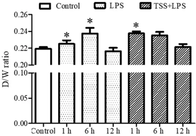
Figure 1. Ratio of wet to dry weight. LPS: Lipopoly-saccharide; TSS: Tanshinone IIA sodium sulfonate. Blank column indicates control group, dot column indicates LPS treatment at all observed time points, diagonal column indicates TSS pretreatment at all observed time points, n=10 in each group. *P<0.05
Results
Effect of TSS on LPS-induced lung edema
To observe the effect of TSS on acute LPS-induce lung edema, wet to dry weight across different time points were investigated. Our results showed that only at 6-hour time point, LPS significantly increased ratio of wet to dry weight compared with control group (P<0.05,
Figure 1), indicating LPS-induced lung edema occur around 6 hours. TSS treatment at 1-hour also markedly increased the ratio compared with control group (P<0.05, Figure 1), indicat-ing TSS may induce early phase of lung edema, while LPS may lead to later phase lung edema (Figure 1).
Effect of TSS on LPS-induced cell infiltration
Histological analysis showed LPS induced increase of cell infiltration at 1-hour and 6-hour compared with control group (P<0.05, Figure 2), but not at 12-hour, indicating LPS induced cell infiltration is time dependent, which is only manifest at the onset of ALI. TSS treatment markedly reduced LPS-induced increase of cell infiltration both at 1-hour and 6-hour compared with LPS group (P<0.05, Figure 2), suggesting TSS has anti-cell-infiltration effect in response to LPS insult.
Effect of TSS on LPS-induced expression of inflammatory factors
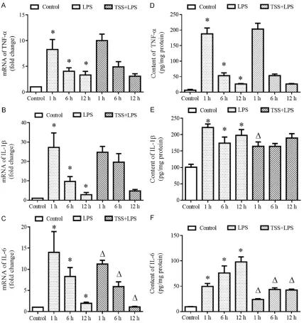
To investigate the effects of TSS on
inflamma-in lung tissue were determinflamma-ined by RT-PCR and ELISA. Data demonstrated that LPS increased both mRNA and protein expressions of TNFα, IL-1β and IL-6 compared with the control group at all observed time points (P<0.05, Figure 3). TSS treatment had no effects on LPS-induced increase of TNFα mRNA and protein expres-sion. TSS inhibited LPS-induced increase of IL-1β protein expression only at 1-hour time point. However, Pretreatment with TSS signifi-cantly suppressed LPS-induced increase of IL-6 mRNA and protein expression at all observed time points compared with the LPS group (P<0.05, Figure 3C, 3F). These results suggest that TTS may exert certain anti-inflammatory effect.
Effect of TSS on LPS-induced tissue autopha-gic level
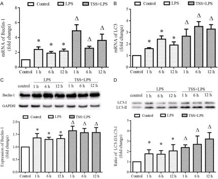
LPS induced increase of mRNA and protein expressions of autophagic markers Beclin-1 and LC3 at all observed time points compared with control group (P<0.05, Figure 4). Pre- treatment with TSS further increased the mRNA and protein expressions of Beclin-1 and LC3 at all observed time points compared with LPS group (P<0.05, Figure 4).
group which (P<0.05, Figure 5). These results indicate that TSS could further activate the tis-sue autophagic level.
Discussion
The major finding of our present study is that TSS has anti-cell infiltration, anti-inflammation effects, as well as activation of autophagy in
response to LPS insult, but has no effect on edema.
[image:5.612.91.519.71.525.2]sary treatment. Among all of the drugs, TSS has been explored to protect LPS-induced lung injury in rats [32]. Later, it has been reported to played an important role in the prevention against LPS-induced ALI in mice [21]. As evi-denced by reduction of lethality and the improvements of lactate dehydrogenase (LDH) content (one index for lung injury), as well as significantly suppressed LPS-induced lung edema [21]. However, our present study dem-onstrated that LPS-induced lung edema is time dependent under present ALI model, which occurs only at the early time (less than 12 hours). Under such circumstance, our results showed that TSS did not exert any protective effect on LPS-induced lung edema, on the
[image:6.612.92.523.72.426.2]con-trary, it could accelerate the occurrence of lung edema in response to LPS stimulation (Figure 1). This discrepancy between our observation with previous may be due to the different mod-els (female Kun-Ming mice in previous study and male C57 mice in our observation) and dif-ferent degree of severity (50 mg/kg in previous study and 10 mg/kg in our observation) of ALI induced by LPS. Therefore, effects of TSS on ALI, especially on the lung edema is still ques-tionable, further investigation is still needed. It is well-known that the pathologic process of ALI involves the infiltration of inflammatory cells into lungs, which released large amounts of inflammatory cytokines, leading to increased Figure 4. Expression of autophagic markers Beclin-1 and LC3. A. mRNA expression of Beclin-1 in each group. B. mRNA expression of LC3 in each group. C. Protein expression of Beclin-1 in each group, upper is the representative blots of Beclin-1 and GAPDH; lower is the densitometric analysis of Beclin-1 expression normalized to GAPDH. D. Protein expression of LC3 in each group, upper is the representative blots of LC3; lower is the densitometric analy-sis of LC3 expression normalized to GAPDH. LPS: Lipopolysaccharide, TSS: Tanshinone IIA sodium sulfonate. Blank column indicates control group, dot column indicates LPS treatment at all observed time points, diagonal column indicates TSS pretreatment at all observed time points, n=10 in each group. Each bar presents the mean ± SEM.
capillary permeability and interstitial edema [2, 3]. Solid evidence has demonstrated that inhibition of release of inflammatory cytokines could ameliorate the development of ALI. Since TSS has been reported to have anti-inflamma-tory property [33, 34], based on the pathogen-esis of ALI, it is reasonable to apply TSS as an anti-inflammatory drug. Previous study all dem-onstrated that TSS could inhibit LPS-induced cell infiltration, expression of inflammatory effects both in rat [32] and mice model [21], and TSS may promote hypoxia-inducible fac- tor-1α protein degradation via the proteasom- al pathway in LPS-stimulated macrophages, thereafter suppress expression of inflammato-ry factors, such as TNF-α, IL-6 and IL-1β [22]. Our present study also demonstrated that TSS could markedly suppress LPS induced cell infil-tration in lung tissue at all observed time points, which is consistent with previous stud-ies. However, although our results also showed LPS increased both mRNA and protein expres-sions of inflammatory factors, such as TNF-α, IL-6 and IL-1β at all observed time points, but marked inhibitory effects of TSS only was observed on the expression of IL-6, but not expression of inflammatory factors, such as TNF-α and IL-1β. This discrepancy may be due to different tissues or models. In the present study we used male C57 mice and other stud-ies used rat or female Kun-Ming mice, and we checked these inflammatory factors in the lung tissues and other study checked these factors
in macrophages or serum. Our present study at least supported that TSS has certain anti-inflammatory in LPS-induced ALI.
Autophagy is one of the innate immune de- fense mechanisms against microbial challeng-es. Previous in vitro and in vivo models of sep-sis demonstrated that autophagy was activat- ed initially in sepsis, followed by a subsequent phase of impairment [35]. Role of autophagy in sepsis is well reviewed recently [36], and authors proposed that autophagy plays a pro-tective role in sepsis according to the exist- ing evidence [37, 38], although contradictory findings also have been reported [39, 40], and they concluded that autophagy increases tran-siently upon encounter of septic insult, this ini-tial increase is followed by a prolonged decline of autophagic flux, contributing to organ dys- function.
[image:7.612.96.519.73.233.2]tophagy with prolonged time. The reason may be due to dosage of LPS applied in the present is mild, or due to rich blood supply of lung tis-sue (while we observed suppression of autoph-agy in intestine with prolonged time, data not shown). Importantly, our present study clearly demonstrated that TSS could further activate autophagic level in response to LPS stimula-tion, which is consistent with our previous study [44] observed that TSS ameliorates ischemia-reperfusion induced myocardial injury may be via enhancing autophagy.
It should be note that in the present study we do not obtain the data of the improvement of TSS on LPS-induced lung edema, which may be due the limitation of observing time. Long term treatment with TSS on LPS induced lung injury should be carried out in the future observation. Especially, our present study does not observe the effects of TSS on LPS-induced lung cell apoptosis. In addition, mechanism of TSS to activate autophagy needs to be investigated and the precise role of elevated autophagy lev-els in the present model also needs to be clarified.
In conclusion, our present study demonstrates that protective effects of TSS in LPS-induced ALI may via certain anti-inflammation and enhancing autophagy.
Disclosure of conflict of interest
None.
Address correspondence to: Chunhua Ling, Depart- ment of Respiratory and Critical Care Medicine, The First Affiliated Hospital of Soochow University, No. 188 Shizi Road, Suzhou 215006, Jiangsu Province, China. Tel: +86-13962146558; E-mail: lingchunhua13sm@163.com; Guoxing Zhang, De- partment of Physiology, Medical College of Sooch- ow University, No. 199 Ren’ai Road, Suzhou 215- 123, Jiangsu Province, China. Tel: +86-0512-65- 880127; Fax: +86-0512-68222218; E-mail: zhang -guoxing46se@163.com
References
[1] Ashbaugh DG, Bigelow DB, Petty TL, Levine BE. Acute respiratory distress in adults. Lancet 1967; 2: 319-323.
[2] Umbrello M, Formenti P, Bolgiaghi L. Current concepts of ARDS: a narrative review. Int J Mol Sci 2016; 18.
[3] Mason C, Dooley N, Griffiths M. Acute respira -tory distress syndrome. Clin Med (Lond) 2016; 16 Suppl 6: s66-s70.
[4] Ferguson ND, Fan E, Camporota L. The berlin definition of ARDS: an expanded rationale, jus -tification, and supplementary material. Inten -sive Care Med 2012; 38: 1573-1582.
[5] Miller AC, Ferrada PA, Kadri SS. High-frequency ventilation modalities as salvage therapy for smoke inhalation-associated acute lung injury: a systematic review. J Intensive Care Med 2018; 33: 335-345.
[6] Slutsky AS, Ranieri VM. Ventilator-induced lung injury. N Engl J Med 2013; 369: 2126-2136. [7] Matthay MA, McAuley DF, Ware LB. Clinical tri
-als in acute respiratory distress syndrome: challenges and opportunities. Lancet Respir Med 2017; 5: 524-534.
[8] Baron D, Drugeon H, Nicolas F, Courtieu A. The use of tombramycin in the management of se-vere infections. Clinical and pharmacological data. Eur J Intensive Care Med 1976; 2: 89-96. [9] Piao C, Park JH, Lee M. Anti-inflammatory ther -apeutic effect of adiponectin gene delivery us-ing a polymeric carrier in an acute lung injury model. Pharm Res 2017; 34: 1517-1526. [10] Zeng M, Sang W, Chen S. 4-PBA inhibits
LPS-induced inflammation through regulating ER stress and autophagy in acute lung injury mod-els. Toxicol Lett 2017; 271: 26-37.
[11] Umapathy NS, Gonzales J, Fulzele S. β-Nicoti-namide adenine dinucleotide attenuates lipo-polysaccharide-induced inflammatory effects in a murine model of acute lung injury. Exp Lung Res 2012; 38: 223-232.
[12] Xiang B, Chen L, Wang X. Transplantation of menstrual blood-derived mesenchymal stem cells promotes the repair of LPS-induced acute lung injury. Int J Mol Sci 2017; 18.
[13] Matthay MA, Pati S, Lee JW. Concise review: mesenchymal stem (stromal) cells: biology and preclinical evidence for therapeutic potential for organ dysfunction following trauma or sep-sis. Stem Cells 2017; 35: 316-324.
[14] Zhao R, Su Z, Wu J. Serious adverse events of cell therapy for respiratory diseases: a system-atic review and meta-analysis. Oncotarget 2017; 8: 30511-30523.
[15] Kai G, Xu H, Zhou C. Metabolic engineering tanshinone biosynthetic pathway in Salvia milt-iorrhiza hairy root cultures. Metab Eng 2011; 13: 319-327.
[16] Ho JW, Jie M. Pharmacological activity of car-diovascular agents from herbal medicine. Car-diovasc Hematol Agents Med Chem 2007; 5: 273-277.
[18] Wang J, Lu W, Wang W. Promising therapeutic effects of sodium tanshinone IIA sulfonate to-wards pulmonary arterial hypertension in pa-tients. J Thorac Dis 2013; 5: 169-172.
[19] Mao S, Li X, Wang L. Rationale and design of sodium tanshinone IIA sulfonate in left ven-tricular remodeling secondary to acute myo-cardial infarction (STAMP-REMODELING) trial: a randomized controlled study. Cardiovasc Drugs Ther 2015; 29: 535-542.
[20] Xie XY, Zhang B, Li JH. Sodium tanshinone iia sulfonate attenuates seawater aspiration-in-duced acute pulmonary edema by up-regulat-ing Na(+), K(+)-ATPase activity. Exp Lung Res 2011; 37: 482-491.
[21] Xu M, Dong MQ, Cao FL. Tanshinone IIA reduc -es lethality and acute lung injury in LPS-treated mice by inhibition of PLA2 activity. Eur J Phar-macol 2009; 607: 194-200.
[22] Xu M, Cao F, Liu L. Tanshinone IIA-induced at -tenuation of lung injury in endotoxemic mice is associated with reduction of hypoxia-inducible factor 1alpha expression. Am J Respir Cell Mol Biol 2011; 45: 1028-1035.
[23] Cheng J, Chen T, Li P. Sodium tanshinone IIA sulfonate prevents lipopolysaccharide-induced inflammation via suppressing nuclear factor-kappaB signaling pathway in human umbilical vein endothelial cells. Can J Physiol Pharmacol 2018; 96: 26-31.
[24] Liu QP, Zhou DX, Lin P. Participation of autoph-agy in acute lung injury induced by seawater. Exp Lung Res 2013; 39: 441-452.
[25] Zhang D, Li C, Zhou J. Autophagy protects against ischemia/reperfusion-induced lung in-jury through alleviating blood-air barrier dam-age. J Heart Lung Transplant 2015; 34: 746-755.
[26] Jurkuvenaite A, Benavides GA, Komarova S. Upregulation of autophagy decreases chlorine-induced mitochondrial injury and lung inflam -mation. Free Radic Biol Med 2015; 85: 83-94. [27] Zhang D, Zhou J, Ye LC. Autophagy maintains
the integrity of endothelial barrier in LPS-in-duced lung injury. J Cell Physiol 2018; 233: 688-698.
[28] Sun Y, Li C, Shu Y. Inhibition of autophagy ame-liorates acute lung injury caused by avian influ -enza A H5N1 infection. Sci Signal 2012; 5: ra16.
[29] Liu Y, Zhang J. Saturated hydrogen saline ame-liorates lipopolysaccharide-induced acute lung injury by reducing excessive autophagy. Exp Ther Med 2017; 13: 2609-2615.
[30] Martin GS, Mannino DM, Eaton S. The epide-miology of sepsis in the United States from 1979 through 2000. N Engl J Med. 2003; 348: 1546-1554.
[31] Genga KR, Russell JA. Update of sepsis in the intensive care unit. J Innate Immun 2017; 9: 441-455.
[32] Shi XM, Huang L, Xiong SD. Protective effect of tanshinone II A on lipopolysaccharide-induced lung injury in rats. Chin J Integr Med 2007; 13: 137-140.
[33] Jang SI, Jeong SI, Kim KJ. Tanshinone IIA from Salvia miltiorrhiza inhibits inducible nitric ox-ide synthase expression and production of TNF-alpha, IL-1beta and IL-6 in activated RAW 264.7 cells. Planta Med 2003; 69: 1057-1059.
[34] Fan GW, Gao XM, Wang H. The anti-inflamma -tory activities of Tanshinone IIA, an active com-ponent of TCM, are mediated by estrogen re-ceptor activation and inhibition of iNOS. J Steroid Biochem Mol Biol 2009; 113: 275-280.
[35] Schmid D, Munz C. Innate and adaptive immu-nity through autophagy. Immuimmu-nity 2007; 27: 11-21.
[36] Ho J, Yu J, Wong SH. Autophagy in sepsis: deg-radation into exhaustion? Autophagy 2016; 12: 1073-1082.
[37] Lin CW, Lo S, Perng DS. Complete activation of autophagic process attenuates liver injury and improves survival in septic mice. Shock 2014; 41: 241-249.
[38] Piquereau J, Godin R, Deschenes S. Protective role of PARK2/Parkin in sepsis-induced cardi-ac contrcardi-actile and mitochondrial dysfunction. Autophagy 2013; 9: 1837-1851.
[39] Unuma K, Aki T, Funakoshi T. Extrusion of mito -chondrial contents from lipopolysaccharide-stimulated cells: involvement of autophagy. Autophagy 2015; 11: 1520-1536.
[40] Ceylan-Isik AF, Zhao P, Zhang B. Cardiac over -expression of metallothionein rescues cardiac contractile dysfunction and endoplasmic retic-ulum stress but not autophagy in sepsis. J Mol Cell Cardiol 2010; 48: 367-378.
[41] Yen YT, Yang HR, Lo HC. Enhancing autophagy with activated protein C and rapamycin pro-tects against sepsis-induced acute lung injury. Surgery 2013; 153: 689-698.
[42] Takaoka Y, Goto S, Nakano T. Glyceraldehyde-3-phosphate dehydrogenase (GAPDH) pre-vents lipopolysaccharide (LPS)-induced, sep-sis-related severe acute lung injury in mice. Sci Rep 2014; 4: 5204.
[43] Colell A, Ricci JE, Tait S. GAPDH and autophagy preserve survival after apoptotic cytochrome c release in the absence of caspase activation. Cell 2007; 129: 983-997.