The effects of clozapine and haloperidol on
astrocyte function and morphology in schizophrenia
This thesis submitted to University of Dublin, Trinity College for the degree of
MSc (Masters) by Research in Neuroscience
Trisha Ang Li Shan
(2019)
Supervisor: Kumlesh K. Dev
Drug Development,
Discipline of Physiology, School of Medicine, Trinity College Dublin, Ireland
Declaration and Statement of Plagiarism
I declare that this thesis has not been submitted as an exercise for a degree at this or any other university and it is entirely my own work.
I agree to deposit this thesis in the University’s open access institutional repository or allow the library to do so on my behalf, subject to Irish Copyright Legislation and Trinity College Library conditions of use and acknowledgement.
Signed Date
______________________________________ ________________
Acknowledgments
First and foremost, I would like to thank my supervisor Professor Kumlesh K. Dev for allowing me to undertake this research project. He has proven a patient and attentive supervisor, providing continuous guidance and support throughout the year. His scientific advice has been invaluable.
Secondly, I would like to express profound gratitude to past and present members of the KKD laboratory: Dr Steven Fagan, Ms Sybille Bechet, Mr Luke Davison, Dr Maria Velasco, Dr Kapil Sharma, Ms Christiana Bils and Ms Adryana Clementino for their unwavering support and friendship.
I would also like to thank my students Maebh O’Brien, Shane O’Neill, Amy Murray, Subash Raj, and Dylan O’Toole for their contribution to the data presented herein.
I am thankful for the staff in the Department of Physiology and members of the Donnelly and Downer laboratory for their continuous encouragement and enthusiasm throughout the year.
Finally, I would like to thank my family and friends, without whom the undertaking of this project would have been considerably more difficult.
Table of Contents
Table of Contents
Declaration and Statement of Plagiarism ... i
Acknowledgments ... ii
Table of Contents ... iii
List of Figures and Tables ... vi
List of Abbreviations ... vii
Scientific Abstract ... viii
Lay Abstract ... ix
Aims and Hypothesis ... x
Value of Research ... xi
Outputs ... xii
Introduction ... 1
1. Schizophrenia – General Overview ... 2
1.1.
Broad overview on symptomatology and epidemiology ... 2
1.2.
Aetiology – gene and environment ... 2
1.3.
Gross and microscopic changes in the brain ... 3
1.4.
Current hypotheses on pathophysiology ... 3
1.4.1.
Dopamine ... 3
1.4.2.
Glutamate ... 4
1.4.3.
Other neurotransmitters ... 4
1.5.
Neuroinflammation ... 4
2. Astrocyte dysfunction in schizophrenia ... 5
2.1.
Introduction to astrocyte morphology ... 5
2.2.
Introduction to astrocyte function ... 5
2.2.1.
The role of astrocytes in neurotransmitter homeostasis ... 5
2.2.2.
The tripartite hypothesis ... 6
2.2.3.
Possible role for maintaining the Blood Brain Barrier (BBB) ... 6
2.3.
Alterations in astrocyte function and morphology seen in schizophrenia ... 6
2.3.1.
Evidence of change in astrocyte morphology ... 6
2.3.2.
Alteration in astrocyte energy metabolism ... 7
2.3.3.
Alteration in synaptic transmission of glutamate ... 7
2.4.
The emerging role of astrocytes in neuroinflammation ... 8
2.4.1.
Reactive astrogliosis ... 8
2.4.2.
S100B as marker of astroglial activation in schizophrenia ... 8
3. Dopamine ... 9
3.1.
Introduction to the dopamine receptor ... 9
3.2.
Dopamine receptor expression in astrocytes, CNS, and PNS ... 9
3.3.
Dopamine receptor structure and function ... 9
3.3.1.
Structure ... 9
3.3.2.
Function ... 10
[image:5.595.81.527.128.785.2]
3.4.1.
Mechanisms of GPCR signalling ... 10
3.4.2.
Regulation of dopamine receptor signalling ... 10
3.4.3.
Role of Akt in D2R-‐mediated psychosis and cognitive dysfunction in schizophrenia ... 11
3.5.
Interactions between dopamine and other signalling pathways ... 11
3.5.1.
Regulation of Mitogen Activated Protein Kinases (MAPK) by D1-‐class and ionotropic glutamate receptors ... 11
3.5.2.
Serotonergic regulation of dopamine neurotransmission ... 12
4. Antipsychotics ... 12
4.1.
Neuropharmacology of antipsychotics ... 12
4.1.1.
Typical antipsychotics ... 12
4.1.2.
Atypical antipsychotics ... 13
4.2.
Current evidence on effect of antipsychotics on astrocyte function ... 13
4.3.
Effects of antipsychotics on inflammation ... 14
Methods and Materials ... 23
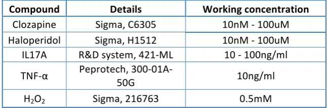
5. Materials ... 24
5.1.
Compounds ... 24
5.2.
Antibodies ... 24
6. Methods ... 24
6.1.
Cell culture ... 24
6.2.
Enzyme Linked Immunoabsorbent Assay (ELISA) ... 25
6.3.
MTT assay ... 25
6.4.
Immunocytochemistry ... 25
6.5.
Analysis of immunofluorescent staining ... 26
6.6.
SDS-‐PAGE and Western immunoblotting ... 26
6.7.
Statistical analysis ... 27
Results ... 31
7. Results ... 32
7.1.
Treatment of human astrocytes with IL-‐17A/TNFα-‐induces expression of IL-‐6. ... 32
7.2.
Clozapine attenuates IL-‐17A/TNFα-‐induced levels of IL-‐6 in human astrocytes. ... 32
7.3.
IL-‐17A/TNFα -‐induced levels of IL-‐6 are decreased by Haloperidol. ... 33
7.4.
Clozapine and Haloperidol do not alter human astrocyte cell viability. ... 33
7.5.
Clozapine and Haloperidol do not significantly alter astrocyte morphology, under conditions of inflammation. ... 33
7.6.
Clozapine and Haloperidol increase ERK phosphorylation in astrocytes. ... 34
Discussion ... 43
8. Summary of findings ... 44
8.1.
TNFα and IL-‐17A induce IL-‐6 expression in human astrocytes. ... 44
8.2.
Clozapine and haloperidol decrease IL-‐6 expression in human astrocytes ... 45
8.3.
The interaction between IL-‐6 and dopamine receptor signalling ... 45
8.4.
Clozapine and haloperidol do not alter human astrocyte morphology and survival ... 46
8.5.
Changes in ERK 1/2 phosphorylation induced by clozapine as compared to haloperidol . 46
9. Future studies and limitations ... 47
References ... 50
Appendix ... 70
Appendix 1 ... 69
Appendix 2 ... 70
List of Figures and Tables
Introduction
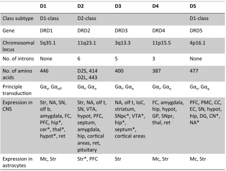
Table 1.1 Summary of major genes implicated in schizophreniaTable 1.2 Properties of each dopamine receptor class subtype
Table 1.3 Pharmacological effects of clozapine and haloperidol
Figure 1.1 Epigenetic model of schizophrenia
Figure 1.2 Model of the tripartite synapse
Figure 1.3 Representation of the progression of different stages of reactive astrogliosis
Figure 1.4 Early and late dopamine receptor signalling
Figure 1.5 Regulation of ERK 1/2 by D1, D2 and glutamate receptors
Material and Methods
Table 1.1 List of CompoundsTable 1.2 List of Antibodies
Figure 1.1 Cellular extension quantification
Results
Figure 3.1 Figure 3.2 Figure 3.3
Figure 3.4 Figure 3.5 Figure 3.6 Figure 3.7
Treatment of human astrocytes with IL17A/TNFα-‐induces expression of IL-‐6
Clozapine attenuates TNFα/IL17A-‐induced IL-‐6 expression in human astrocytes
Haloperidol attenuates the expression of IL-‐6 induced by IL17A/TNFα in human
astrocytes
Clozapine and haloperidol do not alter astrocyte cell survival Astrocyte morphology is not altered by clozapine
Astrocyte morphology is not altered by haloperidol
Clozapine and Haloperidol increase ERK phosphorylation in astrocytes
Discussion
Figure 1.1 Molecular pathway linking dopamine, TNFα, IL17A receptors with IL-‐6
List of Abbreviations
Abbreviation Definition
Akt Protein kinase B
ATP Adenosine triphosphate
BBB Blood brain barrier
BMI Body mass index
CNS Central nervous system
COMT Catechol-‐O-‐methyltransferase
CSF Cerebrospinal fluid
DISC-‐1 Disrupted in schizophrenia-‐1 gene
GABA Gamma-‐amino butyric acid
GDP Guanosine 5'-‐diphosphate
GRK G-‐protein coupled receptor kinase
GTP Guanosine triphosphate
HRP Horseradish peroxidase
Ig Immunoglobulin
IL Interleukin
INF Interferon
L-‐DOPA L-‐3,4-‐dihydroxyphenylalanine
MAPK Mitogen activated protein kinase
NF-‐ κB Nuclear factor kappa-‐light chain enhancer of activated B cells
NGF Nerve growth factor
OPD o-‐Phenylenediamine dihydrochloride
PKA Protein kinase A
PP2A Protein phosphatase 2
PYGM Glycogen phosphorylase
RH Rel homology domain
S100B S-‐100 calcium binding protein B
TGF Transforming growth factor
TNF Tumour necrosis factor
Scientific Abstract
Schizophrenia is a chronic debilitating psychiatric disorder affecting approximately 1% of the population worldwide. The disorder is characterised by positive symptoms, negative symptoms, and clinical deficits in cognition. Current therapeutic agents, known as antipsychotics, all display some level of dopamine antagonism thereby reducing symptoms of psychosis. It has been suggested that abnormal astrocyte function is involved in aberrant neurotransmitter signalling and neuroinflammation seen in schizophrenia. The effect of antipsychotics on astrocyte cell function is still being explored. Using a human astrocyte culture model, this study aims to determine the effect of typical and atypical antipsychotics, haloperidol and clozapine, on cytokine levels, the intracellular signalling molecule Extracellular Signal-‐Regulated kinase 1/2 (ERK 1/2), astrocyte morphology and survival. Human astrocytes were cultured and pre-‐treated with clozapine or haloperidol for 1h, then
stimulated with TNFα/IL-‐17A for 18h. Cytokine analysis was carried out using Enzyme Linked
Immunoabsorbent Assay (ELISA). Changes in astrocyte morphology was analysed using immunocytochemistry, with glial fibrillary acidic protein (GFAP) and vimentin protein expressions labelling astrocytes. The results of this study showed that both clozapine and haloperidol attenuated
TNFα/IL-‐17A induced expression of IL-‐6 in human astrocytes. Under pro-‐inflammatory conditions,
pre-‐treatment with both clozapine and haloperidol did not change astrocyte morphology. Furthermore, an MTT assay showed that both clozapine and haloperidol did not affect cell viability. To quantify changes in extracellular signal-‐regulated kinases 1 and 2 (ERK 1/2), astrocytes were exposed to clozapine or haloperidol for 15 mins and processed for western immunoblotting. This study demonstrated that haloperidol, but not clozapine, significantly increased ERK 1/2 phosphorylation in human astrocytes. Taken together, we are able to ascertain that clozapine and
haloperidol change the role of astrocytes in inflammation and cellular signalling in schizophrenia.
Lay Abstract
Schizophrenia is a chronic severe mental disorder that affects approximately 1% worldwide population. The disorder is characterised by distortions in perception, emotions and language, which manifest into hallucinations (i.e. hearing voices) and erratic behaviour. The exact cause of schizophrenia is unknown, although altered levels of the neurotransmitter dopamine is implicated. The treatment used in schizophrenia, known as antipsychotics, work by changing levels of neurotransmitter dopamine in the brain, but their exact mechanism is unknown. It has been suggested that astrocytes, a specialised cell type in the brain that controls essential complex functions and supports neurotransmission in the brain, may be involved in the development of schizophrenia. Therefore, this study aims to examine the effect of certain antipsychotics, clozapine and haloperidol, on astrocyte function. The results of this current study showed that both clozapine and haloperidol have a protective effect, where both antipsychotics induce an anti-‐inflammatory effect on astrocytes. Addtionally, this study found that antipsychotics do not change the shape of astrocytes and do not adversely affect their survival. Taken together, we suggest that antipsychotics may have a beneficial effect on astrocytes, which may influence the treatment of symptoms associated with schizophrenia.
Aims and Hypothesis
We hypothesise that both clozapine (an atypical antipsychotic) and haloperidol (a typical antipsychotic), are able to alter astrocyte morphology as well as astrocyte function, in terms of its role in neuroinflammation and regulation of neurotransmitter signalling molecules.
Based on the above hypothesis, the aims of this study are as follows:
• To examine the direct effects of clozapine and haloperidol on the pro-‐inflammatory cytokine
release in astrocytes
• To investigate the impact of clozapine and haloperidol on astrocyte survival
• To identify the effects of clozapine and haloperidol on astrocyte cellular processes
• To explore the role of clozapine and haloperidol on intracellular dopamine signalling
molecules in astrocytes
Value of Research
Schizophrenia has a high prevalance and incidence rate, affecting more than 23 million individuals worldwide according to current WHO statistics. Additionally, schizophrenia is associated with considerable disability for the individual, often affecting access to employment, housing and healthcare. The aetiopathophysiology of schizophrenia is complex, and largely unknown. It is likely that there is a complex interplay between genetic and environmental factors that contribute to altered neurotransmission, of which dopamine overactivity is thought to play a major role during symptoms of psychosis. Although it is established that antipsychotics exert its therapeutic effects via dopamine antagonism, its effects on glial cells is still being elucidated. Astrocytes are specialised macroglia and are the most abundant cell type in the CNS. Astrocytes are in constant communication with neurons; increased neuronal activity leads to homeostatic changes in astrocytes, which include augmented metabolic activity and synthesis of lactate, clearance of neurotransmitters and buffering extracellular K+. Furthermore, astrocytes play a role in regulating cytokines and other immune molecules in the brain, of which are also thought to be dysregulated in schizophrenia. Although there has been some evidence of astrocyte dysfunction in schizophrenia, there has been limited studies on the effect antipsychotics have on astrocyte function and morphology. This study aims to create a model of neuroinflammation in order to investigate the effect of clozapine and haloperidol
on astrocytes and cellular signalling in schizophrenia.
Outputs
Papers
Ang, T., Dev, K.K., Sharma, K. The effects of antipsychotics on astrocyte function and morphology in
schizophrenia. Journal of Neuropharmacology, 2019 (in preparation)
Presented Posters
Ang, T., Dev, K.K., The role of astrocytes in schizophrenia. 7th Annual Meeting Frontiers in Neurology
Ireland, Dublin, Ireland, November 2017. (Appendix 1)
Ang, T., Dev, K.K., The effect of antipsychotics clozapine and haloperidol on astrocyte function and morphology. 11th FENS Forum of Neuroscience, Berlin, Germany, July 2018. (Appendix 2)
Introduction
1.
Schizophrenia – General Overview
1.1. Broad overview on symptomatology and epidemiology
Schizophrenia is a psychiatric disorder characterised by its phenomenology: positive symptoms (i.e. thought disorder, hallucinations, delusions), negative symptoms (i.e. blunted affect, reduced motivation, poverty of thought) and clinically significant deficits in cognition (including memory,
attention and executive functions) (Yamamuro et al., 2015). In addition, symptoms need to be
associated with reduced occupational and social functioning in order to fulfil diagnostic criteria, as
per DSM-‐5 (Diagnostic and statistical manual of mental disorders, Fifth Edition) (American
Psychiatric Association, 2013). The prevalence of schizophrenia reaches approximately 1%
worldwide, with incidence approaching 1.5 per 10,000 per annum (McGrath et al., 2008). Incidence
is often higher in men, at a ratio of 1.4:1 (Abel et al., 2010). Typically, disease onset occurs during
late adolescence (18-‐25 years), with females diagnosed slightly later (American Psychiatric
Association, 2013). Regrettably, schizophrenia is associated with a high suicide rate, responsible for
10% of all completed suicides (Arsenault-‐Lapierre et al., 2004) and affects approximately 5% of those
with schizophrenia (Hor and Taylor, 2010).
1.2.Aetiology – gene and environment
Schizophrenia is a highly heritable disease, with a higher monozygotic than dizygotic concordance
rate (40-‐50% vs. 10-‐15%), suggesting a significant genetic component to overall risk (Cannon et al.,
1998; Kringlen, 2000). Genome-‐wide association studies (GWAS) have found that schizophrenia is a polygenetic disorder, where an accumulation of multiple risk alleles leads to increased risk in this
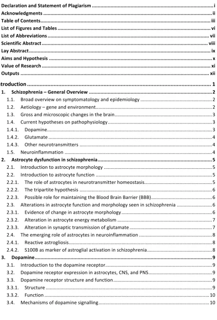
disease entity (Harrison and Weinberger, 2005). Pre-‐GWAS candidate gene studies have identified
specific genes linked to schizophrenia (Table 1.1). Their roles in influencing pathophysiology have
been linked to altered synpatic function, disordered dopamine and glutamate transmission, and
abnormal metabolic and immune functions (Schwab and Wildenauer, 2013). Nevertheless, a meta-‐
analysis suggested that historical candidate genes have not proven to be of particular value in
determining the genetic basis of schizophrenia, largely owing to poor statistical power (Lewis and
Moghaddam, 2006). The largest GWAS to date has identified 108 schizophrenia-‐associated loci that
include genes coding for the dopamine receptor (DRD2) as well as glutamatergic neurotransmission
and synaptic plasticity (e.g. GRM3, GRIN2A, SRR, GRIA1), of which are notable due to their
implications in aetiology and treatment of schizophrenia (Schizophrenia Working Consortium, 2014).
Copy number variants (CNVs) are structural alterations in genes that contribute to human variability,
where lines of DNA can be deleted or duplicated (Thapar and Cooper, 2013). CNVs associated with
schizophrenia are uncommon in the population although carry significant risk (odds ratios 2–60) and
interestingly, overlap with loci contributing to paediatric neurodevelopmental conditions (Marshall
et al., 2017). Most of CNVs accounted are de-‐novo mutations, which would contribute to the
relatively stable prevalence of schizophrenia (Rees et al., 2012). The most frequent CNV being a
deletion on the long arm of chromosome 22 (22q11) (Stefansson et al., 2008). The 22q11
microdeletion occurs at a frequency of 0.3% of all schizophrenia cases.
the table below (Figure 1.1). There have been studies looking at the gene and environment interaction, for example, cannabis use in relation to polymorphisms in genes involved in
dopaminergic pathways (van Winkel, 2011) and polymorphisms in COMT increasing vulnerability
stress-‐induced psychosis (van Winkel et al., 2008). Advances in statistical genetics, using polygenic
profile scoring and Mendelian Randomization scoring tools, will allow us to further elucidate the link
between genetics and environmental risk factors in schizophrenia (Marshall et al., 2017).
1.3.Gross and microscopic changes in the brain
Once known as dementia praecox, schizophrenia is now established as a neurodevelopmental
disorder (Weinberger, 1987). Structural abnormalities likely arise long before first episode psychosis
(Vita et al., 2006). Gross changes in the brain include ventricular enlargement and reduced volume in
the medial temporal lobe (Lewis and Lieberman, 2000). Additionally, decreased grey matter density
and cortical thinning has been found in the cingulate cortex (Heckers et al., 1990; Bouras et al., 2001;
Williams et al., 2014).
Although more prominent changes are seen in grey matter compared to white matter in magnetic
resonance imaging (MRI) studies (Bernstein et al., 2015), other neuroimaging studies and genetic
analyses show alterations in the microstructure of white matter in schizophrenia. These include
decreased anisotropy in the cingulum bundle in chronic schizophrenia (Kubicki et al., 2003), defects
in myelin composition (Du et al.; Bernstein et al., 1992), and decreased dendritic spine density on
prefrontal cortical pyramidal neurons (Glantz and Lewis, 2000). Moreover, hyperpruning of collateral
axons in the prefrontal cortex is seen to contribute to the manifestation of schizophrenia (Nieuwenhuis et al., 2012). These findings are replicated in the hippocampus: decreased pyramidal neuronal size and changes in the dendritic tree contribute to neuronal dysfunction, which is
associated with cognitive decline in schizophrenia (Kolomeets and Uranova, 2010). Whether
abnormal structural connectivity is a primary factor in the pathology of schizophrenia or a
consequence of cortical neuronal dysfunction, remains a question (Bernstein et al., 2015). Increasing
evidence has suggested astroglial involvement in changes in white matter and synaptic abnormalities in schizophrenia, as discussed below.
1.4.Current hypotheses on pathophysiology
Interestingly, schizophrenia appears to be a condition that affect humans exclusively, thus limiting
use of animal models (Taylor A, 2009). Therefore, our understanding of the pathophysiology of
schizophrenia has been largely based on post-‐mortem studies, clinical observational studies and genetic studies.
1.4.1. Dopamine
Altered neurotransmitter levels have been found in schizophrenia. Excess dopamine signalling in the striatal and/or mesolimbic areas of the brain is thought to be responsible for the positive symptoms
seen in schizophrenia (Carlsson, 1988). This hypothesis was born from the success of antipsychotic
medication, most of which act to block dopaminergic D2 receptors. Dopamine excess is contributed
development of psychosis (Kapur et al., 2005; Howes et al., 2009). Interestingly, dopamine release
increases in patients who develop psychosis, and rises further with progression of psychosis (Howes
et al., 2012). Contrastingly, deficits in D1 receptor signalling in the prefrontal cortex is associated
with negative symptoms (Goldman-‐Rakic et al., 2004). Dopamine is not the only neurotransmitter
involved in the pathophysiology of schizophrenia. This is suggested by the fact that clozapine, a weak
D2 antagonist, has superior efficacy to typical antipsychotics, which are potent D2 antagonists
(Wahlbeck et al., 2000). Furthermore, the hypothesis does not explain the entire clinical picture of schizophrenia, as current antipsychotics have limited effect on negative and cognitive symptoms.
1.4.2. Glutamate
Another major neurotransmitter implicated in schizophrenia is glutamate. N-‐methyl-‐D-‐aspartate (NMDA) antagonists such as phencyclidine (PCP) and MK-‐801 (dizocilpine) have been shown to induce and exacerbate acute psychotic and negative symptoms, by preventing flow of ions such as Ca2+ (Steinpreis, 1996; Stone, 2009). Furthermore, studies have found increased levels of kynurenic
acid, an endogenous NMDA receptor antagonist, in both cerebrospinal fluid and certain areas of the
brain of schizophrenic patients (Schwarcz et al., 2001; Erhardt et al., 2007). This suggests deficient
glutamate signalling is involved in schizophrenia, although there is debate as to whether this is due
to abnormal glutamate pathway signalling or a defect in the glutamate receptor (Katsel et al., 2011).
1.4.3. Other neurotransmitters
Abnormalities in levels of other neurotransmitters have been proposed to affect cognitive functioning in schizophrenia. Decreased GABA in the dorsolateral prefrontal cortex (DLPFC) leads to
impaired synchronisation in pyramidal cells, which is linked to deficits in working memory (Lewis and
Moghaddam, 2006).
There is also recent evidence that the cholinergic system is involved in schizophrenia. Reduced
cortical levels of muscarinic M1 receptors have been found in schizophrenic patients, hypothesised
to affect working memory by disrupting information between the cortex and hippocampus (Scarr
and Dean, 2008; Karam et al., 2010). Interestingly, there is a higher proportion of smokers in schizophrenia patients than the average population, possibly as a method to self-‐medicate and
amend deficits in sensory and cognitive processing (Mobascher and Winterer, 2008). This is
supported by up-‐regulation of high affinity neuronal nicotinic acetylcholine receptors (nAChRs) and
reduced levels of α7-‐nAChR in the hippocampus and frontal cortex found in schizophrenia (Ochoa
and Lasalde-‐Dominicci, 2007).
1.5.Neuroinflammation
There is emerging interest in neuroinflammation and its involvement in the pathophysiology of
schizophrenia. Early stresses, including maternal infection with certain viruses (Figure 2), have been
shown to induce an inflammatory response, where oxidative stress, release of pro-‐inflammatory
cytokines and priming of glial cells, ultimately disrupts normal brain maturation (Meyer, 2013).
Fitting with this model, altered secretion patterns of cytokines is thought to occur in schizophrenia,
including an increase in pro-‐inflammatory IL-‐6 levels (van Kammen et al., 1999; O' Connell and Dev,
2014). Additionally, an increased prevalence of positive autoantibody titres are seen in patients with first-‐episode psychosis compared to controls, including anti-‐cardiolipin IgG and anti-‐NMDA receptor,
contribute to psychosis by cross-‐reacting directly with CNS antigens (e.g. anti-‐NMDA receptor) or via
cytokine mediated aberrant dopaminergic transmission (Zalcman et al., 1994; Ezeoke et al., 2013).
2.
Astrocyte dysfunction in schizophrenia
2.1.Introduction to astrocyte morphology
In terms of anatomic distribution, astrocytes are mapped out in a well-‐organised, non-‐overlapping
manner, where no part of the CNS is without astrocytes or similar cells (Sofroniew and Vinters,
2010). Historically, astrocytes have been divided into protoplasmic or fibrous cell types. Protoplasmic astrocytes have a globoid distribution of finely branching processes from numerous
stem branches, whereas fibrous cell types are characterised by long fibre-‐like processes (Ramón y
Cajal, 1909). In the grey matter, interconnecting processes of protoplasmic astrocytes form gap junctions. Branching processes from a single protoplasmic astrocyte in the hippocampus or cortex is estimated to be in contact with over 100,000 different synapses, forming exclusive territories of
functional islands (Bushong et al., 2002; Halassa et al., 2007).
2.2.Introduction to astrocyte function
Astrocytes are the most abundant cell type in the brain, making up five times the population of
neurons in the brain (Sofroniew and Vinters, 2010). They are specialised macroglia, which form a
group of glial cells that arise from the ectoderm cell line. Astrocytes are considered non-‐neuronal neurons. They express K+ and Na+ ion channels that induce inward currents, but do not propagate
action potentials like neurons (Nedergaard et al., 2003; Sofroniew and Vinters, 2010). However,
astrocytes are able to display excitability through regulated increases in intracellular Ca2+, via release
from intracellular stores or triggered by neurotransmitter release (Figure 1.2) (Charles et al., 1991;
Sofroniew and Vinters, 2010). This enables astrocytes to communicate with neighbouring astrocytes,
via gap junctions, or neurons (Nedergaard et al., 2003). Astrocytes are in constant communication
with neurons; increased neuronal activity leads to homeostatic changes in astrocytes, which include augmented metabolic activity and synthesis of lactate, clearance of neurotransmitters and buffering
extracellular K+ (Yamamuro et al., 2015).
2.2.1. The role of astrocytes in neurotransmitter homeostasis
Astrocytes supply energy for neuronal activity (Sofroniew and Vinters, 2010; Elsayed and Magistretti,
2015). Astrocytic processes come into contact with blood vessels, take up glucose, and feed this to
various neural elements (axons, synapses, neural perikarya) in grey and white matter (Sofroniew and
Vinters, 2010). Lactate is produced as an end product of aerobic glycolysis during glucose uptake
from blood vessels; it is released by astrocytes to be taken up by neurons (Chuquet et al., 2010;
Magistretti and Allaman, 2015). Astrocyte-‐neuron lactate transport is shown to be involved in
induction of memory formation and maintain long-‐term potentiation (Suzuki et al., 2011; Yang et al.,
2.2.2. The tripartite hypothesis
Astrocytes are involved in synaptic development and plasticity. The relationship between astrocytes and neurons during synaptic transmission has been conceptualised as the “tripartite synapse”
(Figure 1.2A) (Newman, 2003; Perea et al., 2009; Sofroniew and Vinters, 2010; Verkhratsky et al., 2013). There are three elements to this model: the pre-‐synapse, the post-‐synapse and the glial cell
(Figure 1.2A) (Mitterauer, 2005). In this model, astrocytes act as regulators of neuronal activity, where they can up-‐ or down-‐regulate neurotransmitters. Astrocytes can also directly affect the post-‐
synaptic neuron, causing an excitatory or inhibitory response (Newman, 2003; Mitterauer, 2005). In
addition, a specific group of astrocytes uses exocytosis, via vesicular glutamate transporter (vGluT),
to release glutamate into the synaptic cleft upon activation (Jourdain et al., 2007). In this way,
astrocytes are able to modify neuronal activity and influence synaptic plasticity during learning and
maintaining memories (Araque and Perea, 2004; Yang et al., 2009). In the glutamatergic synapse,
removal of excess glutamate by glutamate transporters on astrocytic processes prevents build up of excitotoxic neurotransmitter in the synaptic cleft, thereby delivering a system of neuroprotection (Choi, 1987; Rothstein et al., 1996).
2.2.3. Possible role for maintaining the Blood Brain Barrier (BBB)
The BBB consists of cerebral capillary endothelial tight junctions that act as a diffusion barrier to exclude certain blood-‐bourne molecules based on polarity and size, the surface of which are covered
mostly by astrocytic end-‐feet (Kacem et al., 1998). The mechanism by which astrocytes maintain BBB
structural integrity is controversial. Some in vitro studies have suggested astrocytes to have barrier
properties by influencing polarity of the BBB (Beck et al., 1984; Takano et al., 2006). Astrocytes are
also able to control cerebral blood flow by changing vessel diameter in response to increased neural activity, via molecular mediators such as prostaglandin, arachadonic acid and nitrous oxide (NO) (Takano et al., 2006; Iadecola and Nedergaard, 2007). Taking into account its role in BBB function, dysfunction in astrocytes causes increased permeability in the endothelial barrier, resulting in the
influx of peripheral immune cells and pathogens (Sofroniew, 2015). This demonstrates the role of
astrocytes in regulation of inflammation in the CNS, as implicated in schizophrenia.
2.3.Alterations in astrocyte function and morphology seen in schizophrenia
2.3.1. Evidence of change in astrocyte morphology
An overall decrease in glial cell density is seen in schizophrenia, in contrast to an increase in glial cell
density in bipolar disorder and major depressive disorder (Cotter et al., 2001). Studies have shown
significantly decreased astrocyte anisotropy in the anterior cingulate cortex (ACC) and subgenual
cingulate cortex (SCC) involved in mood and cognition (Haznedar et al., 1997; Kubicki et al., 2003).
Interestingly, reduction in astrocyte density in this region is contributed only by a decrease in fibrillary astrocytes, with no change in gemistocytic astrocytes. This implies that dysfunction of the SCC may be primarily or partially due to the specific role of fibrillary astrocytes, which is now
understood to involve glutamate regulation (Williams et al., 2014). The morphology of astrocytes is
also altered. A study using electron microscopy found swollen and dystrophic astrocytes in areas of
neuronal loss in the prefrontal cortex of schizophrenic patients (Oifa and Uranova, 1991). Increased
upregulated autophagy in schizophrenia, which has a role in synaptic and dendritic function (Bernstein et al., 2015; Merenlender-‐Wagner et al., 2015). There are also abnormalities in astrocyte cell organelles. Mitochondrial deficits, accumulation of lipofuscin and reduction in vacuoles
contribute to astrocyte dysfunction, and may also progress with duration of illness (Kolomeets and
Uranova, 2010).
2.3.2. Alteration in astrocyte energy metabolism
Impaired working memory, an important clinical feature of the disease process in schizophrenia, is suggested to be mediated by a reduction in glutamatergic activity and energy metabolism in the
DLPFC (Lewis and Moghaddam, 2006; Molina et al., 2009). During periods of high neuronal activity
where energy demand exceeds glucose supply, glycogenolysis in astrocytes provides lactate as a transient source of energy, thus coupling glucose utilisation to glutamatergic neurotransmission in
the brain (Pellerin and Magistretti, 1994; Brown and Ransom, 2007). Glycogenolysis is regulated by
the key enzyme glycogen phosphorylase, of which the glial isoform is PYGM (Pfeiffer-‐Guglielmi et al.,
2003). This is activated by allosteric modification by isoform A of Ras-‐related C3 botulinum toxin
substrate 1 (RAC1) in T cells (Arrizabalaga et al., 2012). Decreased levels of PYGM and RAC1 were
found in post-‐mortem DLPFC of schizophrenia patients. Notably, PYGM protein levels inversely correlated with duration of illness, suggesting it may be a dynamic biomarker of progression of
disease in chronic schizophrenia (Pinacho et al., 2016). RAC1 is the only regulator of PYGM that is
altered, and may be involved in other processes such as regulation of neuronal migration and
growth, thus may affect abnormal neural circuits seen in the DLPFC in schizophrenia (Govek et al.,
2005).
2.3.3. Alteration in synaptic transmission of glutamate
Excitatory amino acid transporters 1 and 2 (EAAT1, EEAT2) are the main regulators of glutamate uptake and transport glutamate into astrocytes for conversion to glutamine by glutamine synthase (Valjent et al.) (Figure 1.2B). EAAT and GS are localised to perisynaptic astrocytes (Katsel et al., 2011). EAAT1 (GLT1) is the main regulator of EEAT2 (GLAST) (Nicholson et al., 2014). There is evidence of decreased expression of glutamate transporters EAAT1 and EAAT2 in the
parahippocampus and DLPFC in schizophrenia (Verkhratsky et al., 2013). GS expression is also
reduced, in the superior temporal gyrus and deep layers of ACC in schizophrenia (Steffek et al., 2008;
Katsel et al., 2011). This correlates to a decrease in astrocyte number in the deep but not superficial
layer of ACC seen in schizophrenia (Katsel et al., 2011). This suggests that a deficiency in astrocyte
proteins may play a role in defective glutamate transmission due to reduced recycling of glutamate.Altered glutamate transmission in schizophrenia could be result of abnormalities in D-‐ serine. D-‐serine is a co-‐agonist to glutamate at the NMDA receptor, fundamental for its activation (Johnson and Ascher, 1987). It is considered a gliotransmitter released by astroyctes and its
availability may be controlled by astrocytes independent of neuronal activity (Papouin et al., 2017).
Decreased plasma levels of D-‐serine are found in schizophrenia patients, suggesting it could be a measurable biomarker for schizophrenia. The percentage of D-‐serine among total serine was significantly lower than healthy controls, therefore suggesting decreased conversion of L-‐serine to D-‐
serine by serine racemase (Hashimoto et al., 2003). Furthermore, polymorphisms have been
identified in serine racemase associated with a risk of paranoid schizophrenia (Morita et al., 2007).
The well-‐established DISC-‐1 mutation disrupts binding to serine racemase and increased
ubiquination and degradation of serine racemase, leading to decreased endogenous levels of D-‐
antagonist and schizophrenia-‐like symptoms (Ma et al., 2013). High dose D-‐serine was studied as a treatment to patients with schizophrenia, and was found to significantly improve persistent positive
and negative symptoms, as well as neurocognitive symptoms (Kantrowitz et al., 2010).
2.4.The emerging role of astrocytes in neuroinflammation
2.4.1. Reactive astrogliosis
Reactive astrogliosis is a progressive, context-‐specific process of potential molecular, cellular and
functional changes that occur to astrocytes in response to CNS insults (Sofroniew, 2009). Reactive
astrocytes change their morphology and gene expression, ultimately expressing major histocompatibility complex antigens (MHA) required for T-‐cell activation, secreting cytokines and
altering BBB permeability (Dong and Benveniste, 2001; Elsayed and Magistretti, 2015). Reactive
astrogliosis is often used as a pathological sign of CNS injury or disease (Sofroniew and Vinters,
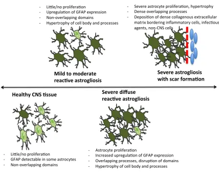
2010). The changes that occur range from mild-‐moderate to severe with glial scar formation (Figure 1.3) (Sofroniew, 2009). Astrogliosis involves proliferation of astrocytes, and can be marked by
increased glial fibrillary acidic protein (GFAP) expression (Sofroniew and Vinters, 2010). Large arrays
of molecular mediators are known to trigger reactive astrogliosis, including growth factors and cytokines. Reactive astrocytes play a pro-‐inflammatory role after CNS damage, while concurrently restricting inflammatory cell spread between areas of CNS damage and healthy tissue, thereby
limiting the inflammatory process (Figure 1.3) (Sofroniew, 2009). There is conflicting evidence for
GFAP-‐ reactive astrocytosis in schizophrenia. The majority of studies show significantly decreased GFAP-‐ reactive astrocytes in the cingulate and prefrontal cortex of patients with schizophrenia (Rajkowska et al., 2002; Webster et al., 2005; Toro et al., 2006), supporting the evidence describing
an overall decreased glial cell density in schizophrenia (Cotter et al., 2001). Contrastingly, a post-‐
mortem study observed increased GFAP levels in the DLPFC of patients with schizophrenia compared to controls, which may be explained by a specific dysfunction in GFAP and/or partial activation of
astrocytes, despite unchanged astrocyte numbers (Feresten et al., 2013). Further studies need to be
undertaken to determine whether changes in GFAP is related to other variables such as duration of illness, stage of illness, and treatment.
2.4.2. S100B as marker of astroglial activation in schizophrenia
S100B is often regarded as a marker of astroglial activation. S100B is a Ca2+ binding protein
expressed by astrocytes in the CNS. At low concentrations, it is thought to regulate proliferation and differentiation of neurons and glia. It also functions as a neurotrophic factor, regulating
synaptogenesis and neurotransmitter function (Rothermundt et al., 2009; Steiner et al., 2011).
Excessive levels of S100B protein promotes an inflammatory response via expression of nitric oxide
synthase or pro-‐inflammatory cytokines leading to neuronal dysfunction and apoptosis (O’Connell et
al., 2013). Increased levels of S100B are seen in the CSF and in serum of patients with schizophrenia
compared to healthy controls (Rothermundt et al., 2004; Rothermundt et al., 2009; Steiner et al.,
2011; O’Connell et al., 2013). This is supported by our previous study which found that higher serum S100B levels was found in females compared to males, possibly due to an increased BMI, which may
result from altered S100B release from adipocytes (O’Connell et al., 2013). It is important to note
and adipocytes, suggesting S100B has a role in altered immune response and metabolic activity (Miuller and Schwarz, 2007; Steiner et al., 2010). Nevertheless, elevated serum levels of S100B correlate closely with CSF levels both in healthy controls and in schizophrenia, suggesting that
increased levels contribute mainly to increased secretion from astrocytes (Rothermundt et al.,
2009). This indicates astroglial activation occuring as part of or in respone to the disease process in schizophrenia, although whether reactive astrogliosis occurs needs to be further deineated.
3.
Dopamine
3.1.Introduction to the dopamine receptor
The unifying feature of all antipsychotics is that they display a varying degree of dopamine antagonism, therefore, inhibit dopamine transmission and dopamine-‐dependent functions. There are four major dopaminergic pathways in the CNS: nigrostriatal, mesolimbic, mesocortical and
tuberoinfundibular (Andén et al., 1964). Each dopamine system is implicated in different CNS
functions; for example, dysfunction in the nigrostriatal pathway is associated with motor deficits in Parkinson’s disease, whereas the mesocortical pathway mediates cognitive processes and the
mesolimbic pathway in reward feedback (Alex and Pehek, 2007). Dopamine is a monoamine
neurotransmitter that exerts its physiological functions via G-‐protein coupled receptors (GPCR) – D1,
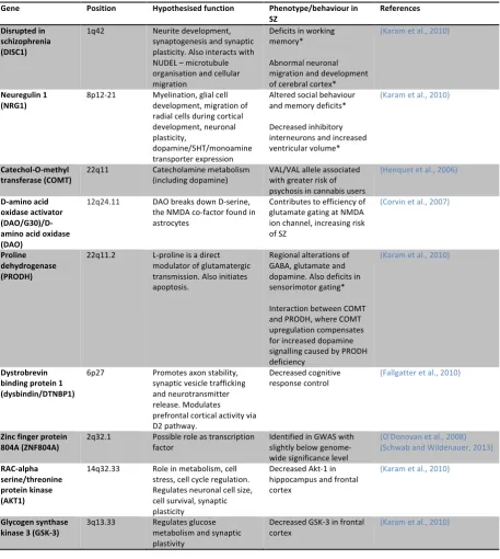
D2, D3, D4, D5 (Table 1.2) (Beaulieu and Gainetdinov, 2011). There are 2 major classes of dopamine
receptors, divided into D1-‐class receptors (includes D1 and D5 subtypes) and D2-‐class receptors (D2,
D3, D4 subtypes) (Tiberi et al., 1991; Clifford et al., 2000). The classification is based on shared
biochemical, pharmacological, structural and genetic properties between each class subtype.
3.2.Dopamine receptor expression in astrocytes, CNS, and PNS
Dopamine receptors are widely expressed in the periphery and CNS. The receptor subtype expressed
in different areas of the CNS is summarised in Table 1.2. D2-‐class receptors are found on both pre-‐
and post-‐synaptic dopaminergic neurons, whereas D1-‐class receptors are found exclusively on post-‐
synaptic dopamine-‐target cells (e.g. GABAergic medium spiny neurons in the striatum) (Sokoloff et
al., 2006; Rankin and Sibley, 2010; Rondou et al., 2010). Previous studies have determined that
dopamine receptors are expressed in astrocytes (Hansson and Ronnback, 1988; Bal et al., 1994;
Zanassi et al., 1999). Direct evidence of D1, D3, D4 and D5 receptors has been shown in astrocytes within the basal ganglia, whereas high expression of D2 receptors is found in astrocytes of the
prefrontal cortex (Table 1.2) (Miyazaki et al., 2004; Mladinov et al., 2010). In the periphery, each
receptor subtype is found in varying degrees in the sympathetic ganglia, adrenal glands, kidneys,
heart, blood vessels and gastrointestinal tract (Beaulieu and Gainetdinov, 2011).
3.3.Dopamine receptor structure and function
3.3.1. Structure
All dopamine receptor subtypes have seven transmembrane-‐spanning domains and are coupled to G-‐proteins, which mediate intracellular signalling. The specific G-‐protein that is coupled to each
subunits that form an inactive trimeric complex, where the α subunit is bound to GDP (Beaulieu and Gainetdinov, 2011). The D1 and D2-‐receptor classes differ in genetic structure. Only D2-‐class
receptors have introns within their genetic coding regions, which leads to the formation of genetic slice variants for each receptor subtype that differ anatomically, pharmacologically and in their signalling properties. Most notably, alternative splicing of D2 receptors leads to the development of 2 major receptor variants – D2S (D2-‐short) and D2L (D2-‐long). Both variants differ in structure, by 29
amino acids in the third intracellular loop, and in receptor function (Clifford et al., 2000; De Mei et
al., 2009). D4 receptors have the highest affinity for antipsychotic drugs, particularly clozapine. Interestingly, polymorphic variants in D4 receptors have varying affinity for clozapine, however,
possession of these variants does not relate to an increased incidence of schizophrenia (Wong and
Van Tol, 2003).
3.3.2. Function
In the central nervous system, dopamine receptor functions include regulation of cognitive function, affect, sleep pattern, feeding, and voluntary movement. Dopamine effect on locomotor activity has
been an area extensively explored, and this is mainly mediated via D1, D2 and D3 receptors (Missale
et al., 1998; Sibley, 1999). These receptors are also involved in reward and reinforcement
mechanisms (Missale et al., 1998; Beaulieu and Gainetdinov, 2011). D1 and D2 receptors in the
prefrontal cortex are critical in learning and executive functions such as maintaining working
memory (Goldman-‐Rakic et al., 2004). As mentioned, clinical effect of antipsychotic drugs on D2
receptors suggest that these receptors are involved in the biochemistry of psychosis (Snyder et al.,
1970; Roth et al., 2004). Little is known about the specific physiological functions of D3-‐5 receptors. In the peripheral nervous system, dopamine has wide-‐ranging effects which include sympathetic,
immune and hormonal regulation (Beaulieu et al., 2015).
3.4.Mechanisms of dopamine signalling
3.4.1. Mechanisms of GPCR signalling
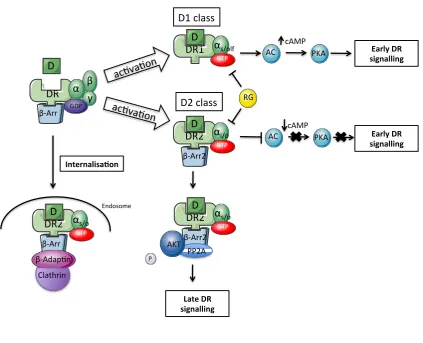
Dopamine receptors are known to be G-‐protein coupled receptors (GPCRs). Upon activation of the trimeric GPCR complex by a dopamine receptor agonist, GDP is released from its guanine nucleotide
binding site on the α-‐subunit, leading to the dissociation of the α-‐subunit from its βγ-‐subunit
counterpart (Figure 1.4) (Beaulieu and Gainetdinov, 2011). This allows the individual subunits to
stimulate separate signalling pathways. Generally, dopamine receptors are classified based on biochemical observations of the receptor’s ability to modulate adenylate cyclase (AC) activity and
subsequent cAMP production. D1-‐class dopamine receptors are known to stimulate AC and
upregulate cAMP production via coupling to Gαs/olf receptors, whereas D2-‐class dopamine receptors
inhibit AC and reduce cAMP production via Gαi/o family of G-‐proteins (Figure 1.4) (Beaulieu and
Gainetdinov, 2011).
3.4.2. Regulation of dopamine receptor signalling
Regulator proteins affect the downstream signalling effects of a DR agonist. The RGS (regulators of
G-‐protein signalling) family of proteins act to reduce G-‐protein signalling (Figure 1.4). This occurs
through GTP hydrolysis, where the RGS protein binds to the GTP-‐bound α-‐subunit via the RH domain
or RGS box, thereby reducing the lifespan of the active GTP-‐α subunit and βy-‐complexes (Dohlman
kinases (GRK)/arrestin system can lead to the promotion of G-‐protein independent signalling or inhibition of the G-‐protein coupled signalling. Desensitisation of the GPCR can occur through phosphorylation by GRK at the -‐COOH terminal of the GPCR, which then recruits and binds the
adaptor protein arrestin, and inhibits GPCR activation (Beaulieu and Gainetdinov, 2011).
Additionally, the GRK system promotes receptor endocytosis through binding of arrestin to clathrin
via adaptor protein β-‐adaptin (Figure 1.4) (Laporte et al., 2002). Alternatively, the GRK/arrestin
system can enhance G-‐protein independent signalling; arrestins serve as signalling mediators that
promote scaffolding of various proteins (Akt, MAPK) to induce cellular signalling (Luttrell and
Lefkowitz, 2002; Shenoy and Lefkowitz, 2005; Beaulieu et al., 2007).
3.4.3. Role of Akt in D2R-‐mediated psychosis and cognitive dysfunction in schizophrenia
Akt, also known as protein kinase B, is a serine/threonine kinase involved in the control of growth factor-‐mediated cell survival and metabolism, including metabolic functions of insulin (see reviews
mentioned in (Chen et al., 2001)). It is activated via phosphatidylinositol-‐mediated signalling to the
plasma membrane and phosphorylation at threonine 308 and serine 473 by phosphatidylinositol-‐
dependent kinase 1 (PDPK1) and rictor-‐mammalian target of rapamycin (mTOR) respectively (Jacinto
et al.,