Int J Clin Exp Pathol 2015;8(1):594-600
www.ijcep.com /ISSN:1936-2625/IJCEP0003103
Original Article
Long noncoding RNA linc-UBC1 is negative prognostic
factor and exhibits tumor pro-oncogenic activity in
gastric cancer
Yiren Hu, Jianghua Pan, Yi Wang, Linjing Li, Yi Huang
Department of General Surgery,Wenzhou People’s Hospital, The Third Clinical College of Wenzhou Medical University, Wenzhou, China
Received October 12, 2014; Accepted December 1, 2014; Epub January 1, 2015; Published January 15, 2015
Abstract: Despite the advances in the management of gastric cancer, the prognosis of advanced gastric cancer remains relatively poor. Thus, it is of urgent need to identify novel prognostic markers and therapeutic targets of gastric cancer. A growing volume of literature has indicated that lncRNAs are differentially expressed in a diverse array of cancer and play an important role in the development of cancer. Linc-UBC1, a recently identified long non-coding RNA, was initially found to be upregulated in bladder cancer. However, the role of linc-UBC1 in gastric cancer remains to be elusive. In this study, we found that linc-UBC1 was significantly upregulated in gastric cancer tissues compared to adjacent normal tissues. Furthermore, high linc-UBC1 expression was associated with lymph-node me-tastasis, tumor size, TNM stage and poorer prognosis. Inhibition of linc-UBC1 suppressed the proliferation, motility and invasion of gastric cancer cells. Our study suggests that linc-UBC1 may represent a novel diagnostic, prognostic biomarker and a potential therapeutic target of gastric cancer.
Keywords: Gastric cancer, long noncoding RNA, linc-UBC1, prognosis
Introduction
Gastric cancer is fourth most common cancer globally and represents the second most fre-quent cause of cancer-related mortality [1]. Advances in early detection techniques, the application of more aggressive surgical strate-gy and postoperative adjuvant therapy have contributed to the survival improvement for early stage gastric cancer, especially in Asian countries [2-4]. Yet, advanced gastric cancer still poses a formidable challenge to the sur-vival of the patients. Therefore, a better under-standing of the molecular mechanism underly-ing the progression of gastric cancer is essen-tial for the development of novel therapeutic strategies.
Recent advances in high-throughput gene sequencing analysis have revealed that the human genome is pervasively transcribed, and the majority (~98%) has limited or no apparent protein-coding capacity, which is defined as noncoding RNAs (ncRNAs) [5, 6]. MiRNAs
(19-25 nucleotides) have dominated the field of ncRNAs research in the past decade in gastric cancer [7-9]. Another class of ncRNAs, long noncoding RNAs, which are commonly defined as transcripts > 200 nucleotides in length, have emerged as another class of vital regulatory RNAs [10]. A growing volume of literature has indicated that lncRNAs are differentially expressed in a diverse array of cancer and play an important role in the development of cancer [11-14].
In this study, we would like to explore the expression pattern of linc-UBC1 and its correla-tion with clinicopathological factors in gastric cancer. Furthermore, the prognostic signifi-cance of linc-UBC1 was assessed. The onco-genic activity of linc-UBC1 was investigated in gastric cancer cell lines.
Materials and methods
Human tissue specimens
The study were undertaken with the under-standing and written consent of each subject. This study was approved by the Ethics Committee of Wenzhou People’s Hospital at Wenzhou Medical University(Wenzhou, China). The study methodologies conformed to the standards set by the declaration of Helsinke. Eighty-five gastric cancer and matched adja-cent normal non-tumor tissues gastric tissues (> 3 cm away from tumor) were obtained from
patients who underwent surgical resection of primary gastric cancer between 2005 and 2009 at Wenzhou People’s Hospital.The diag-nosis was based on histopathological examina-tion by two experienced pathologists of Wenzhou People’s Hospital. The tissues were obtained before chemotherapy and radiation therapy. No selection bias was introduced in sample collection.Upon removal of the surgical specimen, each sample was immediately fro-zen in liquid nitrogen and stored at -80°C prior to RNA isolation and qRT-PCR analysis. All cases were classified according to the World Health Organization’s pathological classifica-tion of tumors.
Cell lines
[image:2.612.92.517.69.452.2]linc-UBC1 and gastric cancer
emistry and Cell Biology of the Chinese Academy of Sciences (Shanghai, China). Cells were cultured in DMEM or RPMI 1640 medium (Invitrogen, Carlsbad, CA, USA) supplemented with 10% fetal bovine serum (FBS) as well as 100 U/ml penicillin and 100 μg/ml streptomy-cin (Invitrogen, Carlsbad, CA, USA). All cells were maintained at 37°C under an atmosphere of 5% CO2. All cell lines have been passaged for fewer than 6 months.
RNA extraction and quantitative real-time PCR
RT-qPCR was performed to determine the expression of linc-UBC1 in gastric cancer tis-sues and cells. Total RNA of cancer tistis-sues or cultured cells was extracted with Trizol reagent (Invitrogen, Carlsbad, CA, USA) according to the manufacturer’s protocol. RNA was reversed transcribed into cDNAs using the Primer-ScriptTM one step RT-PCR kit (TaKaRa, Dalian, China). The resulting cDNA was amplified by PCR using linc-UBC1 and GADPH (endogenous control) specific primers with real-time RT-PCR using the SYBR® Premix Dimmer Eraser kit (TaKaRa, Dalian, China). The quantitative real-time polymerase chain reaction (qRT-PCR) was performed on ABI 7500 system (Applied Bio- systems, CA, USA) according to the
manufac-Cell proliferation assays were conducted using the CCK-8 assay kits as described by the manu-facturer. EdU immunofluorescence staining was performed using an EdU kit (Roche, Mannheim, Germany).
Wound-healing assay
48 h after transfection, gastric cancer cells were scratched in the monolayer and cultured in normal conditions. The mobilized distances were measured at 0, 16 h after scratching for HGC-27 and at 0, 24 h after scratching for SGC-7901, respectively.
Cell invasion assay
48 h after transfection, 1 × 10⁵ cells suspend-ed in 200 μL serum-free msuspend-edia were addsuspend-ed into the upper chamber of an insert pre-coated with 50 μL Matrigel (8.0 μm, Millipore, MA). The chambers were then incubated in culture medi-um supplemented with 10% FBS in the bottom chambers for 24 h before examination. The cells on the upper surface were scraped and washed away, whereas the cells on the lower surface were fixed with 20% methanol and stained with 0.2% crystal violet. Finally, cells were counted under a microscope and the
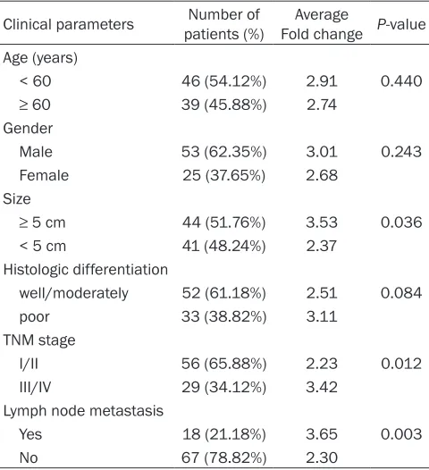
rela-Table 1. Correlation between linc-UBC1 expression and clinicopathological characteristics of gastric cancer
Clinical parameters patients (%)Number of Fold change Average P-value Age (years)
< 60 46 (54.12%) 2.91 0.440 ≥ 60 39 (45.88%) 2.74
Gender
Male 53 (62.35%) 3.01 0.243 Female 25 (37.65%) 2.68
Size
≥ 5 cm 44 (51.76%) 3.53 0.036 < 5 cm 41 (48.24%) 2.37
Histologic differentiation
well/moderately 52 (61.18%) 2.51 0.084 poor 33 (38.82%) 3.11
TNM stage
I/II 56 (65.88%) 2.23 0.012 III/IV 29 (34.12%) 3.42
Lymph node metastasis
Yes 18 (21.18%) 3.65 0.003 No 67 (78.82%) 2.30
turer’s instructions. Specific primers for GADPH: 5’-GTCAACGGATTTGGTCTGTATT- 3’ (forward), 5’-AGTCTTCTGGGTGGCAG- TGAT-3’ (reverse); linc-UBC1: 5’-CCTG- CTTGGAAACTAATGACC-3’ (forward), 5’- AGGCTCAACTTCCCAGACTCA-3’ (reverse). Relative expression values were calculat-ed by the 2-ΔΔCt method using GAPDH as a normalizer.
RNA interference
The nucleotide sequences targeting linc-UBC1: (#1 CCUGUCUACAGACUGAAUATT, #2 CCGGAACAAAUGGCUUCAUTT), and nontargeting siRNAs (UUCUCCGAACGU- GUCACGUTT) were purchased from GenePharma (Shanghai). Cells were grown on six-well plate to 60% confluency and transfected with 75 nM siRNA as well as Lipofectamine 2000 (Invitrogen, Carlsbad, CA, USA) according to the man-ufacturer’s instructions.
[image:3.612.91.331.96.359.2]Figure 2. Linc-UBC1 knockdown attenuates gastric cancer cell proliferation. A. Linc-UBC1 expression level was confirmed by qRT-PCR in multiple gastric cancer cell lines. Mean ± S.D. are shown (n = 3). NC denotes siRNA hav-ing no homology to any known mammalian genes as a negative control. B, C. Linc-UBC1 knockdown attenuated gastric cancer cell line HGC-27 and SGC-7901 proliferation as determined by CCK8 assay. Mean ± S.D. are shown (n = 3). D. Linc-UBC1 knockdown decreased gastric cancer cell line HGC-27 and SGC-7901 in S phase. Blue color represents the nucleus and red color indicates S phase cells (EdU positive). E. Histological analysis of the percent of EdU positive cells in negative control and linc-UBC1 knockdown in gastric cancer cell lines. Mean ± S.D. are shown (n = 3). *, P < 0.05; **, P < 0.01.
[image:4.612.93.518.452.697.2]Representa-linc-UBC1 and gastric cancer
tive number was calculated. Experiments were independently repeated in triplicate.
Statistical analysis
All quantitative data are presented as the mean ± standard deviation (S.D.) from at least three independent experiments. The statistical anal-ysis was performed with SPSS 17.0 software. Unless otherwise specified, the difference between two groups was analyzed with Student’s t test. Metastasis-free survival and overall survival were evaluated using the Kaplan-Meier method, and the long-rank com-parison was carried out to assess differences between stratified survival groups using the median value as the cutoff. The correlation between linc-UBC1 expression and clinico- clinico-pathological characteristics were analyzed using Pearson Chi-square test. A two-sided P value of less than 0.05 was considered to be statistically significant.
Results
Expression of linc-UBC1 in gastric cancer cell lines
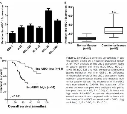
To explore the role of linc-UBC1 in gastric can-cer, we first examined the expression levels of linc-UBC1 in gastric cancer cell lines. As shown in Figure 1A, gastric cancer cell lines expressed higher levels of linc-UBC1 than normal gastric epithelium cell line (GES-1). The expression of linc-UBC1 was most significantly higher in HGC-27 (P < 0.01) and SGC-7901 (P < 0.01) cells. HGC-27 cell line was established from the met-astatic lymph node of gastric cancer patients [16], and SGC-7901 cell was associated with gastric cancer metastasis and invasion, which suggested that the enhanced expression of linc-UBC1 strongly correlated with the migra-tion and invasion of gastric cancer cells. Linc-UBC1 is upregulated in gastric cancer tis-sues
The expression of linc-UBC1 in 85 pairs of matched gastric tumor tissues was analyzed utilizing RT-qPCR. We found that linc-UBC1 was significantly upregulated in gastric cancer
tis-sues compared to non-tumor gastric tistis-sues (Figure 1B). To gain further insights into the observation mentioned above, we analyzed the correlation between linc-UBC1 expression and patient clinicopathological characteristics. As shown in Table 1, linc-UBC1 expression was irrelevant with age, sex and tumor differentia-tion. However, high linc-UBC1 expression was associated with lymph-node metastasis (P < 0.01), tumor size (P = 0.036, P < 0.05) and TNM stage (P = 0.012, P < 0.05).
Enhanced expression of linc-UBC1 was associ-ated with the poor prognosis in gastric cancer
We would like to explore whether linc-UBC1 expression level correlated with outcome of gastric cancer patients after gastrectomy. The median ratio of relative linc-UBC1 expression in gastric cancer (2.81) was used as the cutoff value to stratify high-linc-UBC1 group and low-linc-UBC1-group. Kaplan-Meier survival analy-sis and log-rank tests were conducted. Remarkably, patients with higher linc-UBC1 expression level had poorer overall survival (Figure 1C, P < 0.01).
Linc-UBC1 promoted the proliferation and in-vasion of gastric cancer cells in vitro
As demonstrated in Table 1, higher linc-UBC1 expression was associated larger tumor size and lymph node metastasis, suggesting that linc-UBC1 might promote the proliferation and invasion of gastric cancer. To further assess the biological role in gastric cancer, we knocked down linc-UBC1 expression in HGC-27 and SGC-7901 cells using small interfering RNA (Figure 2A). Cell-counting kit-8 assays indicat-ed that linc-UBC1 depletion resultindicat-ed in decreased tumor cell proliferation in gastric cancer cell line HGC-27 and SGC-7901 (Figure 2B, 2C). Furthermore, the percentage of Edu positive decreased significantly after linc-UBC1 knockdown (Figure 2D, 2E). All these data linc-UBC1 promoted the proliferation of gastric can-cer cells.
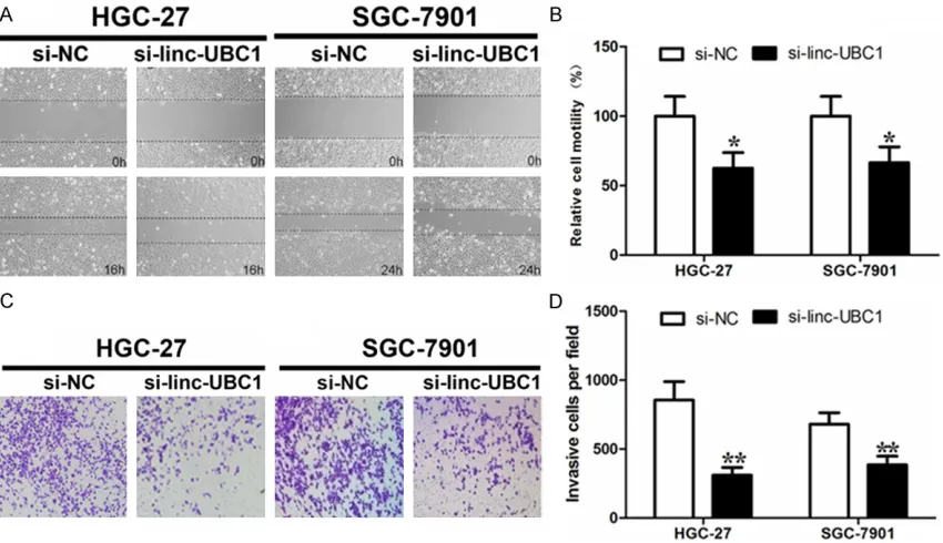
Cancer motility and invasion ability were vital for cancer progression and metastasis. We next examined whether linc-UBC1 had a
tional role in facilitating gastric cancer motility and invasion. We performed wound-healing assay and transwell assay with linc-UBC1 knockdown. As illustrated in Figure 3A and 3B, decreased cell motility was observed in both gastric cancer cell lines with linc-UBC1 deple-tion. Similarly, linc-UBC1 knockdown decreased gastric cancer invasion (Figure 3C, 3D). Taken together, linc-UBC1 may promote the prolifera-tion, motility and invasion of gastric cancer.
Discussion
In recent years, the research on long noncoding RNA has drawn more and more attention. Although thousands of lncRNAs were identified, only a small fraction of them have been charac-terized [5]. However, the current studies have demonstrated that lncRNAs are involved in the pathogenesis of cancer [11-15].
Although previous studies have identified a few cancer-associated lncRNAs, only a small num-ber of gastric cancer-associated lncRNAs have been characterized. Wang et al [17] demon-strated that Long noncoding RNA MRUL con-tributed to the multi-drug resistance of gastric cancer. Zhang et al [18] revealed that lncRNA ANRIL indicates a poor prognosis of gastric cancer. CCAT1, a c-Myc activated lncRNA, pro-motes the progression of gastric cancer [19]. In this study, we found that linc-UBC1 was gen-erally upregulated in gastric cancer and its overexpression correlates with lymph node metastasis, tumor size, TNM stage and a poor-er prognosis of patients with gastric cancpoor-er, which was in agreement with the oncogenic role of linc-UBC1 in bladder cancer [15]. The functional studies showed linc-UBC1 depletion decreased the gastric cancer cell proliferation, motility and invasion. The data highlighted the role of linc-UBC1 in gastric cancer.
In summary, we showed that linc-UBC1 may be gastric cancer-specific lncRNA and may play an important role in the development of gastric cancer. Our study may facilitate the develop-ment of lncRNA-directed diagnostics and thera-peutics against cancers.
Disclosure of conflict of interest
None.
Address correspondence to: Dr. Yiren Hu, De- partment of General Surgery, Wenzhou People’s
Hospital, The Third Clinical College of Wenzhou Medical University, 57 Canghou Road, Wenzhou 325000, People’s Republic of China. Fax: +86-577-88059166; E-mail: [email protected]
References
[1] Shah MA, Kelsen DP. Gastric cancer: a primer on the epidemiology and biology of the disease and an overview of the medical management of advanced disease. J Natl Compr Canc Netw 2010; 8: 437-47.
[2] Allum WH, Blazeby JM, Griffin SM, Cunningham D, Jankowski JA, Wong R. Guidelines for the management of oesophageal and gastric can-cer. Gut 2011; 60: 1449-72.
[3] Saka M, Morita S, Fukagawa T, Katai H. Present and future status of gastric cancer surgery. Jpn J Clin Oncol 2011; 41: 307-13.
[4] Tan HT, Low J, Lim SG, Chung MC. Serum auto-antibodies as biomarkers for early cancer de-tection. FEBS J 2009; 276: 6880-904. [5] Guttman M, Amit I, Garber M, French C, Lin MF,
Feldser D, Huarte M, Zuk O, Carey BW, Cassady JP, Cabili MN, Jaenisch R, Mikkelsen TS, Jacks T, Hacohen N, Bernstein BE, Kellis M, Regev A, Rinn JL, Lander ES. Chromatin signature re-veals over a thousand highly conserved large non-coding RNAs in mammals. Nature 2009; 458: 223-7.
[6] Kapranov P, Cheng J, Dike S, Nix DA, Duttagupta R, Willingham AT, Stadler PF, Hertel J, Hackermüller J, Hofacker IL, Bell I, Cheung E, Drenkow J, Dumais E, Patel S, Helt G, Ganesh M, Ghosh S, Piccolboni A, Sementchenko V, Tammana H, Gingeras TR. RNA maps reveal new RNA classes and a possible function for pervasive transcription. Science 2007; 316: 1484-8.
[7] Wu Q, Yang Z, An Y, Hu H, Yin J, Zhang P, Nie Y, Wu K, Shi Y, Fan D. MiR-19a/b modulate the metastasis of gastric cancer cells by targeting the tumour suppressor MXD1. Cell Death Dis 2014; 5: e1144.
[8] Ma J, Liu J, Wang Z, Gu X, Fan Y, Zhang W, Xu L, Zhang J, Cai D. NF-kappaB-dependent microR-NA-425 upregulation promotes gastric cancer cell growth by targeting PTEN upon IL-1β induc-tion. Mol Cancer 2014; 13: 40.
[9] He XP, Shao Y, Li XL, Xu W, Chen GS, Sun HH, Xu HC, Xu X, Tang D, Zheng XF, Xue YP, Huang GC, Sun WH. Downregulation of miR-101 in gastric cancer correlates with cyclooxygen-ase-2 overexpression and tumor growth. FEBS J 2012; 279: 4201-12.
[10] Kugel JF, Goodrich JA. Non-coding RNAs. key regulators of mammalian transcription. Trends Biochem Sci 2012; 37: 144-151.
non-linc-UBC1 and gastric cancer
coding RNA-LET by histone deacetylase 3 con-tributes to hypoxia-mediated metastasis. Mol Cell 2013; 49: 1083-96.
[12] Huang JF, Guo YJ, Zhao CX, Yuan SX, Wang Y, Tang GN, Zhou WP, Sun SH. Hepatitis B virus X protein (HBx)-related long noncoding RNA (ln-cRNA) down-regulated expression by HBx (Dreh) inhibits hepatocellular carcinoma me-tastasis by targeting the intermediate filament protein vimentin. Hepatology 2013; 57: 1882-92.
[13] Gupta RA, Shah N, Wang KC, Kim J, Horlings HM, Wong DJ, Tsai MC, Hung T, Argani P, Rinn JL, Wang Y, Brzoska P, Kong B, Li R, West RB, van de Vijver MJ, Sukumar S, Chang HY. Long non-coding RNA HOTAIR reprograms chromatin state to promote cancer metastasis. Nature 2010; 464: 1071-6.
[14] Prensner JR, Chen W, Iyer MK, Cao Q, Ma T, Han S, Sahu A, Malik R, Wilder-Romans K, Navone N, Logothetis CJ, Araujo JC, Pisters LL, Tewari AK, Canman CE, Knudsen KE, Kitabayashi N, Rubin MA, Demichelis F, Lawrence TS, Chinnaiyan AM, Feng FY. PCAT-1, a Long Noncoding RNA, Regulates BRCA2 and Controls Homologous Recombination in Cancer. Cancer Res 2014; 74: 1651-60.
[15] He W, Cai Q, Sun F, Zhong G, Wang P, Liu H, Luo J, Yu H, Huang J, Lin T. linc-UBC1 physically as-sociates with polycomb repressive complex 2 (PRC2) and acts as a negative prognostic fac-tor for lymph node metastasis and survival in bladder cancer. Biochim Biophys Acta 2013; 1832: 1528-37.
[16] Akagi T, Kimoto T. Human cell line (HGC-27) de-rived from the metastatic lymph node of gas-tric cancer. Acta medica Okayama 1976; 30: 215-219.
[17] Wang Y, Zhang D, Wu K, Zhao Q, Nie Y, Fan D. Long noncoding RNA MRUL promotes ABCB1 expression in multidrug-resistant gastric can-cer cell sublines. Mol Cell Biol 2014; 34: 3182-93.
[18] Zhang EB, Kong R, Yin DD, You LH, Sun M, Han L, Xu TP, Xia R, Yang JS, De W, Chen JF. Long noncoding RNA ANRIL indicates a poor progno-sis of gastric cancer and promotes tumor growth by epigenetically silencing of miR-99a/ miR-449a. Oncotarget 2014; 5: 2276-92. [19] Yang F, Xue X, Bi J, Zheng L, Zhi K, Gu Y, Fang